

Abstract
This article reports an innovative and unprecedented use of Holmium: YAG laser in the extraction of a foreign body impacted in the upper oesophagus without complications. Hence, Holmium-YAG laser can be a safe, efficient and successful aid to fragment the impacted foreign bodies to assist in their removal.
Keywords: Oesophagus, Foreign body removal, Oesophageal foreign body, Lamb bone, Holmium-YAG laser
Introduction
Foreign body in the oesophagus is not an uncommon clinical condition for an Otolaryngologist in the outpatient department or in the emergency room and is considered to be a serious malady, both in adults and children, due to the risk of possible complications (oesophageal perforation, mediastinitis, fistulisation and airway obstruction) with high mortality and morbidity [1]. Therefore, a rapid and accurate diagnosis, along with subsequent treatment is necessary. In 18–20% of cases, endoscopic or surgical removal is promptly required [2].
Case Report
The case report has been submitted after discussion and review of the case and the draft at the Institutional Ethics committee (IEC).
A 30-year-old male presented to the Emergency Department of a tertiary care centre, with complaints of odynophagia and dysphagia which had begun after a lamb meal.
Antero-posterior, lateral neck and upper chest plain radiograph (Fig. 1) revealed the presence of a radio-opaque foreign body in the upper cervical oesophagus at the level of 5th and 6th cervical vertebra.
Fig. 1.

Anteroposterior and lateral view of neck and chest showing radio opaque foreign body in the oesophagus at the level of 5th and 6th vertebra
The flexible nasopharyngo-laryngoscopy showed pooling of saliva in the pyriform sinuses. A rigid eosophagoscopy approach, under general anaesthesia, was planned. During the procedure, using an oval 30 cm oesophagoscope (12 × 16 mm diameter), the foreign body was identified (Fig. 2) at 18 cm from the upper incisors, lying horizontally in the lumen of the upper oesopahogus like a shelf with its sharp ends impacted into the oesophageal mucosa. The removal manoeuvres failed, due to the shape, the orientation, and the dimensions of this particular foreign body. Any vigorous attempt to remove the foreign body might have led to oesophageal perforation causing life-threatening consequences, therefore, procedure was abandoned.
Fig. 2.

A Rigid oesophagoscope view showing foreign body (lamb bone) impacted in the lumen of oesophagus; B Foreign body (lamb bone) fragmented into two pieces using Holmium: YAG laser
After 24 h, using a dual-channel Gastrointestinal endoscope (Olympus CV-150), the fibre-optic delivery system of Holmium-YAG laser was introduced proximal to the lamb bone and fragmented with energy pulse setting of 0.5 J at 20 Hz (10 watts), leaving a thin bridge of bone between the two pieces which was not accessible. Afterwards, gastrointestinal endoscope was removed and a rigid oesophagoscope was engaged. The leftover thin bridge in the shelf of the lamb bone was broken into two fragments (Fig. 2) using Chevalier-Jackson foreign body removal forceps and dislodged from the oesophageal mucosa. Two fragments were removed separately using the same forceps (Fig. 3).
Fig. 3.
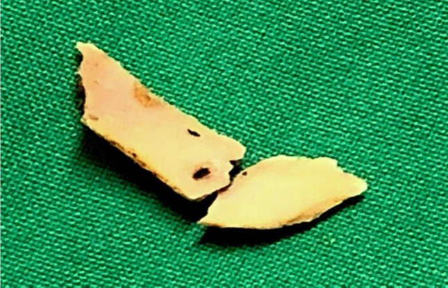
Two fragments of foreign body removed separately using oesophageal foreign body forceps
Post-operative course was uneventful and the oral diet was resumed 8 h after the procedure. The patient was discharged on 2nd post-operative day without any evidence of dysphagia or mediastinitis. Patient was under regular follow-up and there was no clinical evidence of esophageal stricture or obstruction 3 months post-operatively.
Discussion
Although, 80–99% of the ingested foreign objects pass uneventfully through the gastrointestinal tract [2]. Endoscopic and surgical interventions are required in 20 and 1% of cases, respectively [1].
Swallowing of foreign body is more commonly encountered in children and in specific adult risk groups [1]. Structural or functional abnormalities of the oesophagus represent the major risk factors. The foreign body usually lies close to one of the 3 oesophageal anatomical constrictions: the cricopharyngeal ring, the aortic arch narrowing or the oesophagogastric junction. It is also interesting to note that the foreign body position in the oesophagus differs with age [1]. The most common types of impacted objects in the oesophagus are food-related foreign bodies, such as bones, meat bolus, nuts and seeds, or coins, pins and toys. The accidental ingestion of artificial dentures is most common in the elderly [1].
Clinically, oesophageal foreign body ingestion may present with odynophagia, dysphagia, diffuse chest pain, and laryngeal irritation. To confirm the diagnosis of the foreign body of the upper digestive tract, radiological assessment is necessary. Plain radiograph of neck and chest are a very important diagnostic tool, especially in defining the location of the foreign body. A barium-swallow X-ray study could be useful in cases of non-radio-opaque foreign body, but due to possible barium aspiration and\or irritation of the damaged oesophageal mucosa, this procedure is avoided [1]. CT scans can be used to confirm the presence and to study the location of foreign body or to evaluate any eventual damage to the neighbouring structures [2]. In our case, neck X-ray lateral view, already confirmed the presence and the location of the foreign body, and, therefore, no other radiological procedure was necessary.
Endoscopy is the preferred method for oesophageal foreign body extraction with a reported success rate of 83% [3]. Today, either rigid or flexible endoscopy, performed under general anaesthesia or conscious sedation, respectively, is considered to be safe and represent effective methods. For the management of sharp and penetrating foreign body, rigid endoscopy is often the treatment of choice.
The foreign body, in our patient, was located in the upper cervical oesophagus, the site usually presents the greatest difficulties; and may need involvement of otorhinolaryngologist, gastroenterologist and radiologist. A laparoscopic approach is mandatory only in those cases in which an endoscopic approach has failed [3].
Holmium: YAG laser is the workhorse for ureteric and renal calculi fragmentation in lithotripsy. Holmium: YAG laser is a solid-state, pulsed-dye laser that emits light at 2100 nm and the average power from 5 to 80 watts, depending on the intended application.
The laser has been used previously in the treatment of foreign bodies impacted in rectum and the tracheo-bronchial tree to fragment foreign bodies in small pieces [4]. It also has been used to fragment denture in the stomach [5]. We have found Holmium-YAG laser helpful in our patient, and we believe that it may be the first report of the use of the above laser to fragment a lamb bone in the oesophagus.
Conclusion
Management of oesophageal foreign body requires a multidisciplinary approach (gastroenterologist, radiologist, otorhinolaryngologist or even a thoracic surgeon); in the case described herewith, rigid endoscopy and Holmium: YAG laser has been the safe and successful method for the impacted lamb bone extraction. We believe that, with adequate precautions, laser can be proficiently employed in removal of impacted bony foreign body from oesophagus without complication.
Funding
There are no funding sources for the underlying scientific work.
Declarations
Conflict of interest
The authors declares that they have no conflict of interest.
Footnotes
Publisher's Note
Springer Nature remains neutral with regard to jurisdictional claims in published maps and institutional affiliations.
Contributor Information
N. Vishnu Swaroop Reddy, Email: vishnureddyn@hotmail.com
Shubhrendu Shekhar, Email: shekhar.shubho@gmail.com.
Manoj Sharma, Email: dr.sharmamanoj@yahoo.com.
P. B. S. S. Raju, Email: drbhavanis@yahoo.com
References
- 1.Al-Qudah A, Daradkeh S, Abu-Khalaf M. Esophageal foreign bodies. Eur J Cardio Thorac Surg. 1998;13:494–499. doi: 10.1016/S1010-7940(98)00068-2. [DOI] [PubMed] [Google Scholar]
- 2.Henderson CT, Engel J, Schlesinger P. Foreign body ingestion: review and suggested guidelines for management. Endoscopy. 1987;19(2):68–71. doi: 10.1055/s-2007-1018238. [DOI] [PubMed] [Google Scholar]
- 3.Furihata M, Tagaya N, Furihata T, Kubota K. Laparoscopic removal of an intragastric foreign body with endoscopic assistance. Surg Laparosc Endosc Percutan Tech. 2004;14:234–237. doi: 10.1097/01.sle.0000136682.69871.db. [DOI] [PubMed] [Google Scholar]
- 4.Hayashi AH, Gillis DA, Bethune D, Hughes D, O’Neil M. Management of foreign-body bronchial obstruction using endoscopic laser therapy. J Paediatr Surg. 1990;25(11):1174–1176. doi: 10.1016/0022-3468(90)90757-Z. [DOI] [PubMed] [Google Scholar]
- 5.Lam YH, Ng EKW, Chung SCS, Li AKC. Laser-assisted removal of a foreign body impacted in the esophagus. Lasers Surg Med. 1997;20(4):480–482. doi: 10.1002/(SICI)1096-9101(1997)20:4<480::AID-LSM16>3.0.CO;2-F. [DOI] [PubMed] [Google Scholar]