Abstract
Angina bullosa haemorrhagica (ABH) is characterized by the recurrent appearance of haemorrhagic bullae on the oropharyngeal mucosa which rupture spontaneously leading to complete recovery within a weeks’ time without any scarring. We report the clinical features of six cases of ABH. A cross-sectional observational study was performed. A total of six cases of ABH fulfilling the Ordioni et. al. criteria for diagnosis of ABH were enrolled.The age of our patients were 65, 25, 20, 35, 55 and 48 years. Four of them were females (67%), whereas two were males (33%).Retromolar trigone involvement was most common.The chief complaint in all was reddish bulla(e) in the oral cavity of 1 day duration. Five of the patients had solitary lesions, while one had multiple lesions. The lesions measured from 1 to 3 cm in diameter. Complete blood counts and clotting factors were normal in all patients. All cases healed within a week’s time. ABH is not a very common disorder encountered by ENT surgeons, dermatologists, general practitioners, and the lack of knowledge of its normal presentation can lead to unnecessary anxiety and incorrect treatment. The typical hemorrhagic bulla(e) usually erupt after eating hard, hot, or spicy food. These lesions heal spontaneously and treatment is not necessary except for reassurance and mild anxiololytics.
Keywords: Angina bullosa haemorrhagica, Reassurance, Oral mucosa blisters, Coagulation profile
Introduction
Angina bullosa haemorrhagica (ABH) was described in 1967 by Badham as an alteration causing recurrent haemorrhagic bullae of the oropharyngeal mucosa [1].This entity is not limited to the pharynx but may occur anywhere in the entire oral cavity [2]. ABH is characterized by sudden appearance of hemorrhagic bulla(e) not attributable to blood dyscrasias, vesiculobullous disorders, systemic diseases, or any known causes. They either present with mild symptoms of pain, tingling, foreign body sensation or are asymptomatic. The lesions heal spontaneously without scarring within a week’s time. It tends to occur in adults between the fifth and the seventh decades of life, with a mean age of 55.4 years [2]. The exact cause and pathogenesis are not clear. Trauma caused by hot drink, restorative dentistry, periodontal therapy, dental procedure, shouting, diabetes and inhaled long-term steroids are found to have variable degree of association with this entity [3].
The differential diagnosis includes dermatoses that present as mucocutaneous bullous lesions and bloodborne diseases (leukaemia, thrombocytopenia, von Willebrand disease) [3]. Although the diagnosis of ABH can be based on clinical examination, history, and follow-up, there is significant variability in the diagnostic approach and management of the disease in the literature [2]. Ordioni et al. [4] proposed a nine point diagnostic criteria for ABH, the combination of at least six of their nine diagnostic criteria, with criteria I and II being systematically present, should lead to the diagnosis of ABH.Treatment is not necessary in most cases. Nonsteroidal anti-inflammatory drugs (NSAIDs), antimicrobial mouth washes, and anxiolytics can be given in some cases for symptomatic relief. We present our case series of six patients with ABH.
Materials and Methods
This is an observational case series of the patients attending our Outpatient/Emergency Department of ENT. Patients that met the diagnostic criteria for ABH proposed by Ordioni et al. (2019) were included.The nine points in the diagnostic criteria of Ordioni et.al. are (I) clinically notable haemorrhagic bulla or erosion with a history of bleeding in the oral mucosa; (II) exclusively oral or oropharyngeal localization; (III) palatal localization; (IV) triggering event or promoting factor (food intake); (V) recurrent lesions; (VI) favourable evolution without a scar within a few days; (VII) painless lesion, or a tingling or burning sensation; (VIII) normal platelet count and coagulation test results; (IX) negative DIF results.
As proposed by Ordioni et.al, patients were included only when they met at least six of their criteria with criteria I and II being systematically present. No patient underwent DIF.
All relevant details of patients, including triggering factors, clinical features, and treatment, are presented.
No ethical approval was necessary for this observational case series.
Results and Observations
A total of six cases seen from January 2021 to January 2023 are being reported. The details of the cases are given in Table 1.
Table 1.
Characteristics of patients
| Age/sex | Triggering factors | Symptoms | Max diameter of largest lesion | Area of involvement | Complete blood counts and coagulation studies | No of lesions | Treatment |
|---|---|---|---|---|---|---|---|
| 65/M | Intake of hot tea about half an hour back | Foreign body sensation with mild pain | 1.5 cm | Hard palate | Normal | Single |
Reassurance Spontaneous rupture on the same day and healing within next 4 days |
| 25/F | None | Mild pain | 1 cm | Retromolar trigone | Normal | Single |
Reassurance Spontaneous rupture on second day and healing within next 3 days |
| 35/M | Intake of hot spicy potato chips about an hour back | Foreign body sensation | 1 cm | Retromolar trigone | Normal | Single |
Reassurance Spontaneous rupture on the same day and healing within next 4 days |
| 55/F | Intake of traditional Hot spicy food of Kashmir(Wazwan) about 15 min back | Foreign body sensation with mild discomfort | 1 cm | Uvula | Normal | Single |
Reassurance Spontaneous rupture on the same day and healing within next 3 days |
| 20/F | None | Foreign body sensation and mild pain | 1 cm | Soft and hard palate | Normal | Multiple |
Reassurance Spontaneous rupture on the same day and healing of all lesions within next 5 days Anxiolytics given |
| 48/F | None | Tingling sensation | 3 cm | Retromolar trigone | Normal | Single |
Had to be punctured next day and spontaneous healing within next 5 days Anxiolytics given |
The age of our patients were 65, 25, 20, 35, 55 and 48 years. Four of them were females(67%), whereas two were males(33%).The chief complaint in all was reddish bulla(e) in the oral cavity of 1 day duration. Foreign body sensation, tingling sensation, mild pain /discomfort, and anxiety were other accompanying symptoms.
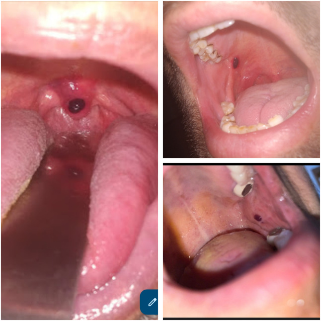
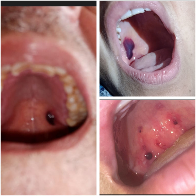
On examination, a hemorrhagic bulla were found in different areas of oral cavity as described in Table 1 and shown in Figs. 1 and 2. Five of the patients had solitary lesions, while one had multiple lesions. The lesions measured from 1 to 3 cm in diameter. There was no history of similar lesions in past or any haematological disorder, leukemia, drug reaction, diabetes, long use of inhalers, frequent epistaxis and gastrointestinal (GI) bleeding. Complete blood counts and clotting factors were normal in all patients.
Fig. 1.

Showing one ABH blisters on Uvula (Left side) and one on retromolar trigones(Right upper and lower pictures)
Fig. 2.

Showing one ABH blisters on Hard palate (Left side picture), right upper one picture showing blister on retromolar trigone and right lower one showing multiple blisters on Hard and Soft Palate
A clear-cut triggering factor was seen in three patients(50%) in the form of appearance of bulla within 15 min to 60 min after drinking hot tea and eating spicy food in the form of hot potato and traditional local cuisine(WAZWAN). All patients were reassured, and anxiolytics were given to two patients in the form of Alprazolam (0.5 mg) once at bedtime for 3 days.
The lesion persisted till evening of the same day in four patients and till next morning in one patient and then spontaneously ruptured in all five patients, leading to ulcer formation which eventually healed in next 2–3 days with no scarring. In one patient, the haemorrhagic bulla persisted till next day evening causing foreign body sensation/mild pain and extreme anxiety and it was eventually punctured at evening of next day.
Discussion
ABH is often described as rare [2] while some authors suggest that ABH is probably underdiagnosed due to the lack of knowledge on the part of clinician, its self-limited nature and spontaneously resolution without scarring [2].
The other terms that have been proposed to define this pathology without consensus among the scientific community are stomatopompholyx hemorrhagica, localized oral purpura, hemorrhagic bullous stomatitis, and traumatic or recurrent oral hemophlyctenosis [4]. The term ABH is the most common and appropriate term used for this condition [4].
A previous history of trauma has been the main finding reported in most cases (> 80%), and it seems to be the most relevant etiological agent [2]. The trauma leads to a loss of cohesion between the epithelium and connective tissue. A possible fragility of the vasculature and/or elastin and/or collagen in some patients could favor subepithelial hemorrhages. However, further studies are needed to confirm this hypothesis [2].
Precipitating factors preceding bulla were seen in three (50%) of our patients in the form of consumption of hot tea and hot spicy food just 15 min to 60 min before the eruption of bulla. Ordioni et al. [4] in their systematic review found that 80% of cases were associated with mastication-related precipitating factors (hard, hot, spicy food, etc.). John Lennon Silva-Cunha et al. [2] also found trauma associated with hard, hot, or spicy food intake as the most frequently reported factor seen in 52.2% of their cases.
No precipitating factor was seen in three(50%) of our patients, which is in accordance with one of the largest series of 30 patients, whereby no precipitating factor was seen [5]. John Lennon Silva-Cunha et al. [2] in their series also could not identify any triggering event or promoting factor in (47.8%) of their cases.
The age of our patients ranged from 20 to 65 years and four of them were females (67%). The age range in systematic review by Ordioni et al. [4] was from 13 to 86 years with male predominance, while slight predominance of females was seen in study by Radovan Slezák [3].Equal distribution with a 1.1:1 male-to-female ratio was seen in the study by john Lennon Silva-Cunha et al. [2]
The retromolar trigone was the most frequently affected site, followed by hard/soft palate and uvula. Ordioni et al. [4] in their review found palate the most frequently affected site, followed by buccal mucosa, pharynx, and labial mucosa area while uvula as the least involved site (3%).
The lesions in our series measured from 1 to 3 cm in diameter, while the lesions in a review by Ordioni et al. [4] varied from 3 mm to 3.5 cm. Size of lesions in one report by Patigaroo et al. [3] ranged from 0.5 to 2 cm, while in another by Horie et al. [6] ranged from 1 cm to 3.5 cm.
These lesions are solitary and are exceptionally multiple. Five of our lesions were solitary and one case had multiple lesions. Patigaroo et al. [3] found a case with multiple lesions Horie et al. [6] and Adrine Maciel da Rosa et al. [7]. in their studies, found all lesions to be solitary. Ordioni et al. [4] in their review also found majority have solitary lesions while multiple lesions were are also seen in their systematic review.
Patients usually present with a hemorrhagic bulla preceded or not by a burning or tingling or foreign body sensation, as seen in our six cases. These bullae break after a few minutes or hours to give way to an erosion, which is often painless or mildly painful or even asymptomatic and heals without scarring within a few days. The isolated nature, rapid healing, and rare recurrence are clues that point to ABH. The characteristic clinical history allows diagnosis confirmation without the need of a biopsy.
Complete blood counts and coagulation profile were normal in all six patients, which is in accordance with a large systematic review by Ordioni et al. [4] Direct immunofluorescence (DIF) for IgA, IgG, IgM, and fibrin though not done in our cases was negative in almost all cases reviewed. ABH has to be differentiated from blood dyscrasias and immunobullous disorders and to exclude these conditions, clinicians should take proper and thorough history and at least do complete clinical examination and basic hematological tests like complete hemogram and coagulation profile. The presence of areas of ecchymosis, epistaxis, antithrombotic treatment, or a positive DIF or gingival bleeding are signs to rule out ABH.[3] Histopathologic examination and immunofluorescence study are sometimes necessary to exclude these conditions.
Treatment of ABH is symptomatic, and the patient should be reassured [4].The prognosis of ABH is uneventful, and spontaneous healing can be expected within 7–10 days. Analgesic drugs and local care (chlorhexidine 0.12–0.2%) can be provided. Incision and drainage of Large intact lesions especially on the soft palate should be done to avoid a possible obstruction of the upper aerodigestive tract [4, 8]. Some authors have suggested combining ascorbic acid and citroflavonoids as a strategy to prevent recurrences [9, 10].
Conclusion
ABH is a poorly understood disorder, and its etiology remains uncertain. It is not a very common disorder encountered by ENT surgeons, dermatologists, general practitioners, and the lack of knowledge of its normal presentation can lead to unnecessary anxiety and incorrect treatment. The typical hemorrhagic bulla(e) usually erupt after eating hard, hot, or spicy food. These lesions, being benign in nature, need to be differentiated from blood dyscrasias and immunobullous disorders. Complete hemogram and coagulation studies are normal. These lesions heal spontaneously and treatment is not necessary except for reassurance and mild anxiololytics.
Declarations
Conflict of interest
The authors have no relevant financial or non-financial interests to disclose. The authors have no competing interests to declare that are relevant to the content of this article. All authors certify that they have no affiliations with or involvement in any organization or entity with any financial interest or non-financial interest in the subject matter or materials discussed in this manuscript. The authors have no financial or proprietary interests in any material discussed in this article.
Footnotes
Publisher's Note
Springer Nature remains neutral with regard to jurisdictional claims in published maps and institutional affiliations.
Contributor Information
Suhail Amin Patigaroo, Email: Dr_suhail_jnmc@yahoo.co.in.
Mehrukh Sarah, Email: emmsarah27@gmail.com.
Rezwana Nafees, Email: rizwanasheikh668@gmail.com.
Showkat A. Showkat, Email: drshowkatent0000@gmail.com
References
- 1.Badham NJ. Blood blisters and the oesophageal cast. J Laryngol Otol. 1967;81:791–803. doi: 10.1017/S0022215100067700. [DOI] [PubMed] [Google Scholar]
- 2.Silva-Cunha JL, Cavalcante IL, da Silva Barros CC, Felix FA, Venturi LB, et al. Angina bullosa haemorrhagica: a 14-year multi-institutional retrospective study from Brazil and literature review. Med Oral Patol Oral Cir Bucal. 2022;27(1):35–41. doi: 10.4317/medoral.24870. [DOI] [PMC free article] [PubMed] [Google Scholar]
- 3.Patigaroo SA, Dar NH, Thinles T, Ul IM. Multiple angina bullosa hemorrhagica-a case report. Int J Pediatr Otorhinolaryngol Extra. 2014;9:125–127. doi: 10.1016/j.pedex.2014.05.003. [DOI] [Google Scholar]
- 4.Ordioni U, Hadj Said M, Thiery G, Campana F, Catherine JH, Lan R. Angina bullosa haemorrhagica: a systematic review and proposal for diagnostic criteria. Int J Oral Maxillofac Surg. 2019;48:28–39. doi: 10.1016/j.ijom.2018.06.015. [DOI] [PubMed] [Google Scholar]
- 5.Rp SM, Tosinawal OP, Singh NN, Verma S. Angina bullosa hemorrhagica with a possible relation to dental treatment, diabetes mellitus, steroid inhaler, and local trauma. Report of three cases. J Indian Acad Oral Med Radiol. 2010;22:S42–S44. doi: 10.5005/jp-journals-10011-1067. [DOI] [Google Scholar]
- 6.Horie N, Kawano R, Inaba J, Numa T, Kato T, Nasu D, et al. Angina bullosa hemorrhagica of the soft palate-a clinical study of 16 cases. J Oral Sci. 2008;50:33–36. doi: 10.2334/josnusd.50.33. [DOI] [PubMed] [Google Scholar]
- 7.da Rosa AM, Pappen FG, Gomes APN. Angina bullosa hemorrhagica: a rare condition? RSBO. 2012;2:190–192. [Google Scholar]
- 8.Pahl C, Yarrow S, Steventon N, Saeed NR, Dyar O. Angina bullosa haemorrhagica presenting as acute upper airway obstruction. Br J Anaesth. 2004;92:283–286. doi: 10.1093/bja/aeh029. [DOI] [PubMed] [Google Scholar]
- 9.Grinspan D, Abulafia J, Lanfranchi H. Angina bullosa hemorrhagica. Int J Dermatol. 1999;38:525–528. doi: 10.1046/j.1365-4362.1999.00682.x. [DOI] [PubMed] [Google Scholar]
- 10.Paci K, Varman KM, Sayed CJ. Hemorrhagic bullae of the oral mucosa. JAAD Case Rep. 2016;2:433–435. doi: 10.1016/j.jdcr.2016.09.015. [DOI] [PMC free article] [PubMed] [Google Scholar]


