

Abstract
Classical bladder exstrophy is a congenital anomaly whose management and outcome has advanced over years. Management and outcome are better when management starts at the newborn period. This was the management of a neglected bladder exstrophy in a male presenting at 16 years of age. We report our challenges, management and outcome to highlight the rarity of this presentation, and the adaptation to the usual protocol of care. The patient presented at 16 years of age with classic bladder exstrophy. The bladder plate was contracted and had cystitis. The patient had a modification of complete primary repair of exstrophy (CPRE) with bilateral pelvic osteotomy stabilised with a 7-hole plate and 4 screws, then bladder neck reconstruction + bladder augmentation + cross-trigonal neocystoureterostomy in a 12-h procedure. He had surgical site infection, superficial wound breakdown and vesicocutaneous fistula that all healed with dressing and prolonged suprapubic cystostomy drainage. He achieved some degree of urinary continence and ability to void, though he still has stress incontinence and frequency at 6 months of follow-up. He has a micturition interval of 60–120 min, and is expected to improve. Presentation and repair of classic bladder exstrophy in the adolescent is very rare in the literature and therefore no known standard of care. This report adds to the body of knowledge. Again, this experience lends credence to the proponents of CPRE in reducing the number of procedures required to treat exstrophy.
Keywords: Adolescent, bladder exstrophy, complete primary repair of exstrophy, exstrophy-epispadias complex, pelvic osteotomy
INTRODUCTION
Classical bladder exstrophy is an entity in the exstrophy-epispadias complex. It is the commonest anomaly in the spectrum. It is a rare congenital anomaly of the soft tissue, bone and organs of the lower abdomen and pelvis. It is rare occurring in 2–3 in 100,000 live births in the USA, with about equal male-to-female incidence.[1,2] The pathologies in the entity are a lower abdominal wall defect, exposed bladder plate, vesicoureteral reflux, complete epispadias, phallus anomalies, pubic diasthesis, anteriorly displaced genitalia and anus. It basically involves the urinary, genital, abdominal and musculoskeletal systems. The major functional problems are those of urinary incontinence, cosmesis and function of the external genitalia.[2,3] The diagnosis can be made prenatally, and is obvious at birth.[2] Treatment is usually multi-staged endeavor usually commenced at the neonatal stage. However Mitchell had also described the complete primary repair of exstrophy (CPRE), among other repair techniques by other authors.[3] The modern-staged technique has gained wider acceptance and practice because of its good functional outcome.[3] It involves functional bladder closure, pubic approximation + pelvic osteotomy at neonatal age; epispadias repair with urethroplasty at infancy; and bladder neck reconstruction (BNR) + ureteroneocystostomy + bladder augmentation at older age of 4 years and above.[3] The outcome from this multiple surgeries and the pathology have been a subject of several reviews, all geared towards improvement of outcome.
We report a case of neglected classic bladder exstrophy in a male that presented late at adolescent, our management, the initial outcome and the challenges of management.
CASE REPORT
This Case Report was Approved by the Hospital Research Ethics Committee (Hospital/CS/66/VOL 14/VER. 3/287/2021/049).
A 16-year-old male presented to us through the Children Emergency Unit of the Hospital in February 2019 with complaints of fleshy protrusion from the lower abdomen and leakage of urine through the same mass since birth. The external genitalia appeared abnormal. He grew up in this condition. Five years before presentation, he developed a burning painful sensation over the mass. No attempt was made at seeking definite medical attention, due to ignorance on the part of the parents, financial constraint and residence in one of the remotest areas of the state. He has a normal, patent anus and passes stool normally. No other anomaly was reported. He never went to a formal school due to the urine stench around him and the stigma he faces daily. He was never on diapers, and urine was allowed to drench his clothing. He is the 3rd in a family with six children. Both parents were farmers with the highest educational qualification as the primary level for the mother. He was being financially sponsored for care by a philanthropist. The patient hails from and lives at Omor, a remote riverine area of Anambra state in Nigeria.
The boy looked apprehensive, scared and depressed, (forlorn, sad, barely could say his name) with urine stench all over him. He weighed 55 kg. There was a lower midline abdominal defect with an exposed bladder plate [Figures 1 and 2]. The bladder plate was contracted measuring about 6 cm in widest diameter, and a volume of 50 ml (intra-operative assessment), with the ureteric orifices spurting urine intermittently. The exposed bladder was very tender to touch and was continuous with a dorsally split penis up to the tip. The penis was shortened with dorsal chordee [Figure 1]. The scrotum was ventrally displaced and harbours a normal-sized testis in each hemi-scrotum. The scrotal skin has urine dermatitis. There was pubic diasthesis of 8 cm. The anus was normally sited and of normal caliber. He had a waddling gait. Other regions show no abnormality. Diagnosis of neglected classic bladder exstrophy with cystitis was made.
Figure 1.

Pre-operative picture showing classic bladder exstrophy and dorsal penile chordee
Figure 2.

Pre-operative picture exposing the bladder plate and complete epispadias
Further evaluations showed a normal renal function. Abdominal ultrasound shows no abnormality. He also had a pelvic X-ray showing a pubic diasthesis of 8 cm. An intravenous urography shows a normal excretion. The paediatric surgery unit led a multidisciplinary managing team comprising orthopaedic, urology, anaesthetic, paediatric, nursing and public health units. He was placed on oral ciprofloxacin empirically at initial contact. The patient was encouraged to start using adult diapers that covers the bladder to stop the urine stench around him, improve his psychological well-being and ameliorate the bladder plate exposure. A modification of the CPRE was planned for the child. The patient and his parents were counselled on the possible outcomes of the surgery.
Four months after initial presentation, he had a modification of CPRE (consisting of native bladder closure, ileal augmentation cystoplasty, BNR of Young-Dees-Leadbetter, by Ransley Cantwell and pubic apposition) [Figure 3] and bilateral oblique pelvic osteotomies with internal fixation using 7-hole reconstruction plate and 4 screws over the pubis. Stents/drains were used for the ureters, urethra and bladder. Biopsy of the bladder mucosa was taken and revealed stratified squamous epithelium with intestinal-type glands. Wound drain was inserted in the retropubic area. Surgery time was 12 h. He received 4 units of blood intraoperatively.
Figure 3.

Intra-operative picture showing dissected bladder plate, intubated ureters before re-implantation
Postoperatively, he had bilateral skin traction, broad-spectrum antibiotics, anticoagulants and was commenced on isometric muscle exercise of the lower limbs. A check X-ray done after surgery showed good bone-fragment alignment for healing and an intact plate and screws [Figure 4]. He had superficial incisional surgical site infection of the abdominal wound with partial wound breakdown which was managed with wound dressing. The ureteric stents were removed on the 18 day after surgery. The urethral stent/drain was removed on the 21 day after surgery, while the supra-pubic stent was spigotted. He had the urge to urinate and voluntarily urinated, however had dribbling of urine in between. There was the slow progressive improvement of urine dribbling. He was placed on Kiegel’s exercise to help improve the urinary sphincteric action. There was also a vesico-cutaneous fistula (distal to the site of suprapubic cystostomy) that healed spontaneously with prolonged suprapubic bladder drainage. At 6 weeks after surgery, a repeat pelvic X-ray showed a substantial union to allow for mobilisation. The suprapubic drain was removed by the 7th-week post-surgery. He was discharged home on the 58th day after surgery, with persisting urinary frequency and dribbling of urine, but a very happy and vibrant person. There was no limb length disparity.
Figure 4.
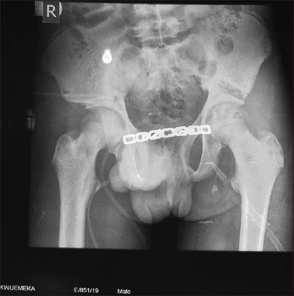
Post-surgical pelvic xray showing osteotomy sites and the plating
He is still on follow-up. Nine months after his discharge, he has had two visits. Micturition interval has improved from every 30–75 min to 60–120 min. He uses Paul’s tubing to care for the dribbling, especially at night and during activities. There is no longer urine stench around him. There is a residual penile torsion [Figure 5].
Figure 5.

A follow-up picture: well healed exstrophy and epispadias repair
DISCUSSION
Bladder exstrophy repair at 16 years of age is not the usual experience of surgeons involved with bladder exstrophy management. Most series of late presentations were in <5 years.[4,5,6,7] The neonatal age or infancy is the usual norm for initial presentation and management.[6] All repair techniques are factored in the first surgery at the neonatal or infancy stage.[2,4] Commencement of repair at this age ensures that the bladder plate is preserved; bladder capacity begins to develop, as well as priming of the patient for continence development. It also obviates the severe psychological affectation of the patient.[8] The index case presenting at 16 years and having first surgery at the same age has its obvious challenges. The bladder plate is not only contracted with reduced capacity but has also undergone metaplasia from urothelial transitional epithelium to stratified squamous epithelium due to exposure to the environmental elements for 16 years. This condition is benign. It is also reversible, but can also undergo further transformation to dysplasia which is pre-malignant.[9] Furthermore, management at this stage was quite tasking in view of the attendant psychologically depressed patient. However, the higher age means we have a relatively lesser anaesthesia risk to the patient. It also gave ample time to evaluate the patient. During the surgery, we have more pliable tissues to work with (such as the BNR) and could conveniently have a more prolonged surgery.
This patient had an opportunity for evaluations of the abdominal viscera with ultrasound, and of the kidneys with intravenous urogram (IVU). He also had pelvic plain radiograph for pelvic bone and sacral bones evaluation. Doing IVU may not be the usual routine when they present at the neonatal age group. These evaluations allow for proper preparation and planning of surgery. In this patient, no other anomalies were noted. The renal function and structure of the kidneys were normal.
This patient had a modification of the CPRE entailing: bladder closure, anterior abdominal wall closure, epispadias repair, pubic apposition, innominate bone osteotomy, pelvic/posterior positioning of the bladder, bladder neck lengthening, ureteral re-implantation and bladder augmentation by ileocystoplasty. The last three procedures are not part of the initial procedures of CPRE. They may be performed at a later time. In this case, all were done in one sitting. There was enough tissue for this. It removed the need for repeated surgeries. Another advantage is that it will allow bladder capacity, urethral resistance, and continence to develop at the same time. This is thought to make a maximal gain incontinence. Again, the upper tracts are protected from ab-initio as micturition is developed. Hafez et al.,[4] in their series, described 30 patients who had CPRE with an age range of 1–8 years. They followed up for 5–64 months. There were 11 cases of late presentation and 19 of failed exstrophy repair. In some of their cases, they did augmentation cystoplasty or BNR or both augmentation and BNR in the same sitting. In their series, all had bilateral iliac osteotomies like in our index case. Seven of their patients, 23% had suprapubic urine leak following removal of the catheter that healed spontaneously, though our index case was on suprapubic drainage to heal. Five out of 30 had concomitant augmentation, of which two persons were continent. Out of the remaining three cases, 2 persons had bladder neck closure and 1 person had BNR to become dry. Out of the other 25, 6 cases were continent without further procedures; 4 were continent after BNR; 1 case was continent after ileocystoplasty + BNR. The remaining 14 were awaiting BNR with or without bladder augmentation. Hence, there was a high rate of second or more procedures to achieve continence. This leads credence to what we did, in which a lot of procedures were done in one sitting to achieve continence. It saves cost. Youssif et al.,[5] in their series did a single-stage repair of exstrophy-epispadias. However, not all the patients had additional procedures to augment the bladder and BNR. In their follow-up, some patients are still incontinent and will require additional procedures. This supports the concept of what we did, that bladder augmentation, and BNR be done at the same time as CPRE, as in the index case.
The choice of this method is informed by the need to conserve the native bladder, maintain a continent micturition as against a non-continent diversion, and have an appropriate pouch to ureteral re-implantation with a view to preventing vesicoureteral reflux, which is common after continent-geared repairs.[4,5,8] There is also an argument for urine diversion in this kind of presentation. This will entail the excision of the bladder because of its contracted nature, irritation and risk of malignancy. However, there is now an improvement in the technique that can allow the retention of the native bladder in reconstruction, achievement of continence, good quality of life and a capacity for adequate follow-up. Also with proper evaluation and endoscopy at follow-up intervals, any suspicious area on the bladder mucosa can be biopsied for histology. The cystoplasty helped expand the bladder capacity. It is thought that the closure of the bladder will ameliorate the irritability with a possibility of reversal of the metaplasia.[9] The progressively improving continence is a testament to these. The native bladder also offered a stable tissue for ureteral re-implantation. Although this is still possible with other continent pouch, it will not be comparable with the native bladder tissue.
Early repair makes correction of bony anomaly easy. Closure of the pubic diastasis is only possible with osteotomy. There is no possibility of apposing the pubis without osteotomy as is done in some neonatal cases. The osteotomy done was a bilateral anterior iliac osteotomy, which is commonly used in late-presenting bladder exstrophy repairs.[8] The osteotomy was quite challenging at this age vis-à-vis the greater force required to break the bones with its attendant risk of injury to pelvic viscera. The blood loss was also substantial, and the conventional means to stabilising the bones could not suffice.[8] The stabilisation was with a 7-hole plate and screw, leaving the middle three holes unscrewed, bilaterally. Shoukry and Shoukry[10] in their series of repair of classic bladder exstrophy in five adults did not use pelvic osteotomies. They did make mention of pubic approximation. However, they have to achieve abdominal wall closure by fasciocutaneous M-plasty.[10]
CONCLUSION
A modification of complete primary repair appears ideal for classic bladder exstrophy repair in an adolescent. Internal fixation for the osteotomy is effective and safe. The outcome is good at the initial stage, and is anticipated will be sustained.
Declaration of patient consent
The authors certify that they have obtained all appropriate patient consent forms. In the form the patient(s) has/have given his/her/their consent for his/her/their images and other clinical information to be reported in the journal. The patients understand that their names and initials will not be published and due efforts will be made to conceal their identity, but anonymity cannot be guaranteed.
Financial support and sponsorship
Nil.
Conflicts of interest
There are no conflicts of interest.
REFERENCES
- 1.Nelson CP, Dunn RL, Wei JT. Contemporary epidemiology of bladder exstrophy in the United States. J Urol. 2005;173:1728–31. doi: 10.1097/01.ju.0000154821.21521.9b. [DOI] [PubMed] [Google Scholar]
- 2.Massanyi EZ, Gearhart JP, Kost-Byerly S. Perioperative management of classic bladder exstrophy. Res Rep Urol. 2013;5:67–75. doi: 10.2147/RRU.S29087. [DOI] [PMC free article] [PubMed] [Google Scholar]
- 3.Woo LL, Thomas JC, Brock JW., 3rd . Bladder and cloacal exstrophy. In: Coran AG, Adzick NS, Krummel TM, Laberge J, Shamberger RC, Caldamone AA, editors. Pediatric Surgery. 7th ed. Philadelphia, USA: Elsevier Saunders; 2012. pp. 1515–29. [Google Scholar]
- 4.Hafez AT, El-Sherbiny MT, Shorrab AA, El-Mowafi H. Complete primary repair of bladder exstrophy in children presenting late and those with failed initial closure:Single center experience. J Urol. 2005;174:1549–52. doi: 10.1097/01.ju.0000179191.45671.3b. [DOI] [PubMed] [Google Scholar]
- 5.Youssif M, Badawy H, Ashraf S, Hanno A, Mokhless I. Single-stage repair of bladder exstrophy in older children and children with failed previous repair. J Pediatr Urol. 2007;3:391–4. doi: 10.1016/j.jpurol.2007.01.193. [DOI] [PubMed] [Google Scholar]
- 6.Hosseini SM, Zarenezhad M, Falahi S, Ahmadi AA, Sabet B, Rasekhi AR. Role of bulking agents in bladder exstrophy-epispadias complexes. Afr J Paediatr Surg. 2013;10:5–8. doi: 10.4103/0189-6725.109373. [DOI] [PubMed] [Google Scholar]
- 7.Bertin KD, Serge KY, Moufidath S, Maxime K, Hervé OK, Baptiste YJ, et al. Complex bladder-exstrophy-epispadias management:Causes of failure of initial bladder closure. Afr J Paediatr Surg. 2014;11:334–40. doi: 10.4103/0189-6725.143149. [DOI] [PubMed] [Google Scholar]
- 8.Inouye BM, Tourchi A, Di Carlo HN, Young EE, Gearhart JP. Modern management of the exstrophy-epispadias complex. Surg Res Pract. 2014;2014:587064. doi: 10.1155/2014/587064. [DOI] [PMC free article] [PubMed] [Google Scholar]
- 9.Xin Z, Zhao C, Huang T, Zhang Z, Chu C, Lu C, et al. Intestinal metaplasia of the bladder in 89 patients:A study with emphasis on long-term outcome. BMC Urol. 2016;16:24. doi: 10.1186/s12894-016-0142-x. [DOI] [PMC free article] [PubMed] [Google Scholar]
- 10.Shoukry AI, Shoukry I. Management of bladder exstrophy in adulthood:Report of 5 cases. J Pediatr Urol. 2013;9:575–8. doi: 10.1016/j.jpurol.2012.06.004. [DOI] [PubMed] [Google Scholar]