Abstract
To investigate whether glucoregulatory neurons in the hypothalamus can sense and respond to physiological variation in the blood glucose (BG) level, we combined continuous arterial glucose monitoring with continuous measures of the activity of a specific subset of neurons located in the hypothalamic ventromedial nucleus that express pituitary adenylate cyclase activating peptide (VMNPACAP neurons) obtained using fiber photometry. Data were collected in conscious, free-living mice during a 1-h baseline monitoring period and a subsequent 2-h intervention period during which the BG level was raised either by consuming a chow or a high-sucrose meal or by intraperitoneal glucose injection. Cross-correlation analysis revealed that, following a 60- to 90-s delay, interventions that raise the BG level reliably associate with reduced VMNPACAP neuron activity (P < 0.01). In addition, a strong positive correlation between BG and spontaneous VMNPACAP neuron activity was observed under basal conditions but with a much longer (∼25 min) temporal offset, consistent with published evidence that VMNPACAP neuron activation raises the BG level. Together, these findings are suggestive of a closed-loop system whereby VMNPACAP neuron activation increases the BG level; detection of a rising BG level, in turn, feeds back to inhibit these neurons. To our knowledge, these findings constitute the first evidence of a role in glucose homeostasis for glucoregulatory neurocircuits that, like pancreatic β-cells, sense and respond to physiological variation in glycemia.
Article Highlights
By combining continuous arterial glucose monitoring with fiber photometry, studies investigated whether neurons in the murine ventromedial nucleus that express pituitary adenylate cyclase activating peptide (VMNPACAP neurons) detect and respond to changes in glycemia in vivo.
VMNPACAP neuron activity rapidly decreases (within <2 min) when the blood glucose level is raised by either food consumption or glucose administration.
Spontaneous VMNPACAP neuron activity also correlates positively with glycemia, but with a longer temporal offset, consistent with reports that hyperglycemia is induced by experimental activation of these neurons.
Like pancreatic β-cells, neurons in the hypothalamic ventromedial nucleus appear to sense and respond to physiological variation in glycemia.
Introduction
Glucose homeostasis is achieved through complex interactions between the endocrine pancreas and glucoregulatory circuits in the brain (1,2). Fundamental to this process is glucose sensing by pancreatic β-cells, which couples detection of rising blood glucose (BG) levels to insulin secretion, which, in turn, lowers the BG level. In the current work, we investigate whether brain control of glycemia involves a similar glucose sensing mechanism.
This work focuses on a specific subset of neurons located in the hypothalamic ventromedial nucleus (VMN) that express pituitary adenylate cyclase activating peptide (VMNPACAP neurons). These neurons were selected because, when they are activated experimentally, BG levels rise, while, conversely, PACAP deletion from this brain area causes obesity, hyperinsulinemia, and glucose intolerance (3–6). Together, these findings support a model in which activation of these neurons leads to mobilization of stored fuel in response to fuel depletion (low BG levels), whereas rising BG levels (e.g., following a meal) inhibit these neurons, which, in turn, promotes fuel storage.
We questioned whether VMNPACAP neurons can sense and respond to physiological variation in glycemia in vivo. To this end, we used fiber photometry to continuously monitor the calcium activity of VMNPACAP neurons while simultaneously measuring arterial BG levels over time using an implantable continuous glucose monitoring (CGM) system. To our knowledge, our findings constitute the first evidence of a role in glucose homeostasis for glucoregulatory neurocircuits that, like pancreatic β-cells, sense and respond to physiological variation in glycemia.
Research Design and Methods
Animals
All procedures were performed in accordance with the National Institutes of Health Guide for the Care and Use of Laboratory Animals and were approved by the Institutional Animal Care and Use Committee at the University of Washington, Seattle, WA. Adult male PACAP-2A-Cre mice (JAX mice, stock no. 030155) and Sprague-Dawley rats were individually housed in a temperature-controlled room on a 14:10 light:dark cycle under specific pathogen-free conditions with ad libitum access to drinking water and standard laboratory chow (PMI Nutrition, St. Louis, MO) unless otherwise stated.
Fiber Photometry
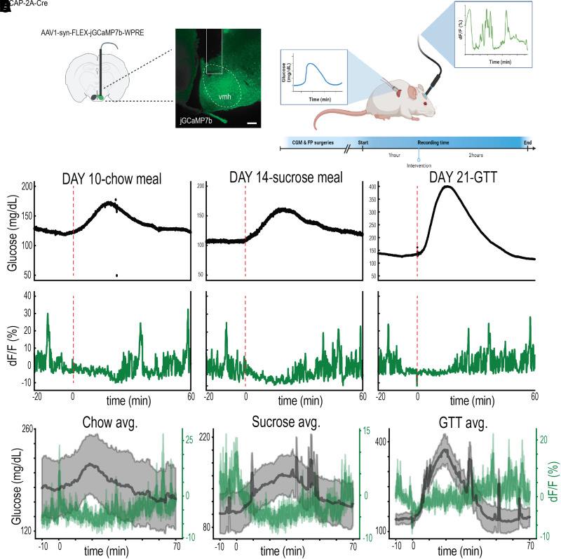
Calcium activity was detected within VMNPACAP neurons by unilateral microinjection of 400 nL pGP-AAV-syn-FLEX-jGCaMP7b-WPRE (Addgene, no. 104493) into the VMN of PACAP-2A-Cre mice (AP: −1.0; ML: −5.7; DV: 0.45) (Fig. 1A and B). During the same surgery, an optical fiber was placed at the injection site as described (7,8). Three weeks later, mice were connected to a fiber photometry system (RZ5P; Tucker-Davis Technology, Alachua, FL) to enable a fluorometric analysis of real-time neuronal activity as described (7,8).
Figure 1.
Methods validation and data acquisition. A: Representative image demonstrating GCaMP7b expression and fiber photometry tract following unilateral microinjection of the AAV containing a Cre-dependent construct for GCaMP7b into the VMN. B: Schematic demonstrating experimental design. C and D: Aligned fiber photometry and continuous glucose traces from a single mouse in response to three paradigms designed to raise glycemia (chow meal, 100 mg sucrose meal, and i.p. glucose injection). E: Temporally aligned mean (±SEM) arterial BG levels and VMNPACAP neuron activity following either a chow meal, a sucrose meal, or an i.p. glucose bolus (at t = 0 min) (n = 4). Panel B was created using Biorender.com.
Custom MATLAB and Python scripts were developed for analyzing fiber photometry data (https://github.com/DeemLab/fiber-photometry), and GCaMP7b fluorescence was determined after adjusting for signal decay and background. Briefly, the isosbestic 405-nm excitation control signal was subtracted from the 470-nm excitation signal to remove movement artifacts from intracellular calcium-dependent GcaMP7b fluorescence. Baseline drift due to slow photobleaching artifacts was evident in the signal, particularly during the first several minutes of each recording session. After baseline correction, dF/F (%) was calculated as individual fluorescence intensity measurements relative to the median fluorescence of the 470-nm channel over the entire session.
Accurate viral targeting and fiber placement were verified post hoc in all animals by immunohistochemical detection of green fluorescent protein in postmortem brain slices using an established method (7,8), and data from animals in which transgene expression or fiber placement was located outside the targeted area were excluded from the analysis. To verify photometric detection of the calcium activity response of VMNPACAP neurons in vivo, we used a novel approach based on evidence that VMN neurons are highly responsive to perceived threats (9). Specifically, we observed that placing an investigator’s hand within the cage is sufficient to reliably and rapidly increase VMNPACAP neuron activity. We therefore used this activation response as an a priori inclusion criteria; animals failing to demonstrate a rapid, >10% increase in dF/F were excluded from further study.
CGM
Adult male PACAP-2A-Cre mice meeting a priori inclusion criteria underwent arterial implantation of a glucose telemetry implant sensor (Data Sciences International, St. Paul, MN) as previously described (10). Following recovery and acclimation, animals were connected to the Tucker-Davis Technology fiber photometry system on a receiver plate to collect CGM data, which were validated by tail bleed with a conventional glucometer (AccuChek; Roche) before and after study interventions.
Study Protocols
Sessions were conducted over 3 h during which CGM and fiber photometry data were collected continuously. During the first hour, no intervention was performed, and food was not available. In both ad libitum fed and in overnight fasted animals, either standard chow or a 100-mg sucrose pellet (Bio-Serv, Flemington, NJ) was provided at the 1-h time point, with monitoring continuing over the next 2 h. For glucose tolerance testing, an i.p. bolus of either glucose (30% d-glucose; 2 g/kg) or saline was given to 4-h fasted mice after a 1-h baseline period, followed again by a 2-h period of monitoring. Each of these protocols was conducted in each mouse.
Time Course of Changing MBH Glucose Levels Following an IV Glucose Bolus
A microdialysis guide cannula was implanted into the mediobasal hypothalamus (MBH) (AP: −2.58 mm; ML: ± 2.6 mm; DV: −8.8 mm; angle: 14°) of male Sprague-Dawley rats (N = 8) following catheterization of the left carotid artery for blood sampling and the right jugular vein for infusion. One week after surgery, the animals underwent a 5-h fast prior to measuring baseline plasma and microdialysate glucose concentrations. At time 0 min, a bolus of 50% dextrose (1 g/kg body weight) was infused intravenously over 15 s. Blood samples (50 μL) were collected every 2 min for the first 20 min and then at 25, 30, 40, 50, 60, 90, and 120 min for plasma glucose measurements. Microdialysate samples were collected at 10-min intervals from t = −30 min through study completion. Following separation of plasma from the blood samples, the remaining red blood cells were resuspended in artificial plasma and reinfused back into the animals to prevent volume depletion and anemia. At the end of the study, the animals were sacrificed, and their brains were collected and frozen for verification of proper probe placement. Only animals with probes correctly targeting the MBH were included in the analysis.
Statistical Analysis
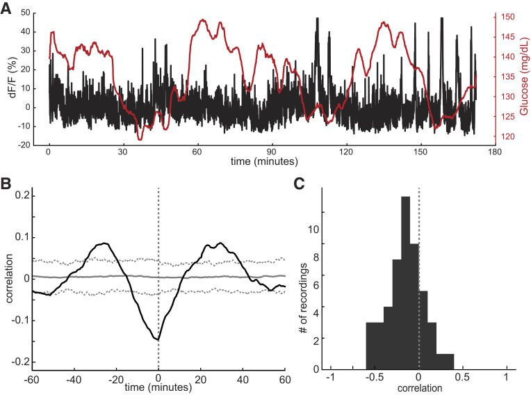
All CGM data were obtained using DSI software. Statistical analyses were performed using GraphPad Prism (version 7.4; GraphPad, San Diego, CA), R (version 3.6.2; R Project for Statistical Computing), python, and MATLAB (MathWorks, Natick, MA). For glucose measurements, the CGM device produces an amperometric signal proportional to glucose concentration. This raw current readout—measured in nanoamperes—was used for all statistical analysis. For fiber photometry, the dF/F signal was used for all statistical analyses. In all instances, P < 0.0.05 was considered significant. Cross-correlations were calculated for each 1-s bin between −60 and +60 min (n = 46 sessions) (Fig. 2B). To generate a null distribution for these correlations, each session was randomly circularly permuted before calculating an average shuffled cross-correlation across sessions. The random value for this permutation was selected from between −30 and +30 min. The average and average ±3 SDs of these cross-correlations are shown in Fig. 2B as the solid and dashed red lines, respectively.
Figure 2.
Relationships between continuous measures of VMNPACAP neuron activity and the BG level. A: Example session showing temporally aligned fiber photometry measurements (black) and detrended glucose concentrations (red). B: Average cross-correlogram across all sessions between neural activity and glucose measurements (N = 46 sessions). The gray line indicates the average correlation of a null distribution where each session is randomly circularly shuffled. Dashed gray lines indicate +3 and −3 SDs from this mean. The negative peak just prior to the zero time point documents the strong negative correlation between glycemia and neuron activity, with the former preceding the latter by 1–2 min. The positive correlation at the 20- to 30-min time indicates that, in the absence of any intervention, increased spontaneous activity of VMNPACAP neurons is associated with a rising glucose level in the circulation. C: Histogram of the cross-correlation values between neural activity and glucose measurements at a −60-s temporal lag (the negative peak in B). Data in A are from a no-intervention, observation-only session, while B and C include all data from all intervention types as well as the no-intervention sessions.
Data and Resource Availability
The data sets and resources generated and analyzed during the current study are available from the corresponding authors upon reasonable request.
Results
To study the relationship between the arterial BG level and VMNPACAP neuron activity over time, we combined CGM with fiber photometry following microinjection into the VMN of PACAP-2A-Cre mice of a cre-inducible GCaMP7b AAV vector. Histochemical detection of GCaMP7b fluorescence was limited to the VMN of PACAP-2A-Cre mice following AAV microinjection into this brain area, confirming proper targeting of VMNPACAP neurons in all mice studied (Fig. 1A). As illustrated schematically in Fig. 1B, both CGM and fiber photometry data were obtained from each of four PACAP-2A-Cre mice during a 1-h baseline monitoring period (without access to food), followed by a 2-h intervention period during which the BG level was elevated by consuming either a chow or a 100 mg sucrose meal. Studies were performed in both ad lib fed and overnight fasted animals, and results were compared with no-intervention controls. To raise the BG level independently of nutrient ingestion, 4-h fasted animals received an i.p. glucose injection, with results compared with saline injection. In total, 46 study sessions were included in the analysis.
Fig. 1C and D show representative photometry and CGM traces from a single PACAP-2A-Cre mouse during each intervention over a 10,000-s (167-min) period. Mean (±SEM) CGM and fiber photometry data obtained from all study animals (n = 4) in response to a chow meal, a sucrose meal, or an i.p. glucose injection are shown in Fig. 1E. When signals are aligned, a consistent decline of VMNPACAP neuron activity is observed as BG levels begin to rise, and a similar relationship is evident in the absence of any intervention (Fig. 2A). Cross-correlation analysis of data from all mice revealed that following a 60- to 90-s delay, interventions that raise the BG level are reliably associated with significantly reduced VMNPACAP neuron activity (Fig. 2B) (P < 0.01). This inverse relationship was consistent irrespective of how the BG level was increased (ingestion of either chow or sucrose; i.p. glucose injection). Variation in cross-correlation values describing the relationship between neural activity and glucose measurements at a −60-s temporal lag (with a change of BG preceding the associated shift in neuron activity) is depicted for all mice as a histogram in Fig. 2C. This analysis shows that, although the correlation coefficient varied between +0.4 and −0.6 at the −60-s time point, the correlation was negative in ∼80% of the mice studied. Therefore, on average, physiological stimuli that raise the BG level in freely moving, conscious mice couple reliably to reduced VMNPACAP neuron activity after a 1- to 2-min delay.
Interestingly, a different relationship was observed between levels of BG and VMNPACAP neuron activity under baseline conditions (i.e., in the absence of any intervention). Specifically, these two variables were significantly positively correlated but with a 20- to 30-min offset (Fig. 2B). In some cases, the change of neuronal activity preceded the change of glycemia, while the reverse was true in others. The association between increased spontaneous VMNPACAP neuron activity and a rising BG level is consistent with evidence that experimental activation of these neurons elicits hyperglycemia (3–6).
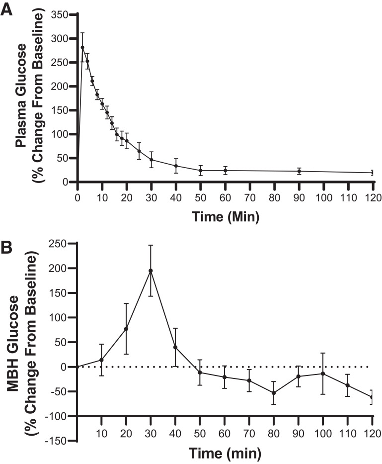
To measure the rate at which glucose appears in the MBH following an i.v. glucose bolus, serial measures of MBH interstitial fluid (ISF) glucose levels were made by microdialysis in adult male Sprague-Dawley rats. Following i.v. glucose injection, plasma glucose levels rose rapidly to ∼300 mg/dL (a 300% increase over baseline) within 2–3 min and dropped rapidly thereafter. A corresponding rise of MBH glucose levels was detectable after a 10-min lag and peaked 20–25 min later (Fig. 3). Although performed in a different species, these data suggest that transfer of glucose from plasma into the MBH is too slow to explain the rapid decrease of VMNPacap neuron activity (<2 min) we have observed following either a meal or i.p. glucose injection (Fig. 2).
Figure 3.
Time course of changing glucose concentrations in A) plasma and B) MBH following an i.v. glucose bolus. A: Plasma glucose levels rose rapidly, peaking 2 min following intravenous glucose administration and rapidly returning to baseline values. B: Extracellular glucose levels in the MBH measured by microdialysis rose detectably within 10 min after intravenous glucose administration and peaked at ∼30 min, before returning to baseline levels by ∼40–50 min. Data are presented as average percentage change from baseline values ± SEM.
Discussion
We investigated whether, like pancreatic β-cells, subsets of hypothalamic glucoregulatory neurons are capable of sensing and responding to physiological changes in glycemia in vivo. By combining fiber photometry with CGM in conscious, free-living mice, we show that the calcium activity of VMNPACAP neurons decreases within 60–90 s of a physiological rise of glycemia (induced by either carbohydrate ingestion or exogenous glucose administration). To our knowledge, these findings constitute the first evidence that the activity of hypothalamic glucoregulatory neurons is regulated in vivo by physiological variation in the circulating glucose level.
One possible mechanism to explain these changes in VMNPACAP neuron activity involves the transfer of glucose across the blood-brain barrier and into the extracellular fluid surrounding the VMN. To explore this possibility, we measured the time course over which glucose appears in the MBH following an i.v. glucose bolus in rats. Consistent with published evidence (11), we report a relatively slow rate of appearance of glucose in the MBH, being barely detectable by 10 min following i.v. glucose and peaking ∼30–35 min after the plasma peak. Such a time course would appear to be too slow to explain the rapidity with which VMNPACAP neuron activity decreases following an induced increase of the BG level (<2 min).
Instead, we favor the hypothesis that afferent information regarding the circulating glucose level is detected by glucose-sensing neurons supplying the vasculature. Such glucose-sensing afferents are well-described in vascular beds such as the hepatic portal and mesenteric veins (12–14), as well as in the median eminence (15), a circumventricular organ that lacks a properly formed blood-brain barrier and is located within the MBH. Once a change of glucose within the vasculature is detected, this afferent input is conveyed to glucoregulatory neurocircuits within the brain, of which VMNPACAP neurons are a part.
Under baseline conditions (i.e., in the absence of any intervention), spontaneous VMNPACAP neuron activity correlated positively with variation in glycemia but with a much longer temporal offset (20–30 min). This suggests that activation of these neurons raises the BG level, consistent with the published effect of chemogenetic activation of these neurons (3). Together, these observations are suggestive of a closed-loop control system whereby VMNPACAP neuron activation raises the BG level which, in turn, feeds back to inhibit the activity of these neurons, functionally the inverse of the canonical coupling of β-cell function to the BG level.
A limitation of this work is that firm conclusions about causality cannot be drawn solely from associations between neuronal activity and BG levels. For example, it is possible that observed neuronal responses are coupled to changing glycemia indirectly (e.g., via “gut-brain signaling” following detection of nutrients in the gastrointestinal tract) and that VMNPACAP neurons are not unique but, instead, are one of many neuronal subsets regulated by changes in glycemia. With these limitations in mind, we interpret our finding that VMNPACAP neuron activity is rapidly reduced following interventions that raise the BG level as the first direct evidence of a neural mechanism that transduces sensory detection of glycemic variation into an adaptive hypothalamic response.
Article Information
Acknowledgments. The authors appreciate technical assistance provided by Dr. Chelsea Faber (Barrow Neurological Institute, Phoenix, AZ) and mouse colony management by Vincent Damian (University of Washington, Seattle, WA). The Metabolic and Cellular Phenotyping Core of the NIDDK-funding Diabetes Research Center at the University of Washington performed CGM implantation.
Funding. This work was supported by National Institutes of Health-National Institute of Diabetes and Digestive and Kidney Diseases (NIH-NIDDK) grants R01DK101997 and R01DK083042 (M.W.S.); R01DK089056 and DK124238 (G.J.M.); R03DK128383 (J.M.S.); the NIH-NIDDK–funded Nutrition Obesity Research Center (P30DK035816) and Diabetes Research Center (P30DK017047); the Diabetes, Obesity and Metabolism (T32DK007247, N.E.R.) and Nutrition, Obesity and Atherosclerosis (T32HL007028, J.D.D.) training grants and the National Institute of Mental Health–funded P50MH106428 at the University of Washington, an American Diabetes Association Innovative Basic Science Award (ADA 1-19-IBS-192, G.J.M.); and a U.S. Department of Defense grant W81XWH2110635 (J.M.S.); and by research funding provided by Novo Nordisk.
Duality of Interest. M.W.S. receives research support from Novo Nordisk and is a consultant for NodThera. A.S. and A.-M.B. are employees at Novo Nordisk and own minor shares in the company. No other potential conflicts of interest relevant to this article were reported.
Author Contributions. Each co-author contributed to developing hypotheses and the research protocols used to test them, along with manuscript composition and editing. J.D.D. was responsible for the conduct of most studies. D.T. supervised cross-correlation analysis. M.W.S. is the guarantor of this work and, as such, had full access to all the data in the study and takes responsibility for the integrity of the data and the accuracy of the data analysis.
References
- 1. Myers MG Jr, Affinati AH, Richardson N, Schwartz MW. Central nervous system regulation of organismal energy and glucose homeostasis. Nat Metab 2021;3:737–750 [DOI] [PubMed] [Google Scholar]
- 2. Mirzadeh Z, Faber CL, Schwartz MW. Central nervous system control of glucose homeostasis: a therapeutic target for type 2 diabetes? Annu Rev Pharmacol Toxicol 2022;62:55–84 [DOI] [PMC free article] [PubMed] [Google Scholar]
- 3. Khodai T, Nunn N, Worth AA, et al. PACAP neurons in the ventromedial hypothalamic nucleus are glucose inhibited and their selective activation induces hyperglycaemia. Front Endocrinol (Lausanne) 2018;9:632. [DOI] [PMC free article] [PubMed] [Google Scholar]
- 4. Hawke Z, Ivanov TR, Bechtold DA, Dhillon H, Lowell BB, Luckman SM. PACAP neurons in the hypothalamic ventromedial nucleus are targets of central leptin signaling. J Neurosci 2009;29:14828–14835 [DOI] [PMC free article] [PubMed] [Google Scholar]
- 5. Bozadjieva-Kramer N, Ross RA, Johnson DQ, et al. The role of mediobasal hypothalamic PACAP in the control of body weight and metabolism. Endocrinology 2021;162:bqab012. [DOI] [PMC free article] [PubMed] [Google Scholar]
- 6. Resch JM, Boisvert JP, Hourigan AE, Mueller CR, Yi SS, Choi S. Stimulation of the hypothalamic ventromedial nuclei by pituitary adenylate cyclase-activating polypeptide induces hypophagia and thermogenesis. Am J Physiol Regul Integr Comp Physiol 2011;301:R1625–R1634 [DOI] [PMC free article] [PubMed] [Google Scholar]
- 7. Faber CL, Matsen ME, Meek TH, Krull JE, Morton GJ. Adaptable angled stereotactic approach for versatile neuroscience techniques. J Vis Exp 2020;159:e60965. [DOI] [PMC free article] [PubMed] [Google Scholar]
- 8. Deem JD, Faber CL, Pedersen C, et al. Cold-induced hyperphagia requires AgRP neuron activation in mice. eLife 2020;9:e58764. [DOI] [PMC free article] [PubMed] [Google Scholar]
- 9. Kunwar PS, Zelikowsky M, Remedios R, et al. Ventromedial hypothalamic neurons control a defensive emotion state. eLife 2015;4:e06633. [DOI] [PMC free article] [PubMed] [Google Scholar]
- 10. Evers SS, Kim KS, Bozadjieva N, et al. Continuous glucose monitoring reveals glycemic variability and hypoglycemia after vertical sleeve gastrectomy in rats. Mol Metab 2020;32:148–159 [DOI] [PMC free article] [PubMed] [Google Scholar]
- 11. Hwang JJ, Jiang L, Hamza M, et al. Blunted rise in brain glucose levels during hyperglycemia in adults with obesity and T2DM. JCI Insight 2017;2:e95913. [DOI] [PMC free article] [PubMed] [Google Scholar]
- 12. Bohland M, Matveyenko AV, Saberi M, Khan AM, Watts AG, Donovan CM. Activation of hindbrain neurons is mediated by portal-mesenteric vein glucosensors during slow-onset hypoglycemia. Diabetes 2014;63:2866–2875 [DOI] [PMC free article] [PubMed] [Google Scholar]
- 13. Watts AG, Donovan CM. Sweet talk in the brain: glucosensing, neural networks, and hypoglycemic counterregulation. Front Neuroendocrinol 2010;31:32–43 [DOI] [PMC free article] [PubMed] [Google Scholar]
- 14. Fujita S, Bohland M, Sanchez-Watts G, Watts AG, Donovan CM. Hypoglycemic detection at the portal vein is mediated by capsaicin-sensitive primary sensory neurons. Am J Physiol Endocrinol Metab 2007;293:E96–E101 [DOI] [PubMed] [Google Scholar]
- 15. Lynch RM, Tompkins LS, Brooks HL, Dunn-Meynell AA, Levin BE. Localization of glucokinase gene expression in the rat brain. Diabetes 2000;49:693–700 [DOI] [PubMed] [Google Scholar]


