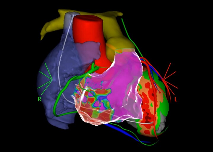
Figure 1.
Co-registered 3D CT of a patient with ARVC. Left ventricle shows septal wall thinning, right atrium (blue), RCA (green), aorta and LAD (red), pulmonary artery (yellow), ICD lead (white), and left phrenic nerve (green). Electroanatomical reconstruction of the RV with infero-basal scar. LAD, left anterior descending coronary artery.

