

Abstract
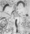
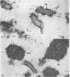
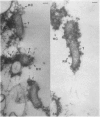
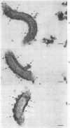
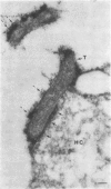
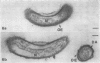
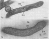
Pathogenic T. pallidum cells possess and extracellular layer when observed in vivo and in vitro after exposure to ruthenium-red. The extracellular layer is partially removed from a pre-fixed cell by repeated washing in vitro. Non-pathogenic treponemes examined in this study do not possess an extracellular layer. It is hoped that the data presented herein will cause our colleagues to take another look at the methods and techniques used for preparing pathogenic T. pallidum cells for practical end-point objectives.
Full text
PDF


Images in this article
Selected References
These references are in PubMed. This may not be the complete list of references from this article.
- CHRISTIANSEN S. Protective layer covering pathogenic treponemata. Lancet. 1963 Feb 23;1(7278):423–425. doi: 10.1016/s0140-6736(63)92309-2. [DOI] [PubMed] [Google Scholar]
- Chen T. H., Elberg S. S., Boyles J., Velez M. A. Yersinia pestis: correlation of ultrastructures and immunological status. Infect Immun. 1975 Jun;11(6):1382–1390. doi: 10.1128/iai.11.6.1382-1390.1975. [DOI] [PMC free article] [PubMed] [Google Scholar]
- Cheng K. J., Costerton J. W. Localization of alkaline phosphatase in three gram-negative rumen bacteria. J Bacteriol. 1973 Oct;116(1):424–440. doi: 10.1128/jb.116.1.424-440.1973. [DOI] [PMC free article] [PubMed] [Google Scholar]
- Costerton J. W., Damgaard H. N., Cheng K. J. Cell envelope morphology of rumen bacteria. J Bacteriol. 1974 Jun;118(3):1132–1143. doi: 10.1128/jb.118.3.1132-1143.1974. [DOI] [PMC free article] [PubMed] [Google Scholar]
- HANSON A. W., CANNEFAX G. R. RECOVERY OF TREPONEMA AND BORRELIA AFTER LYOPHILIZATION. J Bacteriol. 1964 Sep;88:811–811. doi: 10.1128/jb.88.3.811-811.1964. [DOI] [PMC free article] [PubMed] [Google Scholar]
- Jackson S., Black S. H. Ultrastructure of Treponema pallidum Nichols following lysis by physical and chemical methods. I. Envelope, wall, membrane and fibrils. Arch Mikrobiol. 1971;76(4):308–324. doi: 10.1007/BF00408528. [DOI] [PubMed] [Google Scholar]
- Jones H. C., Roth I. L., Sanders W. M., 3rd Electron microscopic study of a slime layer. J Bacteriol. 1969 Jul;99(1):316–325. doi: 10.1128/jb.99.1.316-325.1969. [DOI] [PMC free article] [PubMed] [Google Scholar]
- Jones R. H., Nevin T. A., Guest W. J., Logan L. C. Lytic effect of trypsin, lysozyme, and complement on Treponema pallidum. Br J Vener Dis. 1968 Sep;44(3):193–200. doi: 10.1136/sti.44.3.193. [DOI] [PMC free article] [PubMed] [Google Scholar]
- Luft J. H. Ruthenium red and violet. I. Chemistry, purification, methods of use for electron microscopy and mechanism of action. Anat Rec. 1971 Nov;171(3):347–368. doi: 10.1002/ar.1091710302. [DOI] [PubMed] [Google Scholar]
- Myers D. B., Highton T. C., Rayns D. G. Ruthenium red-positive filaments interconnecting collagen fibrils. J Ultrastruct Res. 1973 Jan;42(1):87–92. doi: 10.1016/s0022-5320(73)80008-5. [DOI] [PubMed] [Google Scholar]
- Pate J. L., Ordal E. J. The fine structure of Chondrococcus columnaris. 3. The surface layers of Chondrococcus columnaris. J Cell Biol. 1967 Oct;35(1):37–51. doi: 10.1083/jcb.35.1.37. [DOI] [PMC free article] [PubMed] [Google Scholar]
- Springer E. L., Roth I. L. The ultrastructure of the capsules of Diplococcus pneumoniae and Klebsiella pneumoniae stained with ruthenium red. J Gen Microbiol. 1973 Jan;74(1):21–31. doi: 10.1099/00221287-74-1-21. [DOI] [PubMed] [Google Scholar]
- Wiegand S. E., Strobel P. L., Glassman L. H. Electron microscopic anatomy of pathogenic Treponema pallidum. J Invest Dermatol. 1972 Apr;58(4):186–204. doi: 10.1111/1523-1747.ep12539907. [DOI] [PubMed] [Google Scholar]