Abstract
Mitophagy is an important process that participates in mitochondrial quality control. Dysfunctions in this process can be caused by mutations in genes like PRKN and are associated with the development and progression of Parkinson’s Disease (PD). The most used drug in the treatment of PD is levodopa (LD), but it can cause adverse effects, such as dyskinesia. Currently, few studies are searching for biomarkers for an effective use of lLD for this disease, especially regarding mitophagy genetics. Thus, this work investigates the association of 14 variants of the PRKN gene with LD in the treatment of PD. We recruited 70 patients with PD undergoing treatment with LD (39 without dyskinesia and 31 with dyskinesia). Genotyping was based on Sanger sequencing. Our results reinforce that age at onset of symptoms, duration of PD, and treatment and dosage of LD can influence the occurrence of dyskinesia but not the investigated PRKN variants. The perspective presented here of variants of mitophagy-related genes in the context of treatment with LD is still underexplored, although an association has been indicated in previous studies. We suggest that other variants in PRKN or in other mitophagy genes may participate in the development of levodopa-induced dyskinesia in PD treatment.
Keywords: Parkinson’s disease, mitophagy, levodopa
1. Introduction
Mitochondria are organelles that perform important cellular functions, including ATP synthesis through oxidative phosphorylation (OXPHOS), control of reactive oxygen species (ROS), regulation of oxidative stress, and intracellular signaling [1,2,3]. Alterations in the quality control system of mitochondria are related to several neurodegenerative diseases, due to the accumulation of aged and dysfunctional mitochondria, which may increase ROS production and oxidative stress and generate synaptic and neuronal loss [2,4].
Among the cellular mechanisms that maintain mitochondrial homeostasis, mitophagy stands out, consisting of the process of degradation and removal of mitochondria considered dysfunctional, to prevent them from being in excess [5,6]. The impairment of the mitophagy process has already been widely described as a factor related to different diseases, including Parkinson’s Disease (PD) [3,7,8,9]. Because mitochondrial dysfunction resulting from alterations in genes related to mitophagy, such as LRRK2, DJ-1, PINK1, and PRKN, can lead to the production of ROS and aggregation of α-synuclein (major component of Lewy bodies, pathological hallmarks, and typically found in PD), these genes might be involved in promoting the neurodegeneration observed in PD [10]. Mitophagy is carried out mainly by the PINK1 and PRKN genes, which, when mutated, can lead to irregularities in mitophagy and consequently the selective death of neurons, such as dopaminergic neurons in PD.
In this study, we highlight the PRKN gene (parkin RBR E3 ubiquitin protein ligase), also known as PARK2 or Parkin, being the gene most associated with autosomal recessive PD of early onset [11,12,13]. It encodes Parkin, a protein that regulates mitophagy and mitochondrial biogenesis, participating in the quality control system of mitochondria [5,11]. A study carried out with Drosophila revealed that deficiencies in PRKN lead to the accumulation of dysfunctional mitochondria in several regions, including in dopaminergic neurons. Furthermore, an association between mitophagy failure and inflammation is suggested, through activation of the STING pathway (interferon gene stimulator), which may lead to the loss of dopaminergic neurons [14,15]. Therefore, defects in Parkin-mediated mitophagy are pertinent to PD [5,11,14].
Parkinson’s disease is characterized by the progressive loss of dopaminergic neurons in the substantia nigra of the central nervous system and by the accumulation of misfolded intracellular a-synuclein, leading to impaired muscle function, which causes symptoms such as bradykinesia, tremors, postural instability, and muscle rigidity, as well as non-motor symptoms such as depression, cognitive impairment, dementia, and anxiety [16,17,18,19]. Considered the second most common neurodegenerative disorder worldwide, it is estimated that 1% to 2% of the world’s population over 65 years of age is affected with PD [20].
Despite PD being widely studied, there are still no biomarkers that could indicate the early onset of PD and there are currently few forms of treatment for this disease, such as anticholinergics, amantadine (NMDA antagonist), monoamine oxidase inhibitors (MAOIs), catechol-O-methyl transferase inhibitors (COMTIs), and dopamine agonists [17]. The existing ones are aimed only at reducing the symptoms and not at containing the progression of the disease, in addition to having lower efficacy as neurodegeneration progresses, which represents an impact on the prognosis and quality of life of patients affected with PD [7,14].
In this context, levodopa (LD), a dopaminergic precursor, is the most used drug in the treatment of people affected by PD. LD is a prodrug, metabolized into dopamine when crossing the blood–brain barrier, thus supplementing the endogenous levels of this neurotransmitter, and, compared to other PD drugs, it is the most effective, as it has significant effects for improving symptoms [7,17]. Importantly, it relieves symptoms but does not prevent disease progression, and the prolonged use is related to adverse reactions, such as motor complications, among them motor fluctuations and dyskinesia. Levodopa-induced dyskinesia (LID) is characterized by movement disorders that include dystonia, myoclonus, chorea, and athetosis [21,22]. Undeniably, this condition also impairs the quality of life of patients with PD, who over time have worsening and debilitating symptoms.
Although there are many studies that suggest that mitochondrial dysfunction is essential in PD, there are many studies about LID in PD, and the relationship between mitophagy and PD is highly recognized, more recently in a therapeutic context [7,23,24,25], no studies are found in the global literature on mitophagy specifically in the treatment of LD. Thus, because genetic alterations can lead to dysfunctions in mitophagy, which in turn are related to PD, it is important to investigate potential biomarkers from the perspective of this mitochondrial process. Thus, the present work aims to investigate variants in PRKN, a gene related to mitophagy, in patients with PD who are treated with LD, seeking possible biomarkers that could be associated with therapeutic success and adverse reactions.
2. Materials and Methods
2.1. Sampling
For this research, 70 individuals with Parkinson’s Disease were selected, recruited from the Hospital Ophir Loyola, in the North region of Brazil. These patients were treated with levodopa in a multidrug therapy, of which 39 did not have LID, composing the control group, and 31 did have LID, composing the case group. All patients were at least 42 years of age at diagnosis and were divided into sex-matched groups. In addition, the use of levodopa in the treatment of these patients is equal to or greater than two years.
2.2. Selection of Variants
Through the public databases of the National Center for Biotechnology Information (NCBI) and a literature search, we selected the intronic variants rs9458609 and rs6935164 of the PRKN gene with relevant minor allele frequencies (MAF = 0.3 and MAF = 0.5, respectively) and with the potential to influence the development of diseases [26,27,28] but which have not yet been studied in relation to PD. From the gnomAD browser platform [29], closely located variants were selected for rs9458609 (rs1446954927, rs1562397973, rs1188406822, rs897123960, rs1469130556, rs1037288938 and rs1453267522) and for rs6935164 (rs1460805773, rs972410489, rs1372726016, rs187267734, rs182928580), all present in the PRKN gene.
2.3. DNA Extraction and Quantification
DNA was extracted from peripheral blood samples using the phenol-chloroform method, based on [30]. Quantification of the extracted DNA was performed using the NanoDrop 1000 spectrophotometer (Thermo Fisher Scientific Inc., Wilmington, DE, USA), to ensure quality of the extracted DNA.
2.4. Genotyping
Sequences of interest from the extracted DNA were amplified by PCR using specific primer pairs: for rs9458609 and close variants, 5′GAGTGATTCAGTCCAAGGCT3′ 5′TCAACAGTCAGTAAAAGGCAAC′, and for rs6935164 and close variants, 5′AAAGATTGCAGTGATGTGGC3′ and 5′AGAACTTCCACTTGAGCTGA3′. Table 1 presents the 14 analyzed variants, with their allelic changes and population frequency.
Table 1.
Description of the investigated PRKN gene variants.
| Variant | Alleles | MAF |
|---|---|---|
| rs9458609 | A>G | 0.30678 |
| rs1446954927 | A>G | 0.00007 |
| rs1562397973 | G>A | 0.000007 |
| rs1188406822 | G>A | 0.000007 |
| rs897123960 | T>C | 0.000014 |
| rs1469130556 | A>G | 0.00007 |
| rs1037288938 | G>A | 0.000021 |
| rs1453267522 | T>G | 0.000014 |
| rs6935164 | A>G | 0.497471 |
| rs1460805773 | T>G | 0.000007 |
| rs972410489 | G>C | 0.000007 |
| rs1372726016 | G>A | 0.000007 |
| rs187267734 | C>A | 0.000007 |
| rs182928580 | G>A | 0.003313 |
The PCR protocol was based on the work by Ferraz et al. (2022) [31]. For both primer pairs, the PCR was performed from a reaction with a volume of 20 µL, considering dNTP, 0.2 μL of Taq polymerase, 3.0 μL of DNA, and 11.3 μL of water. A Veriti thermal cycler (Thermo Fisher Scientific) was used for the reaction, through the program of 1 cycle of denaturation at 95 °C for 10 min, 35 cycles of denaturation at 95 °C for 15 s, the annealing temperature adjusted by the primer for 30 s, and extension at 72 °C for 1 min and 30 s.
Then, the PCR products were sequenced using the BigDye Terminator Sequencing Kit version 3.1 (Thermo Fisher Scientific) in an ABI PRISM 3130 Genetic Analyzer (Thermo Fisher Scientific). Nucleotide sequences generated by the DNA sequencer were traced by Sequencing Analysis Software version 5.2 (Thermo Fisher Scientific). The submitted genotyping was read using Clustal Omega [32,33] and the chromatogram analysis was performed with Chromas software version 2.6.6 (https://technelysium.com.au/wp/).
2.5. Genomic Ancestry
Brazil has a population with diverse contributions of genomic ancestry, especially Native American (NAM), European (EUR), and African (AFR), because of the complex process of the population formation. In this context, to avoid possible bias in the interpretation of results due to population substructuring, we employed a set of ancestry informative markers (AIM) previously developed and established by our research group [34,35,36].
2.6. Statistical Analysis
Statistical analyses and graphs were performed using the JASP [37] and R [38], with p values < 0.05 being considered statistically significant.
3. Results
3.1. Characterization of the Cohort
The studied cohort consisted of 70 individuals with PD undergoing treatment with levodopa, in which the case group represents individuals with dyskinesia and the control group represents individuals without dyskinesia. We found that age at onset of symptoms, duration of PD and treatment, and LD dosage were factors that showed statistically significant differences between the two groups (Table 2). On the other hand, we found no significant differences between sex, genomic ancestry, family history of PD, the primary symptom, and UPDRS.
Table 2.
Data from the case group (PD patients who developed dyskinesia) and the control group (PD patients without dyskinesia), both undergoing treatment with LD.
| Variable | Case | Control | p-Value |
|---|---|---|---|
| n | 31 | 39 | |
| Age of onset of symptoms, years a | 47.5 ± 1.68 | 55.3 ± 1.55 | 0.001 |
| Sex, % male/female b | 74.2/25.8 | 61.5/38.5 | 0.263 |
| European ancestry c | 0.643 ± 0.036 | 0.588 ± 0.035 | 0.309 |
| Native American ancestry c | 0.214 ± 0.031 | 0.259 ± 0.033 | 0.350 |
| African ancestry c | 0.143 ± 0.029 | 0.154 ± 0.031 | 0.692 |
| Family history, % yes/no b | 17.2/82.8 | 30.6/69.4 | 0.215 |
| Primary Symptom, % tremor/others b | 48.4/51.6 | 66.7/33.3 | 0.123 |
| Duration of PD d | 10.0 ± 0.92 | 6.9 ± 0.62 | 0.0037 |
| Duration of treatment d | 8.1 ± 0.68 | 4.97 ± 0.51 | 8.1 × 10⁻⁵ |
| LEDD d | 1066.1 ± 94.7 | 540.2 ± 44.7 | 1.8 × 10⁻⁶ |
| UPDRS d | 73.2 ± 6.5 | 64.3 ± 4.3 | 0.370 |
a Mean value ± SE (Standard Error of Mean), Student’s t-test; b Values in distribution percentages, chi-squared test c Mean ± SE values, Mann–Whitney test. d Mean ± SE values, Wilcoxon test. LEDD: Levodopa Equivalent Daily Dose. UPDRS: Unified Parkinson’s Disease Rating Scale.
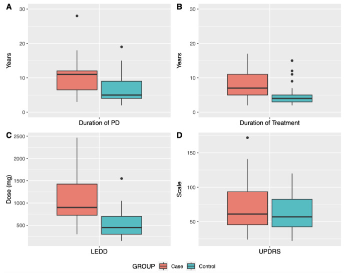
Figure 1 shows that, for patients with dyskinesia, duration of both PD and treatment are longer compared to patients without dyskinesia. Likewise, the dosage of levodopa (LEDD) increases with therapeutic time. These results were expected considering that prolonged treatment with LD can lead to the development of dyskinesia, but these variables were controlled in the statistical analysis. Regarding the UPDRS international scale, there was no statistically significant difference, indicating that both groups would be in similar motor stages of the disease. This scale was proposed in the 1980s and its revised version is the most widely used tool in the world to aid in the diagnosis of PD [39,40].
Figure 1.
Clinical variables related to the treatment of patients with levodopa with and without dyskinesia (case and control, respectively). (A): Duration in years of Parkinson’s Disease for patients in both groups; (B): Duration in years of treatment with levodopa; (C): Dosage in mg of levodopa; (D): UPDRS PD rating scale.
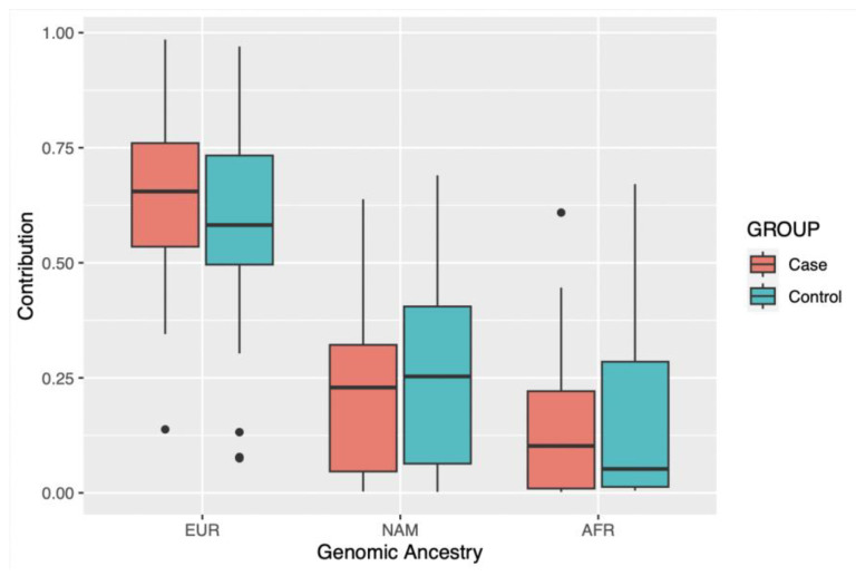
When analyzing the average contribution of genomic ancestry of the study cohort, which is composed of individuals from a population of the Brazilian Amazon, it is observed that the greatest contribution is European, followed by Native American and African, for both groups (Figure 2). This result corroborates previous studies carried out in the same region [31,41].
Figure 2.
Contribution of each genomic ancestry investigated in the studied groups. EUR: European; NAM: Native American; AFR: African.
3.2. Analysis of Variants
Then, the distribution of variants and their allelic and genotypic frequencies between groups were analyzed. All groups are in Hardy–Weinberg Equilibrium (p > 0.05). Only the rs9458609 and rs6935164 variants showed genotype variation in the studied cohort; the other 12 variants presented only the homozygous genotype for the reference allele, reinforcing the previously described MAF. The frequencies of these two variants for our cohort are shown in Table 3. Furthermore, when we analyzed the dependence of the variants in relation to the sex of the patients, we did not observe statistically significant differences.
Table 3.
Distribution of allele and genotypic frequencies of variants rs9458609 and rs6935164 for PD patients with dyskinesia (case) and without dyskinesia (control).
| Variant | Genotype | Case (%) | Control (%) | p-Value a | OR (95%CI) b |
|---|---|---|---|---|---|
| rs9458609 | n = 29 | n = 36 | |||
| AA | 16 (55.2) | 17 (47.2) | 0.773 | 0.817 (0.207–3.229) | |
| AG | 9 (31.0) | 17 (47.2) | 0.807 | 0.847 (0.224–3.203) | |
| GG | 4 (13.8) | 2 (5.6) | 0.330 | 3.306 (0.299–36.601) | |
| A = 0.7/G = 0.3 | A = 0.7/G = 0.3 | ||||
| rs6935164 | n = 28 | n = 30 | |||
| AA | 6 (21.4) | 13 (43.3) | 0.062 | 0.229 (0.049–1.079) | |
| AG | 12 (42.8) | 10 (33.3) | 0.293 | 2.320 (0.483–11.148) | |
| GG | 10 (35.8) | 7 (23.4) | 0.287 | 2.333 (0.491–11.080) | |
| A = 0.4/G = 0.6 | A = 0.6/G = 0.4 |
a p-value obtained by logistic regression with correction for confounding factors: age at onset of symptoms, duration of PD, duration of treatment and LEDD. b Odds Ratio (OR) and 95% Confidence Interval (95%CI) obtained by logistic regression.
4. Discussion
Parkinson’s disease is characterized by the progressive loss of dopaminergic neurons, which leads to motor and non-motor symptoms. Mutations in genes such as PINK1, PRKN, DJ-1, ATP13A2, LRRK2, SNCA, and VPS35 are commonly found in PD, and dysfunctions in these genes affect essential processes in the mitochondrial quality control system that ensure mitochondrial function, like fusion and fission, biogenesis, and mitophagy [5,7]. The role of mitochondria in the neurodegenerative process of PD has been increasingly described. Mitochondrial dysfunction results in multiple effects, such as decreased ATP generation, increased ROS, and dysregulation of Ca2+ levels. These disorders lead to impaired proteostasis in neurons, hence the accumulation of proteins such as a-synuclein and a reduction in axonal transport to vesicles and mitochondria, resulting in neurodegeneration and neuroinflammation [42].
In this sense, mitophagy is a crucial mechanism in mitochondrial quality control and is a key process in neurodegeneration [43] so that mutations in genes responsible for mitophagy, such as PRKN, can lead to the onset and progression of diseases, including PD. The drug most used for the treatment of PD is levodopa, but its prolonged administration can cause adverse effects, such as dyskinesia, characterized by involuntary muscle movements, which affects the quality of life of these individuals [7,21]. One study showed that after 15 years of treatment with levodopa, 94% of patients developed dyskinesia [44], which demonstrates the need for studies that investigate this adverse effect and suggest effective biomarkers to improve quality of life of PD patients.
Among the different genes involved in the complex network of PD, the PRKN gene is most associated with the development of this disease [3,7,10]. However, a study [45] demonstrated that individuals with certain PRKN mutations have a better response to LD treatment, although they are possibly at a higher risk of developing dyskinesia compared to individuals without these PRKN mutations. In fact, recently, it has been suggested that mutations in PRKN could influence the onset of LID at an early stage, although this relationship has not yet been much explored [13,46].
Thus, in the present study, the distribution of 14 variants in the PRKN gene of the mitophagy pathway was investigated in a cohort of PD patients undergoing treatment with LD, divided into individuals with and without dyskinesia. The study looked for two mutations in this gene that are currently understudied but which have relevant MAF (rs9458609 and rs6935164) in addition to 12 closely located variants in PRKN.
No statistically significant association was found for rs9458609 and rs6935164 between the groups with and without dyskinesia, suggesting that these mutations may not be involved with the development of this adverse effect in patients with PD, and these variants do not seem to be dependent on the sex of the patients.
The rs9458609 variant was previously mentioned in [26], who, through a PRKN and PACRG mapping, significantly related this variant to leprosy in Indian patients. More recently, this variant has been reported in linkage disequilibrium with a polymorphism of the same gene, rs1801582 (r2 = 0.06) [28], which, in turn, had already been associated with susceptibility to the development of PD [47], although this result was not observed in a study on PD in another population [48]. As far as we know, the other variants analyzed here had not been reported in the literature until the present study.
In addition, demographic and clinical characteristics related to the treatment of these patients were also investigated. As expected, statistically significant differences were identified between groups comparing age of symptom onset, duration of PD, duration of treatment, and levodopa equivalent daily dose (LEDD). One study highlighted that the age of onset of PD is a determinant aspect of the risk of dyskinesia, whereas patients with 5 years of treatment and age of onset between 40 and 49 years had a 46% higher risk of developing dyskinesia than individuals aged 70 to 79 years at onset of PD [49]. Also, it has already been described that disease progression, younger age at onset of PD, and dose and treatment period are predictive factors for the development of dyskinesia [22,50]. It was demonstrated in a previous study [51] that the female individuals and also those with the most advanced form of the disease, that is, higher scores on the UPDRS, have a greater risk of dyskinesia; however, our analysis did not indicate a statistically significant difference between the groups when observing the sexes and the UPDRS. Regarding genomic ancestry as a risk factor for LID, the same study pointed to individuals from the geographic region of North America as a risk factor compared to Europe. In our study, there were no statistically significant differences when observing the contributions of genomic ancestry (European, Native American, and African) in the investigated population. Currently, there are few studies that assess genomic ancestry as a risk factor for LID, especially in Brazil, which reinforces the relevance of the present work.
5. Conclusions
In short, our results highlight that age at onset of symptoms, duration of PD and treatment, and LD dosage are factors that may influence the occurrence of dyskinesia, but the variants investigated here may not be involved with this adverse effect, suggesting that studies in larger cohorts with the same variants, particularly rs6935164, should be conducted to reinforce our findings, but also that other genetic factors of PRKN and mitophagy in general may play a role in LID in patients with PD.
Acknowledgments
We thank all participants of the study and the funding agencies of the project.
Author Contributions
Conceptualization, A.G.B. and G.C.C.; methodology, A.G.B., C.S.S. and C.S.-d.-S.; formal analysis, A.G.B., C.S.S. and G.C.C.; resources, B.L.S.-L. and Â.R.-d.-S.; data curation, C.S.-d.-S., D.D.M. and B.H.B.K.; writing—original draft preparation, A.G.B. and G.C.C.; writing—review and editing, A.G.B. and G.C.C.; supervision, B.L.S.-L., Â.R.-d.-S. and G.C.C.; project administration, B.L.S.-L., Â.R.-d.-S. and G.C.C.; funding acquisition, B.L.S.-L. and Â.R.-d.-S. All authors have read and agreed to the published version of the manuscript.
Institutional Review Board Statement
The study was conducted in accordance with the Declaration of Helsinki and approved by the Research Ethics Committee of Hospital Ophir Loyola (CAAE 82848118.5.0000.5550).
Informed Consent Statement
Informed consent was obtained from all subjects involved in the study.
Data Availability Statement
The dataset generated and analyzed for this study can be found in the FigShare repository (https://doi.org/10.6084/m9.figshare.21864717), accessed on 26 June 2023.
Conflicts of Interest
The authors declare no conflict of interest. The funders had no role in the design of the study; in the collection, analyses, or interpretation of data; in the writing of the manuscript; or in the decision to publish the results.
Funding Statement
This research was funded by Conselho Nacional de Desenvolvimento Científico e Tecnológico (CNPq), Coordenação de Aperfeiçoamento de Pessoal de Nível Superior (CAPES/Brazil) and Pró-Reitoria de Pesquisa e Pós-Graduação da Universidade Federal do Pará (PROPESP/UFPA). This work is part of Rede de Pesquisa em Genômica Populacional Humana (Biocomputacional–Protocol no. 3381/2013/CAPES) and CNPq/MCTI/FNDCT, grant number 407922/2021-0.
Footnotes
Disclaimer/Publisher’s Note: The statements, opinions and data contained in all publications are solely those of the individual author(s) and contributor(s) and not of MDPI and/or the editor(s). MDPI and/or the editor(s) disclaim responsibility for any injury to people or property resulting from any ideas, methods, instructions or products referred to in the content.
References
- 1.Ng M.Y.W., Wai T., Simonsen A. Quality Control of the Mitochondrion. Dev. Cell. 2021;56:881–905. doi: 10.1016/j.devcel.2021.02.009. [DOI] [PubMed] [Google Scholar]
- 2.Wang Y., Liu H.-H., Cao Y.-T., Zhang L.-L., Huang F., Yi C. The Role of Mitochondrial Dynamics and Mitophagy in Carcinogenesis, Metastasis and Therapy. Front. Cell Dev. Biol. 2020;8:413. doi: 10.3389/fcell.2020.00413. [DOI] [PMC free article] [PubMed] [Google Scholar]
- 3.Dabravolski S.A., Nikiforov N.G., Zhuravlev A.D., Orekhov N.A., Grechko A.V., Orekhov A.N. Role of the MtDNA Mutations and Mitophagy in Inflammaging. Int. J. Mol. Sci. 2022;23:1323. doi: 10.3390/ijms23031323. [DOI] [PMC free article] [PubMed] [Google Scholar]
- 4.Yoo S.-M., Jung Y.-K. A Molecular Approach to Mitophagy and Mitochondrial Dynamics. Mol. Cells. 2018;41:18–26. doi: 10.14348/molcells.2018.2277. [DOI] [PMC free article] [PubMed] [Google Scholar]
- 5.Park J.-S., Davis R.L., Sue C.M. Mitochondrial Dysfunction in Parkinson’s Disease: New Mechanistic Insights and Therapeutic Perspectives. Curr. Neurol. Neurosci. Rep. 2018;18:21. doi: 10.1007/s11910-018-0829-3. [DOI] [PMC free article] [PubMed] [Google Scholar]
- 6.Pickles S., Vigié P., Youle R.J. Mitophagy and Quality Control Mechanisms in Mitochondrial Maintenance. Curr. Biol. 2018;28:R170–R185. doi: 10.1016/j.cub.2018.01.004. [DOI] [PMC free article] [PubMed] [Google Scholar]
- 7.Masaldan S., Callegari S., Dewson G. Therapeutic Targeting of Mitophagy in Parkinson’s Disease. Biochem. Soc. Trans. 2022;50:783–797. doi: 10.1042/BST20211107. [DOI] [PMC free article] [PubMed] [Google Scholar]
- 8.Fonseca Cabral G., Schaan A.P., Cavalcante G.C., Sena-dos-Santos C., de Souza T.P., Souza Port’s N.M., dos Santos Pinheiro J.A., Ribeiro-dos-Santos Â., Vidal A.F. Nuclear and Mitochondrial Genome, Epigenome and Gut Microbiome: Emerging Molecular Biomarkers for Parkinson’s Disease. Int. J. Mol. Sci. 2021;22:9839. doi: 10.3390/ijms22189839. [DOI] [PMC free article] [PubMed] [Google Scholar]
- 9.Liu J., Liu W., Li R., Yang H. Mitophagy in Parkinson’s Disease: From Pathogenesis to Treatment. Cells. 2019;8:712. doi: 10.3390/cells8070712. [DOI] [PMC free article] [PubMed] [Google Scholar]
- 10.Picca A., Guerra F., Calvani R., Romano R., Coelho-Júnior H.J., Bucci C., Marzetti E. Mitochondrial Dysfunction, Protein Misfolding and Neuroinflammation in Parkinson’s Disease: Roads to Biomarker Discovery. Biomolecules. 2021;11:1508. doi: 10.3390/biom11101508. [DOI] [PMC free article] [PubMed] [Google Scholar]
- 11.Li W., Fu Y., Halliday G.M., Sue C.M. PARK Genes Link Mitochondrial Dysfunction and Alpha-Synuclein Pathology in Sporadic Parkinson’s Disease. Front. Cell Dev. Biol. 2021;9:612476. doi: 10.3389/fcell.2021.612476. [DOI] [PMC free article] [PubMed] [Google Scholar]
- 12.Zhang L., Dai L., Li D. Mitophagy in Neurological Disorders. J. Neuroinflamm. 2021;18:297. doi: 10.1186/s12974-021-02334-5. [DOI] [PMC free article] [PubMed] [Google Scholar]
- 13.Chan G.H.-F. The Role of Genetic Data in Selecting Device-Aided Therapies in Patients with Advanced Parkinson’s Disease: A Mini-Review. Front. Aging Neurosci. 2022;14:895430. doi: 10.3389/fnagi.2022.895430. [DOI] [PMC free article] [PubMed] [Google Scholar]
- 14.Miller S., Muqit M.M.K. Therapeutic Approaches to Enhance PINK1/Parkin Mediated Mitophagy for the Treatment of Parkinson’s Disease. Neurosci. Lett. 2019;705:7–13. doi: 10.1016/j.neulet.2019.04.029. [DOI] [PubMed] [Google Scholar]
- 15.Sliter D.A., Martinez J., Hao L., Chen X., Sun N., Fischer T.D., Burman J.L., Li Y., Zhang Z., Narendra D.P., et al. Parkin and PINK1 Mitigate STING-Induced Inflammation. Nature. 2018;561:258–262. doi: 10.1038/s41586-018-0448-9. [DOI] [PMC free article] [PubMed] [Google Scholar]
- 16.Reich S.G., Savitt J.M. Parkinson’s Disease. Med. Clin. N. Am. 2019;103:337–350. doi: 10.1016/j.mcna.2018.10.014. [DOI] [PubMed] [Google Scholar]
- 17.Jankovic J., Tan E.K. Parkinson’s Disease: Etiopathogenesis and Treatment. J. Neurol. Neurosurg. Psychiatry. 2020;91:795–808. doi: 10.1136/jnnp-2019-322338. [DOI] [PubMed] [Google Scholar]
- 18.Simon D.K., Tanner C.M., Brundin P. Parkinson Disease Epidemiology, Pathology, Genetics, and Pathophysiology. Clin. Geriatr. Med. 2020;36:1–12. doi: 10.1016/j.cger.2019.08.002. [DOI] [PMC free article] [PubMed] [Google Scholar]
- 19.Aarsland D., Batzu L., Halliday G.M., Geurtsen G.J., Ballard C., Ray Chaudhuri K., Weintraub D. Parkinson Disease-Associated Cognitive Impairment. Nat. Rev. Dis. Primers. 2021;7:47. doi: 10.1038/s41572-021-00280-3. [DOI] [PubMed] [Google Scholar]
- 20.Brakedal B., Toker L., Haugarvoll K., Tzoulis C. A Nationwide Study of the Incidence, Prevalence and Mortality of Parkinson’s Disease in the Norwegian Population. NPJ Park. Dis. 2022;8:19. doi: 10.1038/s41531-022-00280-4. [DOI] [PMC free article] [PubMed] [Google Scholar]
- 21.Bandopadhyay R., Mishra N., Rana R., Kaur G., Ghoneim M.M., Alshehri S., Mustafa G., Ahmad J., Alhakamy N.A., Mishra A. Molecular Mechanisms and Therapeutic Strategies for Levodopa-Induced Dyskinesia in Parkinson’s Disease: A Perspective through Preclinical and Clinical Evidence. Front. Pharmacol. 2022;13:805388. doi: 10.3389/fphar.2022.805388. [DOI] [PMC free article] [PubMed] [Google Scholar]
- 22.Scarduzio M., Hess E.J., Standaert D.G., Eskow Jaunarajs K.L. Striatal Synaptic Dysfunction in Dystonia and Levodopa-Induced Dyskinesia. Neurobiol. Dis. 2022;166:105650. doi: 10.1016/j.nbd.2022.105650. [DOI] [PubMed] [Google Scholar]
- 23.Xiao B., Kuruvilla J., Tan E.-K. Mitophagy and Reactive Oxygen Species Interplay in Parkinson’s Disease. NPJ Park. Dis. 2022;8:135. doi: 10.1038/s41531-022-00402-y. [DOI] [PMC free article] [PubMed] [Google Scholar]
- 24.Goiran T., Eldeeb M.A., Zorca C.E., Fon E.A. Hallmarks and Molecular Tools for the Study of Mitophagy in Parkinson’s Disease. Cells. 2022;11:2097. doi: 10.3390/cells11132097. [DOI] [PMC free article] [PubMed] [Google Scholar]
- 25.Silvian L.F. PINK1/Parkin Pathway Activation for Mitochondrial Quality Control–Which Is the Best Molecular Target for Therapy? Front. Aging Neurosci. 2022;14:890823. doi: 10.3389/fnagi.2022.890823. [DOI] [PMC free article] [PubMed] [Google Scholar]
- 26.Alter A., Fava V.M., Huong N.T., Singh M., Orlova M., Van Thuc N., Katoch K., Thai V.H., Ba N.N., Abel L., et al. Linkage Disequilibrium Pattern and Age-At-Diagnosis Are Critical for Replicating Genetic Associations across Ethnic Groups in Leprosy. Hum. Genet. 2012;132:107–116. doi: 10.1007/s00439-012-1227-6. [DOI] [PubMed] [Google Scholar]
- 27.Deng N., Zhou H., Fan H., Yuan Y. Single Nucleotide Polymorphisms and Cancer Susceptibility. Oncotarget. 2017;8:110635–110649. doi: 10.18632/oncotarget.22372. [DOI] [PMC free article] [PubMed] [Google Scholar]
- 28.Sartori P.V.U. Ph.D. Thesis. Pontifical Catholic University of Paraná; Curitiba, Brazil: 2019. Mapeamento Fino das Sequências Exônicas de Genes de Susceptibilidade do Hospedeiro à Hanseníase. [Google Scholar]
- 29.Karczewski K.J., Francioli L.C., Tiao G., Cummings B.B., Alföldi J., Wang Q., Collins R.L., Laricchia K.M., Ganna A., Birnbaum D.P., et al. The Mutational Constraint Spectrum Quantified from Variation in 141,456 Humans. Nature. 2020;581:434–443. doi: 10.1038/s41586-020-2308-7. [DOI] [PMC free article] [PubMed] [Google Scholar]
- 30.Sambrook J., Fritsch E.F., Maniatis T. Molecular Cloning: A Laboratory Manual. Cold Spring Harbor Laboratory; Cold Spring Harbor, NY, USA: 1989. [Google Scholar]
- 31.Ferraz R.S., Silva C.S., Cavalcante G.C., de Queiroz N.N.M., Felício K.M., Felício J.S., Ribeiro-dos-Santos Â. Variants in the VDR Gene May Influence 25(OH)D Levels in Type 1 Diabetes Mellitus in a Brazilian Population. Nutrients. 2022;14:1010. doi: 10.3390/nu14051010. [DOI] [PMC free article] [PubMed] [Google Scholar]
- 32.Sievers F., Wilm A., Dineen D., Gibson T.J., Karplus K., Li W., Lopez R., McWilliam H., Remmert M., Soding J., et al. Fast, Scalable Generation of High-Quality Protein Multiple Sequence Alignments Using Clustal Omega. Mol. Syst. Biol. 2014;7:539. doi: 10.1038/msb.2011.75. [DOI] [PMC free article] [PubMed] [Google Scholar]
- 33.Goujon M., McWilliam H., Li W., Valentin F., Squizzato S., Paern J., Lopez R. A New Bioinformatics Analysis Tools Framework at EMBL-EBI. Nucleic Acids Res. 2010;38:W695–W699. doi: 10.1093/nar/gkq313. [DOI] [PMC free article] [PubMed] [Google Scholar]
- 34.Santos N.P.C., Ribeiro-Rodrigues E.M., Ribeiro-dos-Santos Â.K.C., Pereira R., Gusmão L., Amorim A., Guerreiro J.F., Zago M.A., Matte C., Hutz M.H., et al. Assessing Individual Interethnic Admixture and Population Substructure Using a 48-Insertion-Deletion (INSEL) Ancestry-Informative Marker (AIM) Panel. Hum. Mutat. 2010;31:184–190. doi: 10.1002/humu.21159. [DOI] [PubMed] [Google Scholar]
- 35.Ramos B.R.d.A., D’Elia M.P.B., Amador M.A.T., Santos N.P.C., Santos S.E.B., da Cruz Castelli E., Witkin S.S., Miot H.A., Miot L.D.B., da Silva M.G. Neither Self-Reported Ethnicity nor Declared Family Origin Are Reliable Indicators of Genomic Ancestry. Genetica. 2016;144:259–265. doi: 10.1007/s10709-016-9894-1. [DOI] [PubMed] [Google Scholar]
- 36.Andrade R.B., Amador M.A.T., Cavalcante G.C., Leitão L.P.C., Fernandes M.R., Modesto A.A.C., Moreira F.C., Khayat A.S., Assumpção P.P., Ribeiro-dos-Santos Â., et al. Estimating Asian Contribution to the Brazilian Population: A New Application of a Validated Set of 61 Ancestry Informative Markers. G3 Genes Genomes Genet. 2018;8:3577–3582. doi: 10.1534/g3.118.200650. [DOI] [PMC free article] [PubMed] [Google Scholar]
- 37.JASP Team JASP. 2022. version 0.16.3; Computer Software.
- 38.R Core Team R: A Language and Environment for Statistical Computing. R Foundation for Statistical Computing. 2014. [(accessed on 12 November 2022)]. Available online: http://www.R-project.org/
- 39.Ebersbach G., Baas H., Csoti I., Müngersdorf M., Deuschl G. Scales in Parkinson’s Disease. J. Neurol. 2006;253((Suppl. 4)):IV32–IV35. doi: 10.1007/s00415-006-4008-0. [DOI] [PubMed] [Google Scholar]
- 40.Regnault A., Boroojerdi B., Meunier J., Bani M., Morel T., Cano S. Does the MDS-UPDRS Provide the Precision to Assess Progression in Early Parkinson’s Disease? Learnings from the Parkinson’s Progression Marker Initiative Cohort. J. Neurol. 2019;266:1927–1936. doi: 10.1007/s00415-019-09348-3. [DOI] [PMC free article] [PubMed] [Google Scholar]
- 41.Cavalcante G.C., de Moraes M.R., Valente C.M.D., Silva C.S., Modesto A.A.C., de Assumpção P.B., de Assumpção P.P., Santos S., Ribeiro-dos-Santos Â. Investigation of INDEL Variants in Apoptosis: The Relevance to Gastric Cancer. BMC Med. Genet. 2020;21:207. doi: 10.1186/s12881-020-01138-3. [DOI] [PMC free article] [PubMed] [Google Scholar]
- 42.Terron A., Bal-Price A., Paini A., Monnet-Tschudi F., Bennekou S.H., Leist M., Schildknecht S. An Adverse Outcome Pathway for Parkinsonian Motor Deficits Associated with Mitochondrial Complex I Inhibition. Arch. Toxicol. 2017;92:41–82. doi: 10.1007/s00204-017-2133-4. [DOI] [PMC free article] [PubMed] [Google Scholar]
- 43.Grünewald A., Kumar K.R., Sue C.M. New Insights into the Complex Role of Mitochondria in Parkinson’s Disease. Prog. Neurobiol. 2019;177:73–93. doi: 10.1016/j.pneurobio.2018.09.003. [DOI] [PubMed] [Google Scholar]
- 44.Hely M.A., Morris J.G.L., Reid W.G.J., Trafficante R. Sydney Multicenter Study of Parkinson’s Disease: Non-L-Dopa-Responsive Problems Dominate at 15 Years. Mov. Disord. 2005;20:190–199. doi: 10.1002/mds.20324. [DOI] [PubMed] [Google Scholar]
- 45.Lücking C.B., Dürr A., Bonifati V., Vaughan J., De Michele G., Gasser T., Harhangi B.S., Meco G., Denèfle P., Wood N.W., et al. Association between Early-Onset Parkinson’s Disease and Mutations in the Parkin Gene. N. Engl. J. Med. 2000;342:1560–1567. doi: 10.1056/NEJM200005253422103. [DOI] [PubMed] [Google Scholar]
- 46.Sassone J., Valtorta F., Ciammola A. Early Dyskinesias in Parkinson’s Disease Patients with Parkin Mutation: A Primary Corticostriatal Synaptopathy? Front. Neurosci. 2019;13:273. doi: 10.3389/fnins.2019.00273. [DOI] [PMC free article] [PubMed] [Google Scholar]
- 47.Ramakrishnan V., Alphonsa T.A., Husain R.A., Ahmed S.S., Subramaniyan K., Kumar S. Association of Rs1801582 and Rs1801334 PARK2 Polymorphisms with Risk of Parkinson’s Disease: A Case-Control Study in South India and Meta-Analysis. Meta Gene. 2016;10:32–38. doi: 10.1016/j.mgene.2016.09.007. [DOI] [Google Scholar]
- 48.Filatova E., Shadrina M.I., Fedotova E.Y., Ivanova-Smolenskaya I.A., Illarioshkin S.N., Limborska S.A., Slominsky P.A. Analysis of Known Point Mutations and SNPs in Genes Responsible for Monogenic Parkinson’s Disease in Russian Patients. Adv. Park. Dis. 2013;2:28–30. doi: 10.4236/apd.2013.21005. [DOI] [Google Scholar]
- 49.Ku S., Glass G.A. Age of Parkinson’s Disease Onset as a Predictor for the Development of Dyskinesia. Mov. Disord. 2010;25:1177–1182. doi: 10.1002/mds.23068. [DOI] [PubMed] [Google Scholar]
- 50.Kwon D.K., Kwatra M., Wang J., Ko H.S. Levodopa-Induced Dyskinesia in Parkinson’s Disease: Pathogenesis and Emerging Treatment Strategies. Cells. 2022;11:3736. doi: 10.3390/cells11233736. [DOI] [PMC free article] [PubMed] [Google Scholar]
- 51.Warren Olanow C., Kieburtz K., Rascol O., Poewe W., Schapira A.H., Emre M., Nissinen H., Leinonen M., Stocchi F. Factors Predictive of the Development of Levodopa-Induced Dyskinesia and Wearing-off in Parkinson’s Disease. Mov. Disord. 2013;28:1064–1071. doi: 10.1002/mds.25364. [DOI] [PubMed] [Google Scholar]
Associated Data
This section collects any data citations, data availability statements, or supplementary materials included in this article.
Data Availability Statement
The dataset generated and analyzed for this study can be found in the FigShare repository (https://doi.org/10.6084/m9.figshare.21864717), accessed on 26 June 2023.


