Abstract
Background: Individual radiosensitivity is an important factor in the occurrence of undesirable consequences of radiotherapy. The potential for increased radiosensitivity has been linked to highly penetrant heterozygous mutations in DNA repair genes such as BRCA1 and BRCA2. By studying the chromosomal radiosensitivity of BRCA1/2 mutation carriers compared to the general population, we study whether increased chromosomal radiation sensitivity is observed in patients with BRCA1/2 variants. Methods: Three-color-fluorescence in situ hybridization was performed on ex vivo-irradiated peripheral blood lymphocytes from 64 female patients with a heterozygous germline BRCA1 or BRCA2 mutation. Aberrations in chromosomes #1, #2 and #4 were analyzed. Mean breaks per metaphase (B/M) served as the parameter for chromosomal radiosensitivity. The results were compared with chromosomal radiosensitivity in a cohort of generally healthy individuals and patients with rectal cancer or breast cancer. Results: Patients with BRCA1/2 mutations (n = 64; B/M 0.47) overall showed a significantly higher chromosomal radiosensitivity than general healthy individuals (n = 211; B/M 0.41) and patients with rectal cancer (n = 379; B/M 0.44) and breast cancer (n = 147; B/M 0.45) without proven germline mutations. Chromosomal radiosensitivity varied depending on the locus of the BRCA1/2 mutation. Conclusions: BRCA1/2 mutations result in slightly increased chromosomal sensitivity to radiation. A few individual patients have a marked increase in radiation sensitivity. Therefore, these patients are at a higher risk for adverse therapeutic consequences.
Keywords: BRCA1, BRCA2, breast cancer, chromosomal radiosensitivity, FISH assay, radiation oncology, radiotherapy, radiation sensitivity testing
1. Introduction
Breast cancer is the most common cancer worldwide, and the leading cause of cancer death in women [1,2]. Every eighth woman in North America and Northern/Western Europe develops breast cancer during her lifetime [3]. Approximately 15–20% have a familial background [4,5] and 5–10% occur due to mutations in high (e.g., BRCA1, BRCA2, CDH1, PALB2, PTEN, STK11, TP53)- or moderate (e.g., ATM, BARD1, CHEK2, RAD51C/D)-susceptibility genes [4,6]. Mutations in BRCA1/2 are most common, causing a significantly increased risk of breast cancer of about 50–70% (BRCA1) and 40–60% (BRCA2) [4,6,7,8] as well as of ovarian cancer of about 40–50% (BRCA1) and 15–25% (BRCA2) [7,8] by 70–80 years of age. BRCA1/2 are also associated with an elevated risk of male breast cancer, prostate cancer, pancreatic cancer and other entities [9]. Among the genetic variants that are associated with an increased susceptibility to tumors, there is also a fraction that is associated with an increased sensitivity to radiation [10].
BRCA1 located on chromosome 17q21 and BRCA2 located on chromosome 13q12.3 encode essential DNA damage repair proteins that interact with a variety of proteins responsible for genome stability and cell cycle control [11]. They are involved in both the detection and repair of double-strand breaks. The indirect effects on the repair of single-strand breaks and of complex single strands through poly-ADP-ribose polymerase (PARP) are successfully exploited by the use of PARP-inhibitors in different BRCA1/2-mutated cancer entities [11,12]. Genomic alterations in these genes lead to specific molecular types of breast cancer and have implications for therapy planning [12].
According to international guidelines, radiotherapy is the standard of care in all primary breast cancer patients after breast-conserving therapy and also in those after mastectomy with risk factors to reduce the risk of recurrence and death. The German AWMF association recommends conventional irradiation with a total dose between 45 and 50 Gy with single doses of 1.8–2 Gy per session or hypofractionation with a total dose of 40 Gy in 15–16 fractions [13].
Increased radiosensitivity is among factors that are associated with a higher rate of adverse effects from irradiation [10], which can impact quality of life and can be potentially fatal [14,15,16,17,18]. In these cases, monitoring and intensified follow-up are recommended. Thus, above-average-sensitive patients should be identified [19].
The aim of this study was to determine whether pathogenic heterozygous germline variants in the BRCA1/2 genes confer increased chromosomal radiosensitivity compared with measurements of chromosomal radiosensitivity in a cohort of generally healthy individuals, patients with rectal cancer and patients with breast cancer. In addition, we were interested in whether specific BRCA1/2 mutations may be associated with increased chromosomal radiosensitivity.
To assess the individual chromosomal radiosensitivity, a three-color-fluorescence in situ hybridization (FiSH) assay was performed that permits an ex vivo analysis of chromosomal radiosensitivity. In the assay, chromosomal aberrations resulting from 2 Gy ex vivo irradiation with ionizing radiation are counted as breaks per metaphase (B/M) and compared to control groups. We considered B/M above 0.5 as increased chromosomal radiation sensitivity and assumed that it is equal to individual radiation sensitivity. We recommend intensified follow-up care from a B/M above 0.55.
2. Material and Methods
2.1. Patient Recruitment
Venous blood samples of 64 patients with a germline mutation in BRCA1 or BRCA2 were drawn for a three-color FiSH assay. Patients were either consecutively sampled at the department of gynecology and obstetrics of the university hospital of Erlangen-Nürnberg (n = 59), or radiosensitivity testing was requested by various clinics in Germany because of a BRCA1/2 mutation (n = 5). A total of 38 of the BRCA+ patients (BRCA1, n = 26, mean age = 48.8 years, BRCA2, n = 12/51.0) had been diagnosed with breast cancer. There were 26 patients who were identified as BRCA+ (BRCA1, n = 11/37.5, BRCA2, n = 15/38.8) based on their family history, but who did not have an oncogenic disease. Written informed consent was collected from all included patients. Only one sample could not be considered due to low numbers of metaphases. This study was approved by the ethics review committee of the Friedrich-Alexander University Erlangen-Nürnberg (21_19 B). Patients who underwent radiotherapy within the three previous months were excluded. The control cohorts were consecutively sampled at the radiotherapy department of the university hospital of Erlangen-Nürnberg. These include data from healthy individuals (n = 211, mean age 50.1, range 18–81; 121 female, 90 male), rectal cancer patients (n = 379, mean age 57.3, range 28–91; 101 female, 278 male) and breast cancer patients (n = 147), which have been published previously [20,21,22]. The control cohorts were selected to have a healthy cohort with few likely pathogenic genetic variants. The comparison cohort of rectal cancer patients was chosen as a cancer condition mainly induced by noxious agents, while a cohort with breast cancer patients was selected for comparison, in which patients with genetic variants were expected to be more prevalent. In the entire control cohort, the mutation status of BRCA1/2 or other mutations associated with increased chromosomal sensitivity to radiation was not known.
2.2. Chromosome Preparation and the Three-Color Fluorescence In Situ Hybridisation
At least 8 mL of venous blood was drawn in heparin tubes (NH4-Heparin, Sarstedt, Nürnbrecht, Germany). Half of the sample was irradiated ex vivo with 2 Gy in a tissue block by a 6 MV linear accelerator (Mevatron, Siemens, Germany). A dose of 2 Gy corresponds to a fractioned dose per day patients receive during radiotherapy [23]. The other half of the specimen served as a control and was not irradiated. In a culture medium of RPMI, 2.5% phytohemagglutinin, 1% penicillin/streptomycin and 15% fetal calf serum, both portions were incubated for 48 h at a temperature of 37 °C [20]. The peripheral lymphocytes were stimulated by phytohemagglutinin [24]. Lymphocytes were arrested with 0.1 µg/mL of colcemid (Gibco, Wlatham, MS, USA) in the metaphase of the first cell division. After 3.5 h, the culture slides were prepared for isolation of human lymphocytes. Potassium chloride was used to swell the chromosomes. Cultures were fixed by methanol and acetic acid. A clear platelet remained from which the cell suspension was dropped onto slides. Slides were then further prepared.
DNA was hybridized to be able to stain chromosomes #1, #2 and #4. For their staining, fluorescence dyes (Bio-FITC/Dig-Rhodamin; Molecular Probes, Karlsruhe, Germany) were used in the colors red, yellow and green. At the end of the procedure, the chromosomes were counterstained blue with DAPI (Molecular Probes, Karlsruhe, Germany) and covered with Vectashield (Newark, CA, USA) [25,26]. Specimens were visualized by fluorescence microscopy (Zeiss, Axioplan 2, Göttingen, Germany). At least 200 metaphases each of unirradiated blood and irradiated blood were evaluated for chromosomal aberrations and the background was subtracted from those irradiated.
2.3. Image Analysis
Stained chromosomes in the metaphase stage were imaged on a fluorescence microscope (Zeiss, Axioplan 2, Göttingen, Germany) at 630x magnification. The Metasystems software (Metapher 4 V3.10.1, Altlussheim, Germany) was used. First, an automatic detection of chromosomes was performed at 100x amplification. Further, a specified capture of each metaphase in different colors was conducted using the microscope at a magnification of 630x.
The colored images were used for further analyses of chromosomal breaks. An image evaluation software (Biomas 6.1, Erlangen, Germany) served as an input mask to evaluate each metaphase manually. Results were automatically transferred to an Excel spreadsheet (Excel, Microsoft Corporation, Redmond, WA, USA) by Biomas. At least 200 metaphases were assessed each [26].
Translocations, dicentric chromosomes, acentric chromosomes, rings, deletions, insertions and complex chromosomal rearrangements were detected and evaluated in the three stained chromosomes [25,27]. The aberrations were scored by the number of underlying chromosomal breaks according to Savage and Simpson [28]. Therefore, breaks and deletions were counted as one break; translocations, dicentric and rings as two breaks; and insertions as three breaks. Complex aberrations were scored according to how many breaks were theoretically necessary for their formation. Thus, all chromosomal aberrations were considered and summarized in the value breaks per metaphase (B/M). Scores (B/M) were successively projected into an Excel spreadsheet. The B/M value of the irradiated sample was corrected by the B/M value of the non-irradiated control sample [29].
2.4. Statistical Analysis
For statistical analysis, we applied SPSS Statistics 22.0 (IBM, Armonk, NY, USA) [30,31]. Levene’s test and the two-sided T-test were used to test for significant differences between both groups, Pearson’s r correlation was calculated to test possible correlations and Fisher’s exact test to compare the distribution of different groups of radiosensitivity. p values < 0.05 were regarded as significant [23]. For visualizing our data and statistical analysis, GraphPad Prism (2020) was utilized.
3. Results
3.1. Patient Characteristics
The study cohort consisted of blood samples from 37 female patients with a pathogenic heterozygous germline mutation in BRCA1 and 27 female patients with a mutation in BRCA2 according to ACMG criteria. The radiosensitivity of female BRCA1/2 mutation carriers (n = 64, mean age 45.7 years) was compared with that of male and female healthy subjects (n = 211, mean age 50.3 years). In addition, it was compared with that of rectal cancer patients (n = 379, mean age 63.1 years) and breast cancer patients (n = 147, mean age 57.3 years) without a known mutation status. Of the 64 patients with a pathological variant of BRCA1/2, 38 patients were diagnosed with breast cancer. In the remaining individuals, BRCA1/2 status was determined based on family history. The majority of these breast cancers were in an early stage T1 or T2, with a high proportion of triple-negative tumors in both BRCA1/2-mutated groups (Table 1).
Table 1.
Characteristics of patients with BRCA1/2 pathologic variants.
|
BRCA1/2 n = 64 (%) |
BRCA1 n = 37 (%) |
BRCA2 n = 27 (%) |
|||
|---|---|---|---|---|---|
| Mean age (range, years) | 45.7 (25–70) | 45.1 (26–68) | 46.6 (25–70) | ||
| Mean height (m) | 1.65 | 1.66 | 1.65 | ||
| Mean weight (kg) | 70.7 | 71.9 | 69.3 | ||
| Mean BMI (kg/m2) | 25.7 | 26.0 | 25.3 | ||
| Premenopausal | 32 (50.0) | 19 (51.4) | 13 (48.1) | ||
| Postmenopausal | 30 (46.9) | 16 (43.2) | 14 (40.7) | ||
| Not known | 2 (3.1) | 2 (5.4) | 0 | ||
| No breast cancer | 21 (32.8) | 9 (24.3) | 12 (44.4) | ||
| Breast cancer | 38 (59.4) | 26 (70.3) | 12 (44.4) | ||
| Breast cancer status not known | 5 (7.8) | 2 (5.4) | 3 (11.1) | ||
| Tumor stage | Tis | 2 (3.1) | 2 (7.7) | 0 | |
| T1 | 19 (29.7) | 9 (34.6) | 10 (83.3) | ||
| T2 | 12 (18.8) | 10 (38.5) | 2 (16.7) | ||
| T3 | 2 (3.1) | 2 (7.7) | 0 | ||
| T4 | 0 | 0 | 0 | ||
| Not known | 3 (4.7) | 3 (11.5) | 0 | ||
| Regional lymph nodes | N0 | 26 (40.6) | 16 (61.5) | 10 (83.3) | |
| N+ | 9 (14.1) | 7 (27) | 2 (16.7) | ||
| Not known | 3 (4.7) | 3 (11.5) | 0 | ||
| Distant metastasis |
M0 | 33 (51.6) | 21 (80.8) | 12 (100) | |
| M1 | 0 | 0 | 0 | ||
| Mx | 5 (7.8) | 5 (19.2) | 0 | ||
| Receptors | ER or PR positive | 10 (15.6) | 8 (30.8) | 2 (16.7) | |
| Triple negative | 19 (29.7) | 11 (42.3) | 8 (66.7) | ||
| HER2/neu positive | 7 (10.9) | 5 (19.2) | 2 (16.7) | ||
| Not known | 2 (3.1) | 2 (7.7) | 0 | ||
| Grading | G1 | 1 (1.6) | 1 (3.8) | 0 | |
| G2 | 6 (9.4) | 4 (15.4) | 2 (16.7) | ||
| G3 | 27 (42.2) | 17 (65.4) | 10 (83.3) | ||
| Not known | 4 (6.3) | 4 (15.4) | 0 | ||
| Mean B/M values | 0.47 | 0.46 | 0.48 | ||
B/M = Breaks per metaphase; Tis = Tumor in situ.
3.2. Chromosomal Radiosensitivity Testing
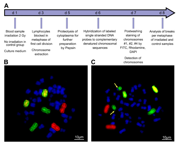
Chromosomal radiation sensitivity of blood lymphocytes was studied using three-color G0 FiSH. Aberrations in chromosomes #1, #2 and #4 induced by ionizing radiation were analyzed for this purpose [26]. Background aberrations and aberrations after irradiation with 2 Gy ionizing radiation (IR) were analyzed and the background was subtracted from the IR-induced aberrations (Figure 1).
Figure 1.
Chromosomal radiosensitivity testing procedure and analysis of chromosomal aberrations scored as breaks per metaphase. (A) Chromosomal radiation sensitivity test procedure of ex vivo blood for irradiation and generation of chromosomal aberrations. (B) Unaffected metaphase with red stained chromosome #1, green chromosome #2 and yellow chromosome #4. (C) Chromosome #2 involved in a translocation depicted by yellow arrows.
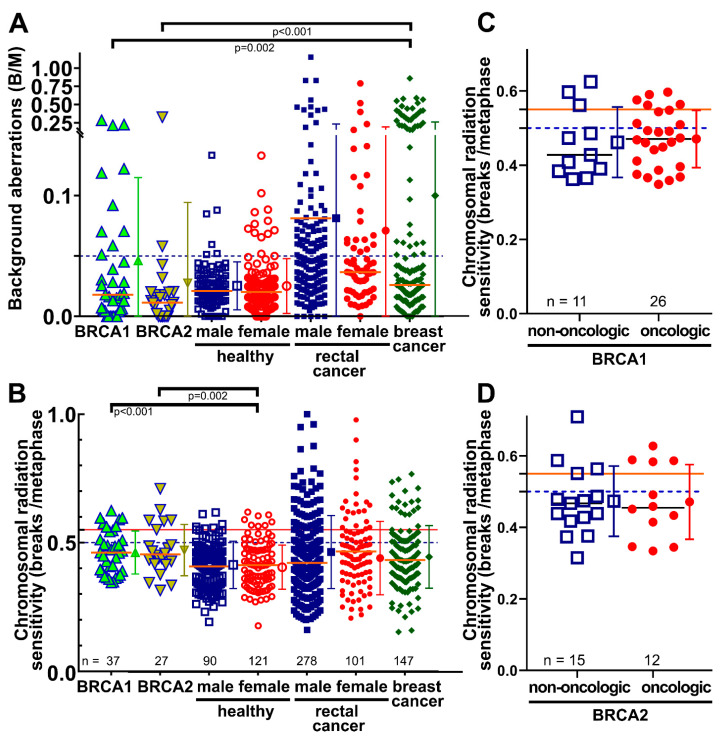
Background aberrations in the cohort with a BRCA1 pathologic variant (0.047 B/M ± standard deviation 0.069) and a BRCA2 pathologic variant (0.012 B/M ± 0.013) were overall comparable as in the control group of healthy female individuals with unknown BRCA1/2 mutation status (0.025 B/M ± 0.023) (p = 0.072 and 0.863). Although there was no difference between BRCA1 and BRCA2 (p = 0.290), the BRCA1-mutated cohort had a markedly higher proportion of 27.8% with elevated background levels above 0.05 B/M (Table 2). This was similar to the group of women with rectal cancer (25.2%) or with breast cancer (30.6%) with unknown mutation status (Figure 2A).
Table 2.
Proportion of individuals in each cohort with chromosomal aberrations above specified thresholds.
| Healthy | Rectal Cancer | Breast | ||||||
|---|---|---|---|---|---|---|---|---|
| Threshold | BRCA1 | BRCA2 | Male | Female | Male | Female | Cancer | |
| Background (%) | ≥0.05 | 27.8 | 8.7 | 6.7 | 9.9 | 36.7 | 25.2 | 30.6 |
| Radiation sensitives (%) |
≥0.5 | 30.6 | 30.4 | 18.9 | 14.9 | 23.9 | 27.3 | 29.9 |
| ≥0.55 | 19.4 | 30.4 | 8.9 | 5.8 | 17.6 | 15.2 | 12.9 | |
| ≥0.6 | 2.8 | 8.7 | 3.3 | 1.7 | 11.9 | 10.6. | 11.6 | |
Figure 2.
Chromosomal aberrations scored as breaks per metaphase in different cohorts. The entire cohort consisted of 37 female patients with pathogenic mutations in BRCA1 and 27 patients with pathogenic mutations in BRCA2, 121 female and 90 male healthy individuals, 101 female and 278 male patients with rectal cancer, and 147 female patients with breast cancer (the latter ones all with unknown BRCA1 mutation status). (A) Background aberrations for breaks per metaphase and (B) chromosomal radiation sensitivity scored as breaks per metaphase after ex vivo irradiation with 2 Gy ionizing radiation and background correction. (C) B/M values in the non-oncologic and oncologic BRCA1 group. (D) B/M values in the non-oncologic and oncologic BRCA2 group. Brackets indicate significant levels of differences between two groups. Horizontal lines depict 0.05 (A), and 0.5 and 0.55 (B) B/M cut-off values. B/M = Breaks per metaphase. Error bars indicate the mean value and the standard deviation.
3.3. B/M Values in Patient Cohorts
After ex vivo irradiation and background subtraction, the chromosomal radiation sensitivity of patients with BRCA1 mutations (0.464 B/M ± 0.083) or BRCA2 mutations (0.476 B/M ± 0.099) was quite similar (p = 0.706) (Table 3). Overall, patients with BRCA1 and BRCA2 pathologic variants had higher mean B/M values compared to the healthy control cohorts (Figure 2B). The BRCA1 cohort and BRCA2 cohort were clearly more chromosomal-radiosensitive than the female (n = 121) healthy cohort (p < 0.001 and p = 0.002) and the total healthy cohort (p < 0.001 and p = 0.002). Elevated chromosomal radiosensitivity above the threshold of 0.5 B/M was found in 18 patients, nine above 0.55 and three above 0.6 B/M (Table 2). There was no difference in B/M values between men and women in the healthy group (p = 0.954).
Table 3.
B/M values in all analyzed cohorts. Cohorts of healthy individuals and patients with rectal cancer and breast cancer have unknown BRCA1/2 mutation status.
| Mean B/M Value (2 Gy) | p-Value | 95% Confidence Interval | |
|---|---|---|---|
| Patients with a BRCA1/2 mutation (n = 64) | 0.468 | --- | 0.445 to 0.491 |
| Healthy individuals (n = 215) | 0.411 | <0.001 | 0.399 to 0.423 |
| Patients with rectal cancer (n = 385) | 0.439 | 0.039 | 0.420 to 0.459 |
| Patients with breast cancer (n = 147) | 0.447 | 0.235 | 0.426 to 0.468 |
There was no difference between the BRCA1 cohort and the BRCA2 cohort and the female cohort with advanced rectal cancer (p = 0.359 and p = 0.187) as well as with the total cohort of advanced rectal cancer (p = 0.190 and p = 0.050). Breast cancer patients were not different from BRCA1- (p = 0.405) or BRCA2- (p = 0.191) mutated patients (Table 3) (Figure 2B). Similarly, healthy individuals and cancer patients with either BRCA1 (p = 0.700) or BRCA2 (p = 0.999) did not differ from each other (Figure 2C,D).
3.4. Radiation Sensitivity among the Non-Oncologic and Oncologic BRCA1/2-Mutated Individuals
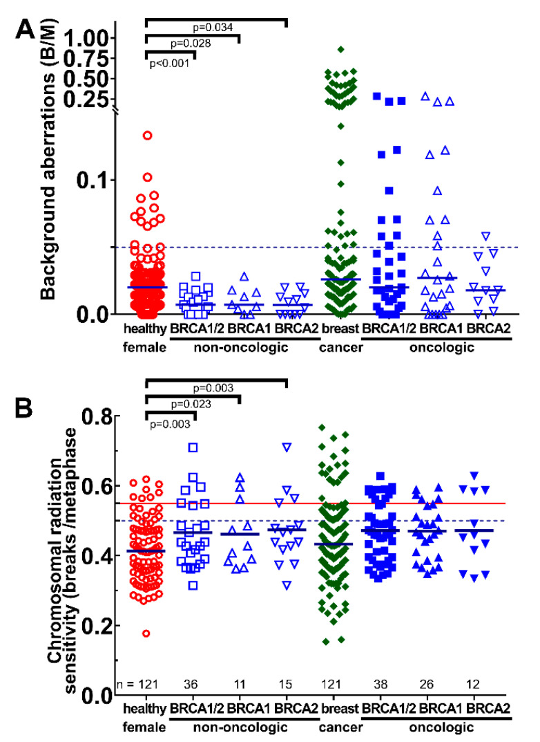
Therefore, next, the non-oncologic BRCA1/2 were compared with the healthy female individuals and the oncologic BRCA1/2 were compared with the patients with breast cancer. These two groups may be better controls because of their cancer and possible mutation status. Non-oncologic individuals with BRCA1/2 had a clearly lower background B/M compared to the healthy female individuals (p < 0.034). Yet, there was no difference between breast cancer patients and oncologic BRCA1/2. What is strikingly very interesting is that the non-oncologic BRCA1/2 have a significantly lower background of B/M compared to the oncologic BRCA1/2 (p < 0.001) (Figure 3A). Similarly, in the ex vivo-irradiated lymphocytes, non-oncologic BRCA1/2 subjects had a distinct increased B/M compared to healthy individuals (p < 0.023). Again, there were no differences between oncologic BRCA1/2 and breast cancer patients (Figure 3B).
Figure 3.
Chromosomal aberrations scored as breaks per metaphase in the non-oncologic and oncologic BRCA1/2 cohorts. Healthy women were compared with all non-oncologic BRCA1/2 or BRCA1 or BRCA2 subjects, and breast cancer patients were compared with oncologic BRCA1/2 or BRCA1 or BRCA2 patients. (A) Background aberrations for breaks per metaphase and (B) chromosomal radiation sensitivity scored as breaks per metaphase after ex vivo irradiation with 2 Gy ionizing radiation and background correction. Horizontal lines depict 0.05 (A), and 0.5 and 0.55 (B) B/M cut-off values. B/M = Breaks per metaphase.
3.5. Radiation Sensitivity with Location or Type of BRCA1/2 Mutations
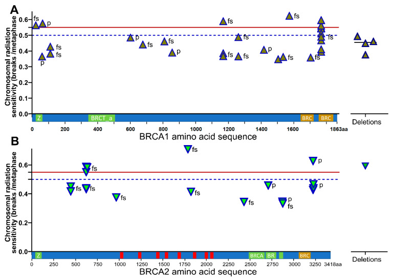
An important question was whether a specific mutation or a specific location of the mutation particularly increases chromosomal radiosensitivity. In the BRCA1 gene, most of the mutations were unique. We had two patients with mutations at positions p.111 (0.406 B/M) and p.1253 (0.427 B/M) and three mutations at position p.1161 (0.448 B/M). The only location of a mutation with a larger number of patients was p.1756 (p.Gln1756Pro fs*74) with ten patients. These ten patients had a significantly higher mean chromosomal radiosensitivity of 0.512 B/M compared to all others with a mean of 0.442 B/M (p = 0.027). Six out of these ten patients had B/M values higher than 0.5. The four patients with deletions in the BRCA1 gene had no increased chromosomal radiosensitivity. Four other patients had values above 0.55 B/M. Three of them had mutations that occurred only once (p.Glu23Valfs*17; p.Cys64Tyr; Tyr1563Stop). One mutation was present three times with one B/M value above 0.55, and the two other B/M values were average (<0.4) (Glu1161Phefs*3) (Figure 4A).
Figure 4.
Chromosomal radiation sensitivity depending on the location of pathologic variants in the amino acid sequence of the BRCA1 and BRCA2 proteins. (A) Location of pathological variants in relation to radiosensitivity in the BRCA1 protein; (B) Location of pathological variants in relation to radiosensitivity in the BRCA2 protein. fs = Frame shift mutation; p = Point mutation.
In the BRCA2 gene, two mutations each were at sites p.433 (0.434 B/M), aa3128 (0.452 B/M) and p.3124 (0.527 B/M). One patient in the latter group had a higher B/M value than 0.55. Four mutations were at p.605 with a high average value of 0.502 B/M, of which two patients again had a higher B/M value than 0.55. Only one patient had a deletion in the BRCA2 gene, but with a high chromosomal radiosensitivity of 0.589 B/M (Figure 4B).
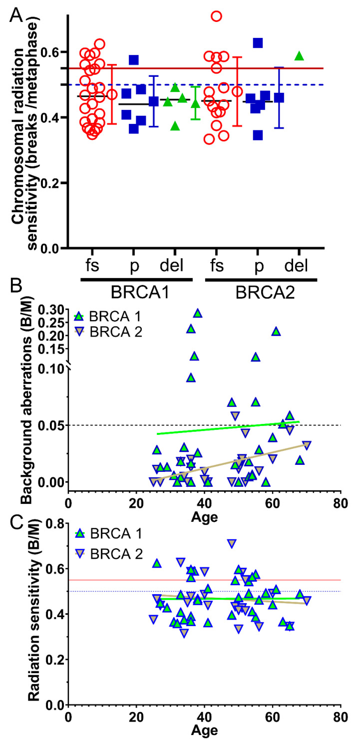
We also investigated whether a particular type of mutation (frameshift mutation, point mutation, deletion) is associated with high chromosomal radiosensitivity. Neither in the BRCA1 nor in the BRCA2 gene was a significantly increased chromosomal radiosensitivity linked to a specific mutation type (Figure 5A).
Figure 5.
Dependence of type of mutation and age from radiation sensitivity. (A) Radiation sensitivity depending on the type of mutation in the BRCA1 and BRCA2 gene. fs = Frameshift mutation; p = Point mutation; del = Deletion. (B) Age-related dependence of background aberrations. (C) Age-related dependence of radiation sensitivity.
Lastly, we studied whether there is an association of radiation sensitivity with age in BRCA1/2-mutated patients. The BRCA1 cohort was 45.1 years old (range 26–68) and the BRCA2 cohort was 46.6 (25–70) years old. Background values for BRCA1 were not associated with age (r2 = 0.044, p = 0.805), and only BRCA2 showed a slight increase with age (r2 = 0.534, p = 0.009) (Figure 5B), while radiation sensitivity did not change with age for BRCA1 (r2 = 0.008, p = 0.963) and BRCA2 (r2 = −0.074, p = 0.736) (Figure 5C).
4. Discussion
In this study, we found that overall BRCA1/2 variants do not confer clearly increased chromosomal radiosensitivity. However, some germline mutations in BRCA1 and BRCA2 lead to increased chromosomal radiosensitivity in the cohort. Individual variants may result in increased radiosensitivity. However, this would need to be confirmed in a study with a larger number of patients. It is difficult to obtain appropriate controls for this cohort. Therefore, we used different control cohorts for comparison. None of the controls have a known mutation status for BRCA1/2 or other variants. This is because the cohorts were established historically, and healthy and tumor patients were not tested and therefore did not know their mutation status. Therefore, we can only estimate the mutation burden in the control cohorts. However, given that the prevalence of BRCA1/2 mutations is estimated to be 1:400–1:500 in the general population [32,33] and 1:30–1:40 in the average breast cancer patient [34], it can be concluded that the vast majority of these patients are BRCA1/2-mutation-negative. Looking at the main breast cancer predisposition genes, 1.6% of the control group and 5.0% of the breast cancer cohort would have a pathologic mutation in a breast cancer gene. This is without knowing the overall effect of these genes on radiation sensitivity [4]. Pathologic variants in colorectal cancer predisposition genes are estimated to be present in 5–16% of colorectal tumors [35].
Our findings confirm the few available data samples on the irradiation of patients with germline BRCA1/2 mutations. However, it has to be noted that these studies used slightly different techniques of analysis after irradiation than in our work [36,37,38,39]. Patients with somatic BRCA1/2 mutations were also found to have above-average radiosensitivity in another retrospective analysis of medical records [40]. In our study, chromosomal radiosensitivity varied greatly from average chromosomal radiosensitivity to very high chromosomal radiosensitivity. Although no correlation with the type of mutation (e.g., frameshift, point mutation, deletion) could be found, there was a wide spectrum of radiosensitivity depending on the specific BRCA1/2 mutation. In the BRCA1 protein, the mutation p.Gln1756Pro fs*74 was associated with a distinct increased chromosomal radiosensitivity. However, even with this common mutation, affected patients showed a wide variety from moderate to high radiation sensitivity. There were four other single mutations in the BRCA1 and BRCA2 genes that were highly sensitive to radiation, but they were only present in a single case. The patient with the p.Glu1734Glyfs*9 mutation in the BRCA2 gene encountered extreme high chromosomal radiosensitivity; yet, this mutation was also present in a single case. Because of this range of variation, other genetic and non-genetic factors could play a role in radiosensitive patients too, including single-nucleotide polymorphisms or mutations in other relevant genes as have been studied in non-BRCA1/2-mutated breast cancer patients [6,41,42].
DNA double-strand breaks, which are the most significant type of DNA damage induced by radiotherapy, are repaired via two distinct repair pathways: the non-homologous end joining (NHEJ) pathway and the HR pathway. Activation of either pathway also depends on the cell cycle phase. NHEJ is dominant in the G0/G1 phase while HR is dominant in the mid-S and mid-G2 phase. Both BRCA1 and BRCA2 play a crucial role in the repair of DNA DSBs [40]. If these DNA double-strand breaks are not repaired or are repaired incorrectly, chromosomal aberrations and mutations occur, which can be analyzed with the ex vivo three-color FiSH assay. Elevated mutation levels in the FiSH assay indicate disruptions in DNA double-strand break repair, signal transduction, cell cycle regulation and cell death control [23,43].
An important advantage of the three-color FiSH assay is its late endpoint, which makes it particularly suitable for determining individual radiation sensitivity. Another advantage is that with only the three colors and the chromosomes 1, 2 and 4, 22% of the total DNA can be screened for aberrations. In addition, the three colors are very easy and intuitive to recognize, making interpretation easier and more reliable [25,44]. After ex vivo irradiation, lymphocytes in G0 must undergo DNA repair and go through the entire cell cycle with all its checkpoints. Although the FiSH has great advantages for radiation sensitivity determination, individual radiation sensitivity is a complex construct that cannot always be addressed by a single test. However, the FiSH assay has been shown to predict radiosensitivity in several previous publications [43,45]. Individual radiosensitivity can be used to predict the risk of undesired side-effects of radiation therapy like fibrosis, skin toxicity or fatigue [19,46,47,48]. In addition, radiation sensitivity is genetically determined, so that the radiation sensitivity of lymphocytes can be extrapolated to all body cells and, in principle, to tumor cells.In a previously published paper, chromosomal radiosensitivity was increased by age; however, this finding was not significant, due to individual variability [21]. Here, among the BRCA1/2 patients, no increase in radiation sensitivity with age was observed. However, non-oncologic BRCA1/2 subjects had significantly fewer background aberrations compared with healthy controls and compared with oncologic BRCA2. Non-oncologic BRCA1 (37.5 years) and BRCA2 (38.8 years) were about 10 years younger than oncologic BRCA1 (48.8 years) and BRCA2 (51.0 years) and healthy females (49.5 years). The chromosomal background aberration increases with age [49] and this certainly explains most of this difference.
Chromosomal aberrations in the ex vivo-irradiated blood of non-oncological BRCA1/2 patients were also significantly higher than in the healthy female control group. This is probably the most comparable group, as both represent non-oncologic subjects who once had BRCA1/2 variants and once were very unlikely to have pathologic BRCA1/2 variants in the healthy group. This clearly suggests that BRCA1/2 contributes to an increase in radiation sensitivity, albeit to a limited extent. In oncologic BRCA1/2 patients, B/M are similarly elevated compared with breast cancer patients, but without statistical significance. The control group of breast cancer patients certainly contains an unknown number of genetic variants of cancer genes and, therefore, probably, no such good separation is possible as in the healthy and the non-oncological BRCA1/2 cohort. We emphasize the importance of measuring radiation sensitivity in individual patients suspected of having increased radiation sensitivity.
In such studies, it is important to have a large, controlled cohort to work with. In this study, there were 211 healthy individuals and 379 patients with rectal cancer as well as 147 patients with breast cancer. The majority of the patients were male. This is probably due to higher background levels in patients with rectal cancer because of their less balanced diet, smoking or other risk factors [50]. However, there were no sex-related differences in inducing chromosomal aberrations by ionizing radiation. Thus, males and females can be used equally as control groups. It is also necessary to determine the cut-off values for increased sensitivity to radiation from the control groups. The threshold for increased radiation sensitivity was set at 0.5 B/M [51]. This is the mean of a Gaussian distribution of a healthy cohort plus three times the standard deviation. However, recognizing that empirically derived therapy doses tend to be geared toward the more radiation-sensitive patients, the threshold above which we recommend dose reduction for patients has been set at 0.55 B/M. Establishing limits for radiation sensitivity is generally difficult. The risk of adverse effects beyond this threshold is still low and increases slowly with dose. Side-effects may not appear for years and are sometimes difficult to objectify. A very important factor in dose reductions is that recurrences do not increase, which would clearly indicate a false limit or dose reduction.
In patients with BRCA1/2 mutations, reports on side-effects from irradiation are relatively rare and primarily retrospective [52]. In most studies, there were no increased late tissue side-effects in the BRCA1/2-mutated vs. non-mutated cohorts [53,54,55,56]. One explanation could be haploinsufficiency, which means that BRCA1/2-mutated patients only have a germline mutation in one allele with a second functional allele [52,57]. This is contradicted by the mutation analysis of our cohort of BRCA1/2 mutation carriers, which clearly reveals a higher incidence of aberrations. Similarly, more chromatid breaks occurred in lymphocytes of heterozygous BRCA1-mutated female patients [58]. This could potentially go along with an increase in tissue damage. However, radiation after breast cancer in general has very little toxicity and is a well-tolerated standard therapy [59]. Therefore, the risk of undesirable side-effects is still low in patients with an increased sensitivity to radiation. For instance, only one patient from 170 accelerated partial breast irradiation patients in the ABPI2 study had a grade 2 pneumonitis [60]. Chromosomal radiosensitivity analysis in this patient showed a significantly increased chromosomal radiosensitivity of 0.79 B/M, presumably due to vitiligo. In another case, a 6-year-old boy with Phelan–McDermid syndrome and an atypical teratoid/rhabdoid tumor (AT/RT), who was measured to have a markedly elevated radiation sensitivity of 0.74 B/M, showed no adverse effects of therapy four years after treatment with a significant dose reduction from 54 Gy to 31 Gy [61]. A case of a three-year-old girl with the same genetic disease and tumor was reported in the literature. She received no dose adjustment and suffers from severe treatment sequelae [62]. This clearly demonstrates the relationship between significantly increased radiation sensitivity and increased risk of side-effects from therapy.
These representative cases illustrate that the risk of side-effects increases dramatically when there is significantly increased radiosensitivity, even with a very low toxic regimen. Another aspect is that when patients are treated with more toxic regimens, such as in head and neck cancer [63], and there is a BRCA1/2 variant with increased radiosensitivity, the risk of adverse therapeutic effects would increase significantly. Consequently, the identification of radiosensitive patients is of interest, especially with regard to specific pathologic variants or specific areas in the associated proteins that are linked to higher radiosensitivity. Such deeper knowledge would avoid the need for time-consuming radiosensitivity testing.
5. Conclusions
Patients with germline BRCA1/2 mutations have very different levels of chromosomal radiosensitivity, ranging from average to clearly elevated chromosomal radiosensitivity in an ex vivo FiSH assay. At present, radiosensitivity cannot be predicted by general types of mutations, e.g., point mutation, frameshift mutation or deletion. However, it appears that certain mutations in BRCA1 and BRCA2 are associated with higher rates of chromosome breaks in the ex vivo FiSH assay. With conservative fractionated breast radiotherapy, the risk of adverse events in these patients appears to be limited [64]. However, there may be an increased risk of using a regimen with higher toxicity that could lead to increased side-effects in patients with certain BRCA1/2 mutations. This study justifies further research on radiosensitivity in patients with mutations in BRCA1/2 and also other genes involved in DNA repair, requiring larger numbers of patients studied.
Acknowledgments
I thank Elisabeth Müller, Doris Mehler and Lukas Kuhlmann for the excellent technical expertise as well as help in conducting experiments, and all individuals for consent to donate blood. We thank the department of Gynecology and Obstetrics at the Friedrich-Alexander University Erlangen-Nürnberg, Germany for providing us with patient data. The present work was performed by the first author in fulfilment of the requirements for obtaining the degree “Dr. med.” At Friedrich-Alexander University Erlangen-Nürnberg (FAU).
Abbreviations
| ACMG | American College of Medical Genetics and Genomics |
| AWMF | Arbeitsgemeinschaft der Wissenschaftlichen Medizinischen Fachgesellschaften e.V. |
| B/M | Breaks per metaphase |
| BRCA1/2 | BReast CAncer 1/2 |
| DAPI | 4′,6-diamidin-2-phenylindol |
| FiSH | Fluorescence in situ hybridization |
| HR | Homologous recombination |
| IR | Ionizing radiation; |
| NHEJ | Non homologous end joining |
| PARP | poly-ADP-ribose polymerase |
| RPMI | Roswell Park Memorial Institute Medium |
Author Contributions
Conceptualization, L.D. and T.Z.K., methodology T.Z.K., L.K. and L.D.; software, L.D.; validation, M.W., M.R., H.H., P.A.F., M.W.B., C.C.H., R.F., T.Z.K. and L.D.; formal analysis, T.Z.K. and L.D.; resources, M.W., M.R., H.H., M.W.B, C.C.H. and P.A.F., data curation, L.D. and T.Z.K., writing—original draft preparation, T.Z.K. and L.D.; writing—review and editing, T.Z.K., M.W., L.D., A.R. and R.F.; visualization, T.Z.K. and L.D.; supervision, L.D., J.H. and A.R. genetic analyses. All authors have read and agreed to the published version of the manuscript.
Institutional Review Board Statement
This study was approved by the ethics review committee of the Friedrich-Alexander University Erlangen-Nürnberg (21_19 B).
Informed Consent Statement
Written informed consent was obtained from all the patients.
Data Availability Statement
All data are available in the main text. Further data can be obtained from the corresponding author on request.
Conflicts of Interest
The authors declare no conflict of interest.
Funding Statement
This research received no external funding.
Footnotes
Disclaimer/Publisher’s Note: The statements, opinions and data contained in all publications are solely those of the individual author(s) and contributor(s) and not of MDPI and/or the editor(s). MDPI and/or the editor(s) disclaim responsibility for any injury to people or property resulting from any ideas, methods, instructions or products referred to in the content.
References
- 1.Arnold M., Morgan E., Rumgay H., Mafra A., Singh D., Laversanne M., Vignat J., Gralow J.R., Cardoso F., Siesling S., et al. Current and future burden of breast cancer: Global statistics for 2020 and 2040. Breast. 2022;66:15–23. doi: 10.1016/j.breast.2022.08.010. [DOI] [PMC free article] [PubMed] [Google Scholar]
- 2.Sung H., Ferlay J., Siegel R.L., Laversanne M., Soerjomataram I., Jemal A., Bray F. Global Cancer Statistics 2020: GLOBOCAN Estimates of Incidence and Mortality Worldwide for 36 Cancers in 185 Countries. CA Cancer J. Clin. 2021;71:209–249. doi: 10.3322/caac.21660. [DOI] [PubMed] [Google Scholar]
- 3.Howlader N., Noone A.M., Miller D., Krapcho M., Brest A., Yu M., Ruhl J., Tatalovich Z., Mariotto A., Lewis D.R., et al. SEER Cancer Statistics Review, 1975–2017; Based on November 2019 SEER Data Submission, Posted to the SEER Web Site; National Cancer Institute: Bethesda, MD, USA, 2020. [(accessed on 11 February 2023)]; Available online: https://seer.cancer.gov/csr/1975_2017.
- 4.Hu C., Hart S.N., Gnanaolivu R., Huang H., Lee K.Y., Na J., Gao C., Lilyquist J., Yadav S., Boddicker N.J., et al. A Population-Based Study of Genes Previously Implicated in Breast Cancer. N. Engl. J. Med. 2021;384:440–451. doi: 10.1056/NEJMoa2005936. [DOI] [PMC free article] [PubMed] [Google Scholar]
- 5.Brewer H.R., Jones M.E., Schoemaker M.J., Ashworth A., Swerdlow A.J. Family history and risk of breast cancer: An analysis accounting for family structure. Breast Cancer Res. Treat. 2017;165:193–200. doi: 10.1007/s10549-017-4325-2. [DOI] [PMC free article] [PubMed] [Google Scholar]
- 6.Dorling L., Carvalho S., Allen J., Gonzalez-Neira A., Luccarini C., Wahlstrom C., Pooley K.A., Parsons M.T., Fortuno C., Wang Q., et al. Breast Cancer Risk Genes-Association Analysis in More than 113,000 Women. N. Engl. J. Med. 2021;384:428–439. doi: 10.1056/NEJMoa1913948. [DOI] [PMC free article] [PubMed] [Google Scholar]
- 7.Chen S., Parmigiani G. Meta-analysis of BRCA1 and BRCA2 penetrance. J. Clin. Oncol. 2007;25:1329–1333. doi: 10.1200/JCO.2006.09.1066. [DOI] [PMC free article] [PubMed] [Google Scholar]
- 8.Kuchenbaecker K.B., Hopper J.L., Barnes D.R., Phillips K.A., Mooij T.M., Roos-Blom M.J., Jervis S., van Leeuwen F.E., Milne R.L., Andrieu N., et al. Risks of Breast, Ovarian, and Contralateral Breast Cancer for BRCA1 and BRCA2 Mutation Carriers. JAMA. 2017;317:2402–2416. doi: 10.1001/jama.2017.7112. [DOI] [PubMed] [Google Scholar]
- 9.Li S., Silvestri V., Leslie G., Rebbeck T.R., Neuhausen S.L., Hopper J.L., Nielsen H.R., Lee A., Yang X., McGuffog L., et al. Cancer Risks Associated With BRCA1 and BRCA2 Pathogenic Variants. J. Clin. Oncol. 2022;40:1529–1541. doi: 10.1200/jco.21.02112. [DOI] [PMC free article] [PubMed] [Google Scholar]
- 10.El-Nachef L., Al-Choboq J., Restier-Verlet J., Granzotto A., Berthel E., Sonzogni L., Ferlazzo M.L., Bouchet A., Leblond P., Combemale P., et al. Human Radiosensitivity and Radiosusceptibility: What Are the Differences? Int. J. Mol. Sci. 2021;22:7158. doi: 10.3390/ijms22137158. [DOI] [PMC free article] [PubMed] [Google Scholar]
- 11.Yoshida K., Miki Y. Role of BRCA1/2 and BRCA1/2 as regulators of DNA repair, transcription, and cell cycle in response to DNA damage. Cancer Sci. 2004;95:866–871. doi: 10.1111/j.1349-7006.2004.tb02195.x. [DOI] [PMC free article] [PubMed] [Google Scholar]
- 12.Gorodetska I., Kozeretska I., Dubrovska A. BRCA Genes: The Role in Genome Stability, Cancer Stemness and Therapy Resistance. J. Cancer. 2019;10:2109–2127. doi: 10.7150/jca.30410. [DOI] [PMC free article] [PubMed] [Google Scholar]
- 13.Wöckel A., Festl J., Stüber T., Brust K., Stangl S., Heuschmann P.U., Albert U.S., Budach W., Follmann M., Janni W., et al. Interdisciplinary Screening, Diagnosis, Therapy and Follow-up of Breast Cancer. Guideline of the DGGG and the DKG (S3-Level, AWMF Registry Number 032/045OL, December 2017)-Part 1 with Recommendations for the Screening, Diagnosis and Therapy of Breast Cancer. Geburtshilfe Frauenheilkd. 2018;78:927–948. doi: 10.1055/a-0646-4522. [DOI] [PMC free article] [PubMed] [Google Scholar]
- 14.Fahrig A., Koch T., Lenhart M., Rieckmann P., Fietkau R., Distel L., Schuster B. Lethal outcome after pelvic salvage radiotherapy in a patient with prostate cancer due to increased radiosensitivity: Case report and literature review. Strahlenther. Onkol. 2018;194:60–66. doi: 10.1007/s00066-017-1207-9. [DOI] [PubMed] [Google Scholar]
- 15.Zhou Y., Yan T., Zhou X., Cao P., Luo C., Zhou L., Xu Y., Liu Y., Xue J., Wang J., et al. Acute severe radiation pneumonitis among non-small cell lung cancer (NSCLC) patients with moderate pulmonary dysfunction receiving definitive concurrent chemoradiotherapy: Impact of pre-treatment pulmonary function parameters. Strahlenther. Onkol. 2020;196:505–514. doi: 10.1007/s00066-019-01552-4. [DOI] [PubMed] [Google Scholar]
- 16.Klement R.J., Schäfer G., Sweeney R.A. A fatal case of Fournier’s gangrene during neoadjuvant radiotherapy for rectal cancer. Strahlenther. Onkol. 2019;195:441–446. doi: 10.1007/s00066-018-1401-4. [DOI] [PubMed] [Google Scholar]
- 17.Scheckenbach K., Wagenmann M., Freund M., Schipper J., Hanenberg H. Squamous cell carcinomas of the head and neck in Fanconi anemia: Risk, prevention, therapy, and the need for guidelines. Klin. Padiatr. 2012;224:132–138. doi: 10.1055/s-0032-1308989. [DOI] [PMC free article] [PubMed] [Google Scholar]
- 18.Taylor A.M., Harnden D.G., Arlett C.F., Harcourt S.A., Lehmann A.R., Stevens S., Bridges B.A. Ataxia telangiectasia: A human mutation with abnormal radiation sensitivity. Nature. 1975;258:427–429. doi: 10.1038/258427a0. [DOI] [PubMed] [Google Scholar]
- 19.Hoeller U., Borgmann K., Bonacker M., Kuhlmey A., Bajrovic A., Jung H., Alberti W., Dikomey E. Individual radiosensitivity measured with lymphocytes may be used to predict the risk of fibrosis after radiotherapy for breast cancer. Radiother. Oncol. 2003;69:137–144. doi: 10.1016/j.radonc.2003.10.001. [DOI] [PubMed] [Google Scholar]
- 20.Auer J., Keller U., Schmidt M., Ott O., Fietkau R., Distel L.V. Individual radiosensitivity in a breast cancer collective is changed with the patients’ age. Radiol. Oncol. 2014;48:80–86. doi: 10.2478/raon-2013-0061. [DOI] [PMC free article] [PubMed] [Google Scholar]
- 21.Schuster B., Ellmann A., Mayo T., Auer J., Haas M., Hecht M., Fietkau R., Distel L.V. Rate of individuals with clearly increased radiosensitivity rise with age both in healthy individuals and in cancer patients. BMC Geriatr. 2018;18:105. doi: 10.1186/s12877-018-0799-y. [DOI] [PMC free article] [PubMed] [Google Scholar]
- 22.Schuster B., Hecht M., Schmidt M., Haderlein M., Jost T., Buttner-Herold M., Weber K., Denz A., Grutzmann R., Hartmann A., et al. Influence of Gender on Radiosensitivity during Radiochemotherapy of Advanced Rectal Cancer. Cancers. 2021;14:148. doi: 10.3390/cancers14010148. [DOI] [PMC free article] [PubMed] [Google Scholar]
- 23.Stritzelberger J., Lainer J., Gollwitzer S., Graf W., Jost T., Lang J.D., Mueller T.M., Schwab S., Fietkau R., Hamer H.M., et al. Ex vivo radiosensitivity is increased in non-cancer patients taking valproate. BMC Neurol. 2020;20:390. doi: 10.1186/s12883-020-01966-z. [DOI] [PMC free article] [PubMed] [Google Scholar]
- 24.Dunst J., Gebhart E., Neubauer S. Can an extremely elevated radiosensitivity in patients be recognized by the in-vitro testing of lymphocytes? Strahlenther. Onkol. 1995;171:581–586. [PubMed] [Google Scholar]
- 25.Neubauer S., Dunst J., Gebhart E. The impact of complex chromosomal rearrangements on the detection of radiosensitivity in cancer patients. Radiother. Oncol. 1997;43:189–195. doi: 10.1016/s0167-8140(97)01932-4. [DOI] [PubMed] [Google Scholar]
- 26.Keller U., Kuechler A., Liehr T., Muller E., Grabenbauer G., Sauer R., Distel L. Impact of Various Parameters in Detecting Chromosomal Aberrations by FISH to Describe Radiosensitivity. Strahlenther. Onkol. 2004;180:289–296. doi: 10.1007/s00066-004-1200-y. [DOI] [PubMed] [Google Scholar]
- 27.Stritzelberger J., Distel L., Buslei R., Fietkau R., Putz F. Acquired temozolomide resistance in human glioblastoma cell line U251 is caused by mismatch repair deficiency and can be overcome by lomustine. Clin. Transl. Oncol. 2018;20:508–516. doi: 10.1007/s12094-017-1743-x. [DOI] [PubMed] [Google Scholar]
- 28.Savage J.R., Simpson P. On the scoring of FISH-"painted" chromosome-type exchange aberrations. Mutat. Res. 1994;307:345–353. doi: 10.1016/0027-5107(94)90308-5. [DOI] [PubMed] [Google Scholar]
- 29.Hecht M., Zimmer L., Loquai C., Weishaupt C., Gutzmer R., Schuster B., Gleisner S., Schulze B., Goldinger S.M., Berking C., et al. Radiosensitization by BRAF inhibitor therapy-mechanism and frequency of toxicity in melanoma patients. Ann. Oncol. 2015;26:1238–1244. doi: 10.1093/annonc/mdv139. [DOI] [PubMed] [Google Scholar]
- 30.Weigert V., Jost T., Hecht M., Knippertz I., Heinzerling L., Fietkau R., Distel L.V. PARP inhibitors combined with ionizing radiation induce different effects in melanoma cells and healthy fibroblasts. BMC Cancer. 2020;20:775. doi: 10.1186/s12885-020-07190-9. [DOI] [PMC free article] [PubMed] [Google Scholar]
- 31.Dobler C., Jost T., Hecht M., Fietkau R., Distel L. Senescence Induction by Combined Ionizing Radiation and DNA Damage Response Inhibitors in Head and Neck Squamous Cell Carcinoma Cells. Cells. 2020;9:2012. doi: 10.3390/cells9092012. [DOI] [PMC free article] [PubMed] [Google Scholar]
- 32.Anglian Breast Cancer Study Group Prevalence and penetrance of BRCA1 and BRCA2 mutations in a population-based series of breast cancer cases. Anglian Breast Cancer Study Group. Br. J. Cancer. 2000;83:1301–1308. doi: 10.1054/bjoc.2000.1407. [DOI] [PMC free article] [PubMed] [Google Scholar]
- 33.Whittemore A.S., Gong G., John E.M., McGuire V., Li F.P., Ostrow K.L., Dicioccio R., Felberg A., West D.W. Prevalence of BRCA1 mutation carriers among U.S. non-Hispanic Whites. Cancer Epidemiol. Biomark. Prev. 2004;13:2078–2083. doi: 10.1158/1055-9965.2078.13.12. [DOI] [PubMed] [Google Scholar]
- 34.Armstrong N., Ryder S., Forbes C., Ross J., Quek R.G. A systematic review of the international prevalence of BRCA mutation in breast cancer. Clin. Epidemiol. 2019;11:543–561. doi: 10.2147/CLEP.S206949. [DOI] [PMC free article] [PubMed] [Google Scholar]
- 35.Rebuzzi F., Ulivi P., Tedaldi G. Genetic Predisposition to Colorectal Cancer: How Many and Which Genes to Test? Int. J. Mol. Sci. 2023;24:2137. doi: 10.3390/ijms24032137. [DOI] [PMC free article] [PubMed] [Google Scholar]
- 36.Sadeghi F., Asgari M., Matloubi M., Ranjbar M., Karkhaneh Yousefi N., Azari T., Zaki-Dizaji M. Molecular contribution of BRCA1 and BRCA2 to genome instability in breast cancer patients: Review of radiosensitivity assays. Biol. Proced. Online. 2020;22:23. doi: 10.1186/s12575-020-00133-5. [DOI] [PMC free article] [PubMed] [Google Scholar]
- 37.Baert A., Depuydt J., Van Maerken T., Poppe B., Malfait F., Storm K., van den Ende J., Van Damme T., De Nobele S., Perletti G., et al. Increased chromosomal radiosensitivity in asymptomatic carriers of a heterozygous BRCA1 mutation. Breast Cancer Res. 2016;18:52. doi: 10.1186/s13058-016-0709-1. [DOI] [PMC free article] [PubMed] [Google Scholar]
- 38.Ernestos B., Nikolaos P., Koulis G., Eleni R., Konstantinos B., Alexandra G., Michael K. Increased chromosomal radiosensitivity in women carrying BRCA1/BRCA2 mutations assessed with the G2 assay. Int. J. Radiat. Oncol. Biol. Phys. 2010;76:1199–1205. doi: 10.1016/j.ijrobp.2009.10.020. [DOI] [PubMed] [Google Scholar]
- 39.Baeyens A., Thierens H., Claes K., Poppe B., Messiaen L., De Ridder L., Vral A. Chromosomal radiosensitivity in breast cancer patients with a known or putative genetic predisposition. Br. J. Cancer. 2002;87:1379–1385. doi: 10.1038/sj.bjc.6600628. [DOI] [PMC free article] [PubMed] [Google Scholar]
- 40.Kim K.H., Kim H.S., Kim S.S., Shim H.S., Yang A.J., Lee J.J.B., Yoon H.I., Ahn J.B., Chang J.S. Increased Radiosensitivity of Solid Tumors Harboring ATM and BRCA1/2 Mutations. Cancer Res. Treat. 2022;54:54–64. doi: 10.4143/crt.2020.1247. [DOI] [PMC free article] [PubMed] [Google Scholar]
- 41.Barnett G.C., Coles C.E., Elliott R.M., Baynes C., Luccarini C., Conroy D., Wilkinson J.S., Tyrer J., Misra V., Platte R., et al. Independent validation of genes and polymorphisms reported to be associated with radiation toxicity: A prospective analysis study. Lancet Oncol. 2012;13:65–77. doi: 10.1016/S1470-2045(11)70302-3. [DOI] [PubMed] [Google Scholar]
- 42.Barnett G.C., Thompson D., Fachal L., Kerns S., Talbot C., Elliott R.M., Dorling L., Coles C.E., Dearnaley D.P., Rosenstein B.S., et al. A genome wide association study (GWAS) providing evidence of an association between common genetic variants and late radiotherapy toxicity. Radiother. Oncol. 2014;111:178–185. doi: 10.1016/j.radonc.2014.02.012. [DOI] [PubMed] [Google Scholar]
- 43.Beaton L.A., Marro L., Samiee S., Malone S., Grimes S., Malone K., Wilkins R.C. Investigating chromosome damage using fluorescent in situ hybridization to identify biomarkers of radiosensitivity in prostate cancer patients. Int. J. Radiat. Biol. 2013;89:1087–1093. doi: 10.3109/09553002.2013.825060. [DOI] [PubMed] [Google Scholar]
- 44.Dunst J., Neubauer S., Becker A., Gebhart E. Chromosomal in-vitro radiosensitivity of lymphocytes in radiotherapy patients and AT-homozygotes. Strahlenther. Onkol. 1998;174:510–516. doi: 10.1007/BF03038983. [DOI] [PubMed] [Google Scholar]
- 45.Mayo T., Haderlein M., Schuster B., Wiesmüller A., Hummel C., Bachl M., Schmidt M., Fietkau R., Distel L. Is in vivo and ex vivo irradiation equally reliable for individual Radiosensitivity testing by three colour fluorescence in situ hybridization? Radiat. Oncol. 2019;15:2. doi: 10.1186/s13014-019-1444-4. [DOI] [PMC free article] [PubMed] [Google Scholar]
- 46.Borgmann K., Hoeller U., Nowack S., Bernhard M., Roper B., Brackrock S., Petersen C., Szymczak S., Ziegler A., Feyer P., et al. Individual radiosensitivity measured with lymphocytes may predict the risk of acute reaction after radiotherapy. Int. J. Radiat. Oncol. Biol. Phys. 2008;71:256–264. doi: 10.1016/j.ijrobp.2008.01.007. [DOI] [PubMed] [Google Scholar]
- 47.Liu L., Yang Y., Guo Q., Ren B., Peng Q., Zou L., Zhu Y., Tian Y. Comparing hypofractionated to conventional fractionated radiotherapy in postmastectomy breast cancer: A meta-analysis and systematic review. Radiat. Oncol. 2020;15:17. doi: 10.1186/s13014-020-1463-1. [DOI] [PMC free article] [PubMed] [Google Scholar]
- 48.Hauth F., De-Colle C., Weidner N., Heinrich V., Zips D., Gani C. Quality of life and fatigue before and after radiotherapy in breast cancer patients. Strahlenther. Onkol. 2021;197:281–287. doi: 10.1007/s00066-020-01700-1. [DOI] [PubMed] [Google Scholar]
- 49.Bolognesi C., Abbondandolo A., Barale R., Casalone R., Dalpra L., De Ferrari M., Degrassi F., Forni A., Lamberti L., Lando C., et al. Age-related increase of baseline frequencies of sister chromatid exchanges, chromosome aberrations, and micronuclei in human lymphocytes. Cancer Epidemiol. Biomark. Prev. 1997;6:249–256. [PubMed] [Google Scholar]
- 50.Lewandowska A., Rudzki G., Lewandowski T., Stryjkowska-Gora A., Rudzki S. Title: Risk Factors for the Diagnosis of Colorectal Cancer. Cancer Control. J. Moffitt Cancer Cent. 2022;29:10732748211056692. doi: 10.1177/10732748211056692. [DOI] [PMC free article] [PubMed] [Google Scholar]
- 51.Keller U., Grabenbauer G., Kuechler A., Sprung C., Müller E., Sauer R., Distel L. Cytogenetic instability in young patients with multiple primary cancers. Cancer Genet. Cytogenet. 2005;157:25–32. doi: 10.1016/j.cancergencyto.2004.05.018. [DOI] [PubMed] [Google Scholar]
- 52.Lazzari G., Buono G., Zannino B., Silvano G. Breast Cancer Adjuvant Radiotherapy in BRCA1/2, TP53, ATM Genes Mutations: Are There Solved Issues? Breast Cancer (Dove Med. Press) 2021;13:299–310. doi: 10.2147/BCTT.S306075. [DOI] [PMC free article] [PubMed] [Google Scholar]
- 53.Huszno J., Budryk M., Kolosza Z., Nowara E. The risk factors of toxicity during chemotherapy and radiotherapy in breast cancer patients according to the presence of BRCA gene mutation. Contemp. Oncol. (Pozn.) 2015;19:72–76. doi: 10.5114/wo.2015.50014. [DOI] [PMC free article] [PubMed] [Google Scholar]
- 54.Park H., Choi D.H., Noh J.M., Huh S.J., Park W., Nam S.J., Lee J.E. Acute skin toxicity in Korean breast cancer patients carrying BRCA mutations. Int. J. Radiat. Biol. 2014;90:90–94. doi: 10.3109/09553002.2013.835504. [DOI] [PubMed] [Google Scholar]
- 55.Pierce L.J., Levin A.M., Rebbeck T.R., Ben-David M.A., Friedman E., Solin L.J., Harris E.E., Gaffney D.K., Haffty B.G., Dawson L.A., et al. Ten-year multi-institutional results of breast-conserving surgery and radiotherapy in BRCA1/2-associated stage I/II breast cancer. J. Clin. Oncol. 2006;24:2437–2443. doi: 10.1200/JCO.2005.02.7888. [DOI] [PubMed] [Google Scholar]
- 56.Shanley S., McReynolds K., Ardern-Jones A., Ahern R., Fernando I., Yarnold J., Evans G., Eccles D., Hodgson S., Ashley S., et al. Late toxicity is not increased in BRCA1/BRCA2 mutation carriers undergoing breast radiotherapy in the United Kingdom. Clin. Cancer Res. 2006;12:7025–7032. doi: 10.1158/1078-0432.CCR-06-1244. [DOI] [PubMed] [Google Scholar]
- 57.Elalaoui S.C., Laarabi F.Z., Afif L., Lyahyai J., Ratbi I., Jaouad I.C., Doubaj Y., Sahli M., Ouhenach M., Sefiani A. Mutational spectrum of BRCA1/2 genes in Moroccan patients with hereditary breast and/or ovarian cancer, and review of BRCA mutations in the MENA region. Breast Cancer Res. Treat. 2022;194:187–198. doi: 10.1007/s10549-022-06622-3. [DOI] [PubMed] [Google Scholar]
- 58.Febrer E., Mestres M., Caballín M.R., Barrios L., Ribas M., Gutiérrez-Enríquez S., Alonso C., Ramón y Cajal T., Francesc Barquinero J. Mitotic delay in lymphocytes from BRCA1 heterozygotes unable to reduce the radiation-induced chromosomal damage. DNA Repair. (Amst.) 2008;7:1907–1911. doi: 10.1016/j.dnarep.2008.08.001. [DOI] [PubMed] [Google Scholar]
- 59.Wang K., Tepper J.E. Radiation therapy-associated toxicity: Etiology, management, and prevention. CA Cancer J. Clin. 2021;71:437–454. doi: 10.3322/caac.21689. [DOI] [PubMed] [Google Scholar]
- 60.Ott O.J., Stillkrieg W., Lambrecht U., Sauer T.O., Schweizer C., Lamrani A., Strnad V., Hack C.C., Beckmann M.W., Uder M., et al. External Beam Accelerated Partial Breast Irradiation in Early Breast Cancer and the Risk for Radiogenic Pneumonitis. Cancers. 2022;14:3520. doi: 10.3390/cancers14143520. [DOI] [PMC free article] [PubMed] [Google Scholar]
- 61.De Rubeis S., Siper P.M., Durkin A., Weissman J., Muratet F., Halpern D., Trelles M.D.P., Frank Y., Lozano R., Wang A.T., et al. Delineation of the genetic and clinical spectrum of Phelan-McDermid syndrome caused by SHANK3 point mutations. Mol. Autism. 2018;9:31. doi: 10.1186/s13229-018-0205-9. [DOI] [PMC free article] [PubMed] [Google Scholar]
- 62.Byers H.M., Adam M.P., LaCroix A., Leary S.E., Cole B., Dobyns W.B., Mefford H.C. Description of a new oncogenic mechanism for atypical teratoid rhabdoid tumors in patients with ring chromosome 22. Am. J. Med. Genet. A. 2017;173:245–249. doi: 10.1002/ajmg.a.37993. [DOI] [PMC free article] [PubMed] [Google Scholar]
- 63.Anderson G., Ebadi M., Vo K., Novak J., Govindarajan A., Amini A. An Updated Review on Head and Neck Cancer Treatment with Radiation Therapy. Cancers. 2021;13:4912. doi: 10.3390/cancers13194912. [DOI] [PMC free article] [PubMed] [Google Scholar]
- 64.Mangesius J., Minasch D., Fink K., Nevinny-Stickel M., Lukas P., Ganswindt U., Seppi T. Systematic risk analysis of radiation pneumonitis in breast cancer: Role of cotreatment with chemo-, endocrine, and targeted therapy. Strahlenther. Onkol. 2022;199:67–77. doi: 10.1007/s00066-022-02032-y. [DOI] [PMC free article] [PubMed] [Google Scholar]
Associated Data
This section collects any data citations, data availability statements, or supplementary materials included in this article.
Data Availability Statement
All data are available in the main text. Further data can be obtained from the corresponding author on request.


