Abstract
Escherichia coli (E. coli) is a ubiquitous microorganism with pathogenic and saprophytic clones. The objective of this study was to evaluate the presence, virulence, antibiotic resistance and biofilm formation of E. coli in three industrial farms in Bulgaria, as well as their adjacent sites related to the utilization of manure (feces, wastewater in a separator, lagoons, means of transport, and soils). The isolation of single bacterial cultures was performed via standard procedures with modifications, and E. coli isolates were identified via matrix-assisted laser desorption/ionization time-of-flight mass spectrometry (MALDI-TOF-MS) and polymerase chain reaction (PCR). The disk diffusion method was used to assess antimicrobial resistance, and PCR was used to detect genes for antibiotic resistance (GAR) (qnr, aac(3), ampC, blaSHV/blaTEM and erm) and virulence genes (stx, stx2all, LT, STa, F4 and eae). The protocol of Stepanović was utilized to measure the biofilm formation of the isolates. A total of 84 isolates from different samples (n = 53) were identified as E. coli. Almost all demonstrated antimicrobial resistance, and most of them demonstrated resistance to multiple antibiotics from different classes. No virulence genes coding the Shiga toxin or enterotoxins or those associated with enteropathogenicity were detected. No GAR from those tested for quinolones, aminoglycosides and macrolides were found. However, all isolates that were resistant to a penicillin-class antibiotic (56) had β-lactamase-producing plasmid genes. All of them had ampC, and 34 of them had blaTEM. A total of 14 isolates formed strongly adherent biofilms. These results in a country where the use of antibiotics for growth promotion and prophylaxis in farms is highly restricted corroborate that the global implemented policy on antibiotics in human medicine and in animal husbandry needs revision.
Keywords: Escherichia coli, pigs, antibiotic resistance, resistance genes, β-lactamase, ESBL, MALDI-TOF-MS, biofilm
1. Introduction
A famous and widely cited report stated that 10 million people will die each year by 2050 due to antimicrobial resistance (AMR), popularly known as antibiotic resistance [1]. However, scientists, who have devoted more studies to predictive modeling, have emphasized that, fortunately, this is not to be expected with the current rate of increasing mortality due to AMR but will occur under a very specific worst scenario if no action is taken [2]. According to our observations, such misunderstandings are to be expected, because there are not many research articles on that topic, and we rely on reports for citing. However, a recent and also much cited article estimated that, in 2019, there were 1.27 million deaths attributable to bacterial AMR and 4.95 million deaths associated (indirectly attributable) with it [3]. Given these numbers, it is absolutely justified that The World Health Organization recognized AMR as a top-priority health threat [4].
Besides unnecessary prescriptions in medicine, antibiotics are used in animal husbandry to specifically promote the growth and/or health maintenance of livestock. This can lead to the development of drug-resistant bacteria in the gut of animals and is considered to be one of the main reasons for the rapid exacerbation of AMR worldwide [5,6]. That phenomenon limits the therapeutic options for severe livestock infectious diseases. The antimicrobials used for food-producing animals are frequently the same or belong to the same classes as those used in human medicine. In addition, the use of one class of antimicrobial may result in the selection of resistance against another, unrelated class (co-resistance) [7].
Drug-resistant bacteria from the intestines of farm animals are considered a potential source of genes for antibiotic resistance (GAR) that can spread horizontally to zoonotic and other bacteria through the food chain but also through water, manure and direct animal contact, and cause illness [7,8]. On a global scale, about 73% of all antimicrobials sold are used in food animals [9], and that figure is increasing. The domestic pig (Sus scrofa domesticus) is one of the main sources of meat for human consumption, as over 40% of all meat consumed in the world comes from this animal species [10]. Some antibiotics, such as fluoroquinolones and tetracyclines, cannot be fully metabolized in the pig intestine, and these residues are found in dust, feces, sewage, soil, surface water and crops. These different groups of antibiotic residues are suitable breeding grounds for resistant bacteria [11,12].
Escherichia coli (E. coli) represents a major reservoir of antimicrobial resistance genes that can spread horizontally to zoonotic and other bacteria. It is actually intrinsically susceptible to almost all clinically relevant antimicrobial agents, but this bacterial species has a great capacity to accumulate resistance genes, mostly through horizontal gene transfer [13]. E. coli also produces extended-spectrum beta lactamase (ESBL), which is also a global health problem. These enzymes are coded by genes such as blaTEM, blaSHV, blaNDM-1, etc. The blaTEM gene is expressed with a high value in livestock nurseries, pigs for fattening, and manure. E. coli has also been found to have an important role in the spread of mcr and/or tet(X3)/(X4). These genes collectively mediate resistance in Gram-negative bacteria to drugs such as penicillins, carbapenems, polymyxins and tigecycline, which may lead to a lack of choice of antimicrobials in both human and veterinary medicine [14,15,16,17]. Plasmid-mediated quinolone resistance (PMQR) genes and 16S rRNA methylases are other problematic genetic determinant classes of AMR in E. coli [18]. It is recommended to monitor commensal E. coli (and enterococci) as a biomarker to monitor AMR in livestock farms, from randomly selected healthy animals, in food and/or in hospitals because their resistance is an indicator of the selective pressure exerted by the use of antimicrobials on intestinal populations of bacteria in food animals [7,19,20,21].
In addition, in 2019, E. coli was considered one of the major pathogens responsible for deaths associated with AMR [3]. Shiga toxin-producing E. coli (STEC) is the fourth most commonly reported foodborne gastrointestinal infection in humans in the EU [22], which determines the importance of testing for virulence genes.
Although E. coli is not as ubiquitous of a pathogen in biofilms found in healthcare as methicillin-resistant Staphylococcus aureus and Pseudomonas aeruginosa are, it can still cause sepsis [23]. Biofilms are one of the mechanisms of AMR, because they protect the inner bacterial cells as a result of reduced permeability. It is established that bacterial AMR (including resistance to host immune factors) increases up to several hundred times in the biofilm. Persister cells also contribute to reduced sensitivity [24]. For those reasons, we included tests for biofilm formation in our research.
In order to continue our work in the field of antimicrobial resistance in Bulgarian farm swine samples, we tested feces and lagoons in the same way as that in our previous work [25], but this time, we include three farms as well as wastewaters, transport vehicles and soils in addition. The results concerning the antimicrobial susceptibility, GAR and biofilm formation of E. coli are presented here.
2. Materials and Methods
2.1. Swine Farms and Sample Collection
The first pig farm for this research was the same as that in our previous study [25] (near Kostinbrod town). Samples were taken in May 2021. Three fecal samples (FK1–FK3), three samples from lagoons (LK1–LK3), three samples from wastewaters from pigs for fattening (WK1–WK3) and five samples from soils from fields adjacent to the farm/lagoons (SK1–SK5) were collected. Additionally, nine soil samples were taken from soils from fertilized fields adjacent to the farm again in November 2022 (SK6–SK14), adding to a total of 23 samples. Sample FK3 was from young pigs, sample FK2 was from lactating sows, and sample FK3 was from pigs for fattening. Samples LK1, LK2 and LK3 were taken at a depth of 10 cm, 50 cm and 70 cm, respectively. WK1, WK2 and WK3 were taken at a depth of 5, 25 and 40 cm, respectively. SK1, SK2, and SK3 were taken at a distance of 20 cm, 1 m and 4 m from a lagoon, respectively, and at a depth of 16 cm, 20 cm and 26 cm. SK4 and SK5 were taken from a surface layer and at a 16 cm depth, respectively, from a fertilized field.
The second swine farm was in Samovodene village (near Veliko Turnovo town). Four fecal samples (FV1–FV4), four samples from wastewaters drained from a separator and at a depth of 1 m (WV1–WV4), one sample from a lagoon with a depth of 1 m (LV1) and three samples from a 25 cm depth from agriculture fields (with tomatoes) within the limits (vicinity) of the farm (S1–S3) were collected (a total of 12 samples). FV1 and FV2 were from pregnant and lactating sows, respectively. FV3 was from pigs for fattening, and FV4 was from young pigs. WV1, WV2, WV3 and WV4 were from pigs for fattening, young pigs, lactating sows and pregnant sows, respectively.
The third pig farm was in Krumovo Gradisthe village (near Karnobat city). The farm was established before 1980 to meet the needs of meat in the Burgas Province, which is the largest province by area and is privately owned. A total of 2560 sows, about 7000 suckling pigs and 8000 growing pigs are reared on the farm. They are raised until the age of 60 days from birth and are then fattened in another pig-rearing complex—the largest in the Burgas region. At the cultivation premises, the fertilizer mass enters a collective shaft, and from there, it is taken to separators for separating the liquid from the solid phase. The solid phase is separated in concrete basins, and after one month, it can be used for fertilizing. The liquid phase goes into clarifiers, and from there, it goes into “earth fill”-type lagoons, where it stays for about six months. From there, it is spread by a tanker as fertilizer after the harvest period. Six fecal samples (F1–F6), two samples from wastewaters (W1–W2), two samples from lagoons (L1–L2), one sample from a transport vehicle (T1) and seven samples from soils from fields adjacent to the farm (S1–S7) were collected in October 2022 (a total of 18 samples). Samples F1 and F2 were from the feces of lactating sows, samples F3 and F4 were from suckling piglets, and samples F5 and F6 were from young pigs. The samples from the wastewaters were a liquid fraction from the border, with a thicker fraction (W1) and a fresh hard sample from a separator (W2). Lagoon samples were from the surface layer at 5 cm (L1) and from the deep layer at 25 cm (L2). T1 was a hard dry sample from a tractor. Soil samples were from different depths and 3 different fields: S1 was taken at 10 cm from field 1; S2–S4 were taken at 10, 30 and 50 cm, respectively, from field 2; and S5–S7 were taken at the same depths from field 3. All samples were collected according to ISO 5667-3:2018 with the permission of the farm owners.
The farms comprise western, eastern and central Bulgaria, and the total number of samples was 53 (n = 53).
2.2. Isolation of Single Bacterial Cultures
Single colonies, suspected to consist of E. coli, were isolated according to ISO 16654:2001/Amd 1:2017 with some modifications. The enriched samples were cultured on HiCrome™ Chromogenic Coliform Agar (CCA) (M1991I) or Endo agar (M029) (HiMedia, Mumbai, India). Because this study was part of research that included culturing in the search for other bacterial genera, single colonies, suspected of having Salmonella spp., were isolated according to ISO 65791:2017. Enriched samples were cultured on XLD agar (M031, HiMedia, Mumbai, India).
For positive controls, we used E. coli ATCC 35218 (American Type Cell Culture Collection, Manassas, VA, USA), as well as E. coli O:157 and E. coli 41 (Collection of the Stephan Angeloff Institute of Microbiology). All isolated colonies were morphologically characterized with the automatic HD colony counter Scan 1200 (INTERSCIENCE, Saint-Nom-la-Bretèche, France).
2.3. Identification of E. coli via Spectral Methods (Matrix-Assisted Laser Desorption/Ionization Time-of-Flight Mass Spectrometry) (MALDI-TOF-MS)
All isolated colonies from Endo and XLD agar were identified via MALDI-TOF mass spectrometer (Bruker Daltonics, Billerica, MA, USA). The essence of the technique is the identification of microorganisms by mapping their unique protein pattern. A small amount of an overnight bacterial mass with a density of 104 to 106 CFU/mL was mixed with 1 μL of a matrix solution—α-cyano-4-hydroxycinnamic acid (HCCA)—and was placed on the corresponding well of the matrix. The mixture thus made was allowed to dry and was later loaded into the apparatus. Mass spectrometry occurred under constant high vacuum values, and each sample was exposed to short pulses of laser rays (acceleration voltage of 20 kV, mass range of 2.6–20 kDa, laser frequency of 60 Hz and pulsed ion extraction delay of 170 ns). With the energy created from the laser ray, ribosomal proteins were ionized. The molecular fingerprints were comparted with a reference database for ID using the MALDI Biotyper software (Bruker Daltonics, Billerica, MA, USA). The strains identified as E. coli were used for further analysis.
2.4. Isolation of DNA of the Bacterial Colonies of E. coli
The strains confirmed for E. coli via MALDI-TOF were recultured, and the total DNA was extracted from single colonies with either the GeneMATRIX Tissue & Bacterial DNA Purification Kit (E3551, EURx Ltd., Gdańsk, Poland) or the GenElute Bacterial Genomic DNA Kit (Sigma-Aldrich, St. Louis, MO, USA) or through crude lysate preparation. The lysates were made by dissolving one bacterial colony in 100 µL of lysis buffer of 0.05 M NaOH and 0.125% sodium dodecyl sulfate (final concentrations), and samples were incubated for 17 min at 90 °C. The DNA concentration and purity were determined with NanoDrop Lite (Thermo Fisher Scientific Inc., Waltham, MA, USA).
2.5. Polymerase Chain Reaction (PCR) Analysis
The extracted DNA from the isolated E. coli strains was subjected to conventional and multiplex PCR with the following:
Gene-specific primers for E. coli (genes uidA, coding β-glucuronidase and yccT, coding a conserved protein with an unknown function);
Primers linked to virulence genes—Shiga toxin (verotoxin)-producing (STEC/VTEC) (stx and stx2all), enterotoxigenic (ETEC) (LT, STa, and F4) and enteropathogenic (EPEC) (eae);
Primers linked to genes for antibiotic resistance (GAR)—quinolones (qnr), aminoglycosides (aac(3)), β-lactamase-producing plasmid genes (ampC and blaSHV/blaTEM) and macrolides (erm) (Table 1);.
BlaSHV/blaTEM codes ESBL, and ampC codes AmpC beta-lactamase.
For PCR amplification, we used the Color perpetual Taq PCR Master Mix (2×) protocol (E2745, EURx Ltd., Gdańsk, Poland) optimized in our laboratory, as follows: 1 cycle of initial denaturation running at 95 °C for 5 min; a total of 30 cycles of denaturation (at 94 °C for 30 s), annealing (depending on the melting temperature of the primer for 30 s) and extension (at 72 °C for 1 min); and 1 cycle of final extension (at 72 °C for 7 min) and cooling (at 4 °C). Where lysates were used, Tween-20 and gelatine were added to the reaction mix to final concentrations of 0.5% and 0.01%, respectively. The PCR products were visualized in 1.5 or 2% agarose gels. For positive controls, we used the following strains: E. coli ATCC 35218 for the detection of E. coli strains (uidA and yccT), E. coli O:157 containing LT and E. coli 41 (Collection of the Stephan Angeloff Institute of Microbiology) for the detection of eae genes. For the other different E. coli, strains from the Collection of the Stephan Angeloff Institute of Microbiology were used.
Table 1.
List of primers with their sequences and temperature of melting (annealing) (Tm).
| Primers | Sequences | Tm | Amplicon | Reference |
|---|---|---|---|---|
| E. coli uidA F | 5′-AAA ACG GCA AGA AAA AGC AG-3′ | 55 °C | 147 bp 1 | [26] |
| E. coli uidA R | 5′-ACG CGT GGT TAC AGT CTT GCG-3′ | |||
| E. coli yccT F | 5′-GCA TCG TGA CCA CCT TGA-3′ | 56 °C | 59 bp | [27] |
| E. coli yccT R | 5′-CAG CGT GGT GGC AAA A-3′ | |||
| stx1-1 F | 5′-TTA GAC TTC TCG ACT GCA AAG-3′ | 60 °C | 531 bp | [28] |
| stx1-1 R | 5′-TGT TGT ACG AAA TCC CCT CTG-3′ | |||
| stx2all F | 5′-TTA TAT CTG CGC CGG GTC TG-3′ | 60 °C | 327 bp | [28] |
| stx2all R | 5′-AGA CGA AGA TGG TCA AAA CG-3′ | |||
| LT F | 5′-TTA CGG CGT TAC TAT CCT CTC TA-3′ | 60 °C | 275 bp | [28] |
| LT R | 5′-GGT CTC GGT CAG ATA TGT GAT TC-3′ | |||
| STa F | 5′-TCC CCT CTT TTA GTC AGT CAA CTG-3′ | 60 °C | 163 bp | [28] |
| STa R | 5′-GCA CAG GCA GGA TTA CAA CAA AGT-3′ | |||
| F4 F | 5′-ATC GGT GGT AGT ATC ACT GC-3′ | 60 °C | 601 bp | [28] |
| F4 R | 5′-AAC CTG CGA CGT CAA CAA GA-3′ | |||
| eae (Intimin) F | 5′-CAT TAT GGA ACG GCA GAG GT-3′ | 60 °C | 791 bp | [28] |
| eae (Intimin) R | 5′-ATC TTC TGC GTA CTG CGT TCA-3′ | |||
| qnrA F | 5′-GGG TAT GGA TAT TAT TGA TAA AG-3′ | 50 °C | 670 bp | [29] |
| qnrA R | 5′-CTA ATC CGG CAG CAC TAT TTA-3′ | |||
| qnrB F | 5′-GAT CGT GAA AGC CAG AAA GG-3′ | 54 °C | 469 bp | [30] |
| qnrB R | 5′-ACG ATG CCT GGT AGT TGT CC-3′ | |||
| aac(3)-IV F | 5′-CTT CAG GAT GGC AAG TTG GT-3′ | 55 °C | 286 bp | [31] |
| aac(3)-IV R | 5′-TCA TCT CGT TCT CCG CTC AT-3′ | |||
| blaSHV F | 5′-TCG CCT GTG TAT TAT CTC CC-3′ | 58 °C | 768 bp | [32] |
| blaSHV R | 5′-CGC AGA TAA ATC ACC ACA ATG-3′ | |||
| blaTEM F | 5′-TCG GGG AAA TGT GCG CG-3′ | 55 °C | 972 bp | [33] |
| blaTEM R | 5′-TGC TTA ATC AGT GAG GCA CC-3′ | |||
| ampC F | 5′-AAT GGG TTT TCT ACG GTC TG-3′ | 58 °C | 191 bp | [34] |
| ampC R | 5′-GGG CAG CAA ATG TGG AGC AA-3′ | |||
| ermB F | 5′-GAA AAA GTA CTC AAC CAA ATA-3′ | 45 °C | 639 bp | [35] |
| ermB R | 5′-AAT TTA AGT ACC GTT AC-3′ |
1 Base pairs.
2.6. Disk Diffusion Method
Antimicrobial susceptibility testing was performed via a standard disk diffusion method, also known as the Kirby–Bauer method, according to the protocols of the CLSI [36]. We again used antibiotics applicable to the treatment of patients, namely meropenem (10 µg, MEM10C Oxoid ltd, Basingstoke, Hampshire, UK), ampicillin (10 µg, SD002-1PK), amoxycillin (25 µg, SD129-1PK), amoxycillin/clavulanic acid (20/10 µg, AUG30C), carbenicillin (100 µg, SD004-1PK), cefamandole (30 µg, SD200-1PK), erythromycin (15 µg, SD013-1PK), streptomycin (10 µg, SD031-1PK), tetracycline (30 µg, SD037-1PK), doxycycline hydrochloride (30 µg, SD012-1PK), chloramphenicol (30 µg, SD006-1PK), nalidixic acid (30 µg, SD021-1PK), ciprofloxacin (5 µg, SD060-1PK), pefloxacin (5 µg, SD070-1PK) and co-trimoxazole (25 µg, SD010-1PK) from HiMedia, India. The results were evaluated according to the cut-off breakpoint values of EUCAST version 12.0, 2022 [37], CLSI, 31st edition [36] and the Manual of BBL Products and Laboratory Procedures [38]. Breakpoint values of erythromycin for other bacterial species were taken for E. coli.
2.7. Test for Biofilm Formation
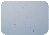
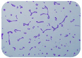
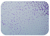
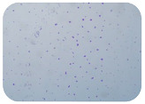
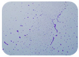
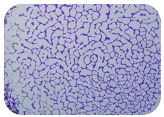
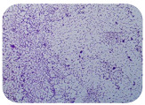
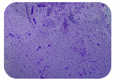
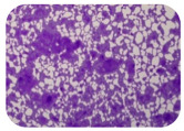
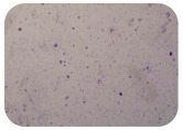
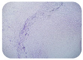
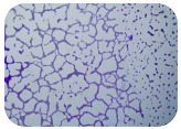
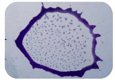
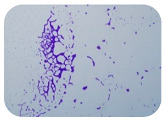
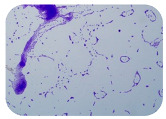
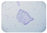
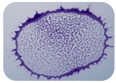
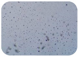
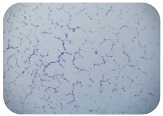
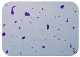
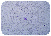
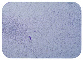
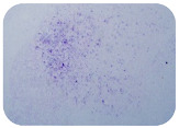
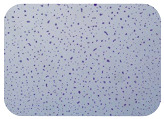
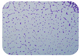
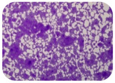
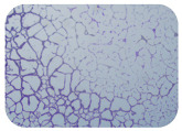
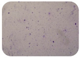
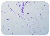
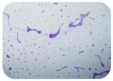
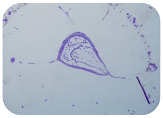
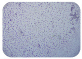
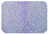
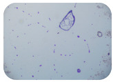
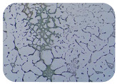
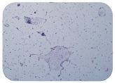
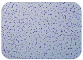
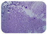
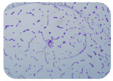
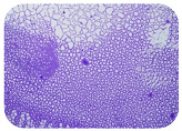
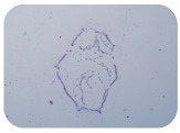
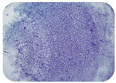
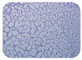
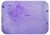
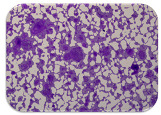
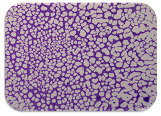
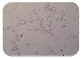
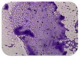
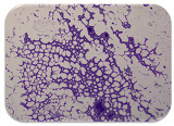
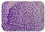
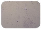
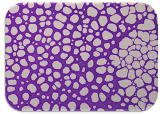
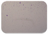
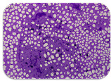
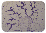
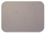
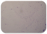
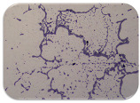
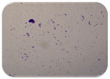
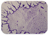
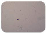
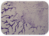
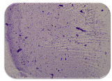
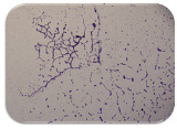
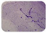
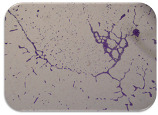
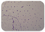
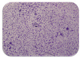
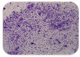
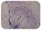
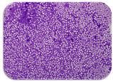
We used the protocol of Stepanović et al. [39] with small modifications, as described in Dimitrova et al. [25]. The biofilms were photodocumented with a microscopic configuration Nikon Eclipse-Ci-L (Nikon Instruments Europe BV, Amstelveen, The Netherlands), and the optical density (OD) was measured at 570 nm by using an ELISA reader ELx800 (BioTek Instruments, Winooski, VT, USA). The classification of Christensen et al. (Table 2) was used again to determine the adherence potential [40].
Table 2.
Correlation between the optical density of samples and bacterial adherence [40].
| Formula | Adherence |
|---|---|
| ODsample ≤ ODblank | non-adherent |
| ODblank < ODsample ≤ 2 × ODblank | weakly adherent |
| 2 × ODblank < ODsample ≤ 4 × ODblank | moderately adherent |
| 4 × ODblank < ODsample | strongly adherent |
3. Results
3.1. Isolation of Single Bacterial Cultures
Selected colonies from CCA, Endo or XLD agar were used for the spectral identification of the bacterial species. Endo agar is recommended for the confirmation of suspected members of the coliform group. E. coli are expected to have a metallic sheen on this agar. As XLD agar is a selective medium for Salmonella spp., no colonies were suspected to be E. coli.
3.2. Identification by MALDI-TOF-MS and PCR
A total of 85 colonies from different samples were identified as E. coli via MALDI-TOF-MS. Later, 84 of them were confirmed with PCR. Isolates that were positive for either the uidA or the yccT gene were accepted as E. coli. Some colonies that did not have a metallic sheen on Endo and that were not suspected for E. coli turned out to be this bacterial species (F4.2 and T1.1), and not all colonies that had a metallic sheen on this agar turned out to be this species. Generally, not all strains of a species isolated with a certain selective nutrient medium have all the expected typical morphological characteristics; therefore, this is not a new phenomenon.
It is interesting that six of the identified colonies were previously suspected to be Salmonella spp., as they were isolated from XLD agar (W2.4, T1.2, T1.3, S6.1, S6.2 and S7.2). Moreover, when recultured on Endo agar, two of them had a metallic sheen (W2.4 and T1.3), but the rest of them did not.
3.3. Antibiotic Resistance from the Disk Diffusion Method
Although erythromycin is not used for E coli, we tested it, because there is the potential horizontal transfer of erythromycin GAR to other bacterial species. As can be seen from Table 3, Table 4, Table 5, Table 6, Table 7 and Table 8, all isolates, except six (WV1.7, WV2.1, WV2.2, WV2.6, SV3.2 and SV3.4), had resistance to at least one agent, and many of them had resistance to multiple antibiotics. The resistance varied in wide ranges—from 0% for cephalosporins to 81% for tetracyclines and other agents. E. coli was isolated from all types of samples. Our results show that almost all isolates had resistance to multiple antibiotic agents, in line with the global tendency of increases in AMR, including in farm animals [5,6]. The antibiotic class that was associated with the greatest developed resistance was tetracycline (81%), followed by penicillins (56%). The percentage of chloramphenicol resistance was very high in the Karnobat farm and much lower in the other two, averaging 42.9%. The resistance to aminoglycoside was 39.3%. The resistance to trimethoprim/sulfamethoxazole followed the same pattern as that of the other unsorted agent, chloramphenicol, averaging 27.4%. Fluoroquinolones, on the contrary, showed much lower resistance in the Karnobat farm, in comparison to the others, and the average was 20.2%. Resistance to the class of macrolides was low (6%), and resistance to the class of carbapenems and cefamandole was absent.
Table 3.
Antibiotic resistances from the disk diffusion method of the isolated E. coli strains from the farm near Kostinbrod (FK3.1–SK3.1).
| Drug Class | Antibiotic/Strain | FK3.1 | FK3.2 | FK3.3 | FK3.4 | FK3.5 | WK1.3 | WK1.4 | WK2.5 | WK2.6 | WK3.3 | LK1.1 | LK1.3 | LK1.6 | LK2.1 | LK2.2 | LK3.1 | LK3.4 | SK3.1 |
|---|---|---|---|---|---|---|---|---|---|---|---|---|---|---|---|---|---|---|---|
| Tetracyclines | Tetracycline | R | R | R | R | R | R | R | R | I | I | R | I | R | R | R | R | R | R |
| Doxycycline hydrochloride | R | R | R | I | R | I | I | R | R | I | I | S | S | I | S | S | R | R | |
| Macrolides | Erythromycin | I | I | I | I | I | I | I | I | I | I | I | I | I | S | I | I | I | I |
| Cephalosporins | Cefamandole | S | S | S | S | S | S | S | S | S | S | S | S | S | S | S | S | S | S |
| Fluoroquinolones | Nalidixic acid | S | S | S | S | S | R | R | R | R | S | S | S | S | S | S | S | S | S |
| Pefloxacin | S | S | S | S | I | R | R | R | R | S | I | R | I | R | R | I | R | S | |
| Ciprofloxacin | S | S | S | S | S | R | I | I | I | S | S | S | S | S | S | S | S | S | |
| Penicillins | Ampicillin | R | R | R | R | R | R | R | R | R | S | S | S | S | S | S | S | S | R |
| Amoxicillin | R | R | R | R | R | R | R | R | R | R | S | S | S | S | S | S | S | R | |
| Amoxicillin/clavulanic acid | R | R | R | R | R | R | R | R | R | S | S | S | S | S | S | S | S | R | |
| Carbenicillin | I | S | S | S | I | R | I | R | I | S | S | S | S | S | S | S | S | I | |
| Carbapenems | Meropenem | I | S | S | S | I | S | S | I | S | S | S | S | S | I | S | S | S | I |
| Aminoglycosides | Streptomycin | R | R | R | R | R | R | R | R | R | S | S | S | S | S | S | S | S | R |
| Other agents | Chloramphenicol | S | S | S | S | S | S | S | S | S | S | S | S | S | S | S | S | S | S |
| Trimethoprim/sulfamethoxazole | S | S | S | S | S | R | R | R | R | R | S | S | S | S | S | S | S | S |
Legend: R, Resistant; I, Intermediate; S, Sensitive; F, Feces; W, Wastewater; L, Lagoon; SK, Soil. The first number is the number of the sample, and the second number is the number of the isolate.
Table 4.
Antibiotic resistances from the disk diffusion method of some of the isolated E. coli strains from the farm near Veliko Turnovo (FV1.1–FV4.3).
| Drug Class | Antibiotic/Strain | FV1.1 | FV2.1 | FV2.2 | FV2.3 | FV2.4 | FV2.5 | FV2.6 | FV2.7 | FV3.1 | FV3.2 | FV3.3 | FV4.1 | FV4.2 | FV4.3 |
|---|---|---|---|---|---|---|---|---|---|---|---|---|---|---|---|
| Tetracyclines | Tetracycline | R | R | R | R | R | I | R | I | R | R | R | I | R | R |
| Doxycycline hydrochloride | R | R | R | I | I | R | I | I | I | R | I | I | R | R | |
| Macrolides | Erythromycin | I | I | I | I | I | I | I | S | I | I | I | I | I | R |
| Cephalosporins | Cefamandole | S | S | S | S | S | S | S | S | S | S | S | S | S | S |
| Fluoroquinolones | Nalidixic acid | S | S | S | I | S | S | S | I | S | S | S | S | S | R |
| Pefloxacin | S | S | I | R | S | S | I | R | S | S | S | S | R | R | |
| Ciprofloxacin | S | S | S | S | S | S | S | S | S | S | S | S | S | I | |
| Penicillins | Ampicillin | R | R | S | R | R | R | R | R | S | S | S | S | R | R |
| Amoxicillin | S | R | S | R | R | R | R | R | S | S | S | S | R | R | |
| Amoxicillin/clavulanic acid | S | S | S | S | S | S | S | S | S | S | S | R | R | S | |
| Carbenicillin | S | I | I | I | I | I | I | I | S | S | S | S | I | I | |
| Carbapenems | Meropenem | S | S | S | S | S | S | S | S | S | S | S | S | I | S |
| Aminoglycosides | Streptomycin | S | S | S | R | S | S | S | S | I | I | R | R | I | R |
| Other agents | Chloramphenicol | S | S | R | S | S | R | R | R | S | S | S | S | S | R |
| Trimethoprim/sulfamethoxazole | S | S | R | R | S | S | R | R | I | S | R | S | S | R |
Legend: R, Resistant; I, Intermediate; S, Sensitive; F, Feces. The first number is the number of the sample, and the second number is the number of isolate.
Table 5.
Antibiotic resistances from the disk diffusion method of some of the isolated E. coli strains from the farm near Veliko Turnovo (FV4.4–WV4.2).
| Drug Class | Antibiotic/Strain | FV4.4 | WV1.2 | WV1.3 | WV1.4 | WV1.5 | WV1.7 | WV2.1 | WV2.2 | WV2.3 | WV2.5 | WV2.6 | WV4.1 | WV4.2 |
|---|---|---|---|---|---|---|---|---|---|---|---|---|---|---|
| Tetracyclines | Tetracycline | R | R | R | R | I | S | I | S | R | R | S | R | R |
| Doxycycline hydrochloride | R | R | R | R | S | S | I | S | S | R | S | R | R | |
| Macrolides | Erythromycin | R | I | I | I | I | I | I | I | I | I | I | I | I |
| Cephalosporins | Cefamandole | S | S | S | S | S | S | S | S | S | S | S | S | S |
| Fluoroquinolones | Nalidixic acid | R | S | S | S | S | S | S | S | S | S | S | S | S |
| Pefloxacin | R | I | I | I | S | S | S | I | S | R | I | S | S | |
| Ciprofloxacin | I | S | S | S | S | S | S | S | S | S | S | S | S | |
| Penicillins | Ampicillin | R | R | R | R | R | S | S | S | S | R | S | R | S |
| Amoxicillin | R | R | R | R | R | S | S | S | S | R | S | R | S | |
| Amoxicillin/clavulanic acid | R | S | S | S | S | S | S | S | S | S | S | R | S | |
| Carbenicillin | I | S | I | I | S | I | I | S | S | S | S | I | I | |
| Carbapenems | Meropenem | S | S | I | S | S | I | S | S | S | S | I | S | S |
| Aminoglycosides | Streptomycin | R | S | S | S | S | S | S | S | S | S | S | I | I |
| Other agents | Chloramphenicol | R | S | S | S | S | S | S | S | S | R | S | S | S |
| Trimethoprim/sulfamethoxazole | R | S | S | R | I | S | S | S | S | R | S | S | S |
Legend: R, Resistant; I, Intermediate; S, Sensitive; F, Feces; W, Wastewater. The first number is the number of the sample, and the second number is the number of the isolate.
Table 6.
Antibiotic resistances from the disk diffusion method of some of the isolated E. coli strains from the farm near Veliko Turnovo (WV4.3–SV3.4).
| Drug Class | Antibiotic/Strain | WV4.3 | WV4.4 | WV4.5 | WV4.6 | LV1.1 | LV1.2 | LV1.3 | LV1.5 | LV1.6 | SV3.1 | SV3.2 | SV3.3 | SV3.4 |
|---|---|---|---|---|---|---|---|---|---|---|---|---|---|---|
| Tetracyclines | Tetracycline | I | R | R | R | R | R | R | R | R | R | S | R | I |
| Doxycycline hydrochloride | I | R | R | R | R | I | R | R | S | R | S | R | S | |
| Macrolides | Erythromycin | I | I | I | I | I | I | I | I | I | I | I | I | I |
| Cephalosporins | Cefamandole | S | S | S | S | S | S | S | S | S | S | S | S | S |
| Fluoroquinolones | Nalidixic acid | S | S | I | S | S | S | S | S | S | S | S | S | S |
| Pefloxacin | S | I | R | I | I | S | I | S | S | S | S | S | S | |
| Ciprofloxacin | S | S | S | S | S | S | S | S | S | S | S | S | S | |
| Penicillins | Ampicillin | R | S | R | R | S | S | R | S | S | S | S | S | S |
| Amoxicillin | R | S | R | R | S | S | R | S | S | S | S | S | S | |
| Amoxicillin/clavulanic acid | S | S | R | I | S | S | S | S | S | S | S | S | S | |
| Carbenicillin | S | S | R | S | I | S | I | S | S | S | I | S | I | |
| Carbapenems | Meropenem | S | S | S | S | I | S | I | S | S | S | S | S | S |
| Aminoglycosides | Streptomycin | S | R | R | S | R | S | S | S | S | S | S | S | S |
| Other agents | Chloramphenicol | R | S | R | R | S | R | S | S | S | S | S | S | S |
| Trimethoprim/sulfamethoxazole | S | S | R | S | S | R | S | S | S | S | S | S | S |
Legend: R, Resistant; I, Intermediate; S, Sensitive; W, Wastewater; L, Lagoon; SV, Soil. The first number is the number of the sample, and the second number is the number of the isolate.
Table 7.
Antibiotic resistances from the disk diffusion method of some of the isolated E. coli strains from the farm near Karnobat (F1.1–W2.4).
| Drug Class | Antibiotic/Strain | F1.1 | F2.1 | F2.2 | F3.1 | F4.1 | F4.2 | F5.1 | F6.1 | F6.2 | W1.1 | W1.2 | W2.1 | W2.2 | W2.3 | W2.4 |
|---|---|---|---|---|---|---|---|---|---|---|---|---|---|---|---|---|
| Tetracyclines | Tetracycline | R | R | R | R | R | R | R | R | R | R | I | S | R | R | R |
| Doxycycline hydrochloride | R | R | R | R | R | R | R | R | R | R | I | R | S | R | R | |
| Macrolides | Erythromycin | I | I | I | R | I | I | I | R | R | I | I | I | I | I | I |
| Cephalosporins | Cefamandole | S | S | S | S | S | S | I | S | S | S | S | S | S | S | S |
| Fluoroquinolones | Nalidixic acid | S | S | S | S | S | S | S | S | S | S | S | S | S | S | S |
| Pefloxacin | S | S | S | S | S | S | S | S | S | S | S | S | R | S | S | |
| Ciprofloxacin | S | S | S | S | S | S | S | S | I | S | S | S | S | S | S | |
| Penicillins | Ampicillin | S | R | R | S | R | R | R | R | R | R | S | R | S | R | S |
| Amoxicillin | S | R | R | S | R | R | R | R | R | R | S | R | S | R | S | |
| Amoxicillin/clavulanic acid | R | S | R | S | R | R | R | R | R | S | S | S | S | S | S | |
| Carbenicillin | I | I | R | S | S | S | I | I | I | S | S | I | S | S | S | |
| Carbapenems | Meropenem | S | S | S | S | S | S | S | S | S | S | S | S | S | S | S |
| Aminoglycosides | Streptomycin | R | R | R | R | R | R | R | S | I | S | S | R | I | I | R |
| Other agents | Chloramphenicol | R | R | R | R | S | S | R | R | R | R | R | R | R | R | S |
| Trimethoprim/sulfamethoxazole | S | R | S | S | R | S | R | S | S | S | R | S | S | S | S |
Legend: R, Resistant; I, Intermediate; S, Sensitive; F, Feces; W, Wastewater. The first number is the number of the sample, and the second number is the number of the isolate.
Table 8.
Antibiotic resistances from the disk diffusion method of some of the isolated E. coli strains from the farm near Karnobat (F1.1–S7.2) and the controls.
| Drug Class | Antibiotic/Strain | L1.1 | L1.2 | L2.1 | L2.2 | T1.1 | T1.2 | T1.3 | S6.1 | S6.2 | S6.3 | S7.2 | E. coli O:157 |
E. coli ATCC 35218 |
|---|---|---|---|---|---|---|---|---|---|---|---|---|---|---|
| Tetracyclines | Tetracycline | S | S | R | R | I | I | R | R | R | R | R | R | S |
| Doxycycline hydrochloride | S | I | R | R | R | R | R | R | R | R | R | S | S | |
| Macrolides | Erythromycin | I | S | I | I | I | I | I | I | I | I | I | S | S |
| Cephalosporins | Cefamandole | S | S | S | S | S | S | S | S | S | S | S | S | S |
| Fluoroquinolones | Nalidixic acid | S | S | S | S | R | S | S | S | S | I | S | S | R |
| Pefloxacin | S | S | S | S | R | S | S | S | S | S | S | S | S | |
| Ciprofloxacin | S | S | S | S | R | S | S | S | S | S | S | S | S | |
| Penicillins | Ampicillin | S | S | S | R | S | R | R | S | S | S | R | S | R |
| Amoxicillin | S | S | S | R | S | R | R | S | S | S | R | - | R | |
| Amoxicillin/clavulanic acid | S | S | S | S | S | R | R | S | S | S | R | S | S | |
| Carbenicillin | S | S | S | S | I | S | S | S | S | I | S | S | S | |
| Carbapenems | Meropenem | S | S | S | S | S | S | S | S | S | S | S | S | S |
| Aminoglycosides | Streptomycin | I | R | R | R | S | S | I | R | R | R | I | S | R |
| Other agents | Chloramphenicol | R | R | R | R | R | R | R | R | R | R | R | R | R |
| Trimethoprim/sulfamethoxazole | S | S | S | S | R | S | R | S | S | R | S | S | S |
Legend: R, Resistant; I, Intermediate; S, Sensitive; L, Lagoon; T, Transport vehicle; S6 and S7, Soil. The first number is the number of the sample, and the second number is the number of isolate.
Multidrug resistance (MDR), defined as resistance to three or more antimicrobial classes of the panel tested, was found for 25 isolates (29.8%).
Although there was a variation in the patterns between farms, fecal samples were resistant to the greatest number of antibiotics (e.g., tetracyclines, penicillins and streptomycin). Likely, the fecal bacteria were subjected to more selective pressure due to the direct consumption of antibiotics, and/or the environmental factors could play a role in losing GAR in some other environments. The fewest E. coli were isolated from lagoons and soils, and they had resistance to fewer antibiotics. However, some of them still showed consistent patterns, such as resistance to tetracyclines or, in the case of Karnobat, to chloramphenicol in addition.
3.4. Detection of Antibiotic Resistance Genes
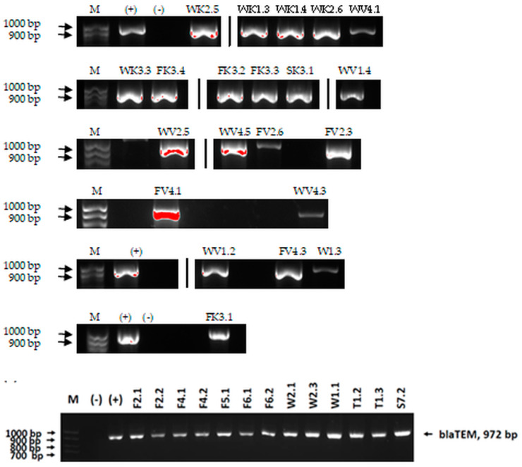
We sought GAR for a certain antibiotic class only in the isolates that showed resistance to an antibiotic from this class (including to antibiotics not presented in this study). The results (Figure 1) show that, from the group of GAR in this study, the isolates were positive only for β-lactamase-producing genes. They were ampC and blaTEM. Out of 56 tested isolates, all the samples had the ampC GAR, and there were 34 isolates that were positive for blaTEM. These results corroborate that ESBL and AmpC β-lactamase production are important resistance mechanisms in members of the Enterobacteriaceae family [18].
Figure 1.
Gel electrophoresis for blaTEM β-lactam resistant gene. The gel electrophoresis from the farm near Karnobat is confirmative. Black lines designate non-adjacent samples. Legend: M, marker; (+), positive control; (−), negative control.
3.5. Detection of Virulence Genes
No virulence genes from the panel STEC/VTEC (stx and stx2all), ETEC (LT, STa, and F4) and EPEC (eae) were detected among the isolates.
3.6. Test for Biofilm Formation
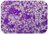
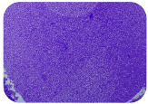
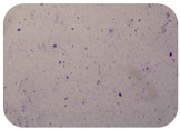
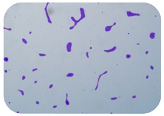
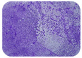
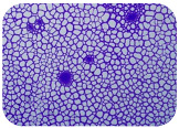
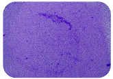
Strongly adherent E. coli (14 isolates) was found among all types of samples except the transport vehicle ones (Table 9, Table 10, Table 11 and Table 12). Moderately adherent E. coli was present among all types of samples. The Karnobat farm had the most strongly adherent isolates (8), whereas the Veliko Turnovo farm had the least adherent ones (2). Apart from that, there was no correlation in concern to the type of sample and the farm.
Table 9.
Adherence of isolated E. coli from the pig farm near Kostinbrod, compared with that of the controls.
| Strain | OD550 nm | Adherence | Biofilm | Strain | OD550 nm | Adherence | Biofilm |
|---|---|---|---|---|---|---|---|
| ATCC 35218 | 0.676 | SA |
|
WK2.6 | 0.711 | SA |
|
| O157 | 0.321 | MA |
|
WK3.3 | 0.239 | WA |
|
| Blank | 0.156 | - |

|
LK1.1 | 0.224 | WA |
|
| FK3.1 | 0.196 | WA |

|
LK1.3 | 0.188 | WA |

|
| FK3.2 | 0.237 | WA |

|
LK1.6 | 0.174 | WA |
|
| FK3.3 | 0.254 | WA |
|
LK2.1 | 0.242 | WA |
|
| FK3.4 | 0.229 | WA |

|
LK2.2 | 0.222 | WA |
|
| FK3.5 | 0.293 | WA |
|
LK3.1 | 0.215 | WA |

|
| WK1.3 | 0.859 | SA |
|
LK3.4 | 0.287 | WA |
|
| WK1.4 | 0.864 | SA |
|
SK3.1 | 0.580 | MA |
|
| WK2.5 | 0.680 | SA |
|
Table 10.
Adherence of a part of the isolated E. coli from the pig farm near Veliko Turnovo, compared with that of the controls.
| Strain | OD550 nm | Adherence | Biofilm | Strain | OD550 nm | Adherence | Biofilm |
|---|---|---|---|---|---|---|---|
| ATCC 35218 | 0.676 | SA |
|
FV3.1 | 0.208 | WA |

|
| O157 | 0.321 | MA |
|
FV3.2 | 0.143 | NA |

|
| Blank | 0.156 | - |

|
FV3.3 | 0.262 | WA |
|
| FV1.1 | 0.382 | MA |
|
FV4.1 | 0.193 | WA |
|
| FV2.1 | 0.269 | WA |
|
FV4.2 | 0.293 | WA |
|
| FV2.2 | 0.246 | WA |
|
FV4.3 | 0.204 | WA |
|
| FV2.3 | 0.313 | MA |
|
FV4.4 | 0.323 | MA |
|
| FV2.4 | 0.238 | WA |

|
WV1.2 | 0.192 | WA |
|
| FV2.5 | 0.330 | MA |
|
WV1.3 | 0.283 | WA |
|
| FV2.6 | 0.351 | MA |
|
WV1.4 | 0.253 | WA |

|
| FV2.7 | 0.200 | WA |
|
WV1.5 | 0.229 | WA |
|
Table 11.
Adherence of the other part of the isolated E. coli from the pig farm near Veliko Turnovo, compared with that of the controls.
| Strain | OD550 nm | Adherence | Biofilm | Strain | OD550 nm | Adherence | Biofilm |
|---|---|---|---|---|---|---|---|
| ATCC 35218 | 0.676 | SA |
|
WV4.4 | 0.275 | WA |
|
| O157 | 0.321 | MA |
|
WVT4.5 | 0.231 | WA |
|
| Blank | 0.156 | - |

|
WV4.6 | 0.264 | WA |
|
| WV1.7 | 0.168 | WA |

|
LV1.1 | 0.300 | WA |
|
| WV2.1 | 0.499 | MA |
|
LV1.2 | 0.442 | MA |
|
| WV2.2 | 0.314 | MA |
|
LV1.3 | 0.202 | WA |
|
| WV2.3 | 0.302 | WA |
|
LV1.5 | 0.232 | WA |
|
| WV2.5 | 0.175 | WA |
|
LV1.6 | 0.261 | WA |
|
| WV2.6 | 0.258 | WA |

|
SV3.1 | 0.550 | MA |
|
| WV4.1 | 0.207 | WA |
|
SV3.2 | 0.747 | SA |
|
| WV4.2 | 0.192 | WA |
|
SV3.3 | 0.996 | MA |
|
| WV4.3 | 0.406 | MA |
|
SV3.4 | 0.780 | SA |
|
Table 12.
Adherence of the isolated E. coli from the pig farm near Karnobat, compared with that of the controls.
| Strain | OD550 nm | Adherence | Biofilm | Strain | OD550 nm | Adherence | Biofilm |
|---|---|---|---|---|---|---|---|
| ATCC 35218 | 0.680 | SA |
|
W2.2 | 0.645 | SA |
|
| O157 | 0.317 | MA |
|
W2.3 | 0.975 | SA |
|
| Blank | 0.156 | - |

|
W2.4 | 0.532 | MA |
|
| F1.1 | 0.469 | MA |

|
L1.1 | 0.737 | SA |
|
| F2.1 | 0.461 | MA |
|
L1.2 | 0.759 | SA |
|
| F2.2 | 0.235 | WA |
|
L2.1 | 1.084 | SA |
|
| F3.1 | 0.278 | WA |
|
L2.2 | 0.190 | WA |
|
| F4.1 | 0.229 | WA |
|
T1.1 | 0.423 | MA |
|
| F4.2 | 0.323 | MA |
|
T1.2 | 0.461 | MA |
|
| F5.1 | 0.306 | WA |
|
T1.3 | 0.502 | MA |
|
| F6.1 | 0.650 | SA |
|
S6.1 | 0.266 | WA |
|
| F6.2 | 0.686 | SA |
|
S6.2 | 0.295 | WA |
|
| W1.1 | 0.317 | MA |
|
S6.3 | 0.455 | MA |
|
| W1.2 | 0.568 | MA |
|
S7.2 | 0.546 | MA |
|
| W2.1 | 0.949 | SA |
|
Legend: NA, non-adherent confirmed E. coli strains; WA, weakly adherent confirmed E. coli strains; MA, moderately adherent confirmed E. coli strains; SA, strongly adherent confirmed E. coli strains.
4. Discussion
The use of antibiotics as growth promoters and for prophylaxis in farms is a highly debated issue. However, there is enough evidence not only for the spread of AMR from livestock, such as swine, to humans (pig feces and wastewater are one of the hotspots for the spread and circulation of AMR and GAR) but also for genetic similarity (clonal types) between resistant bacteria in animals and in humans, some of which are given in a comprehensive review by Sirichokchatchawan et al., 2021 [41]. For example, four sequence types of ESBL producing E. coli shared between humans and pigs were found in Thailand [41]. Plasmid (sub)types and, again, ESBL genes such as blaCTX-M-1 were found to be shared between Dutch pigs and pig farmers [42]. Therefore, the general agreement among policy makers and society is that the disadvantage of creating bacterial AMR outweighs the benefits of antibiotics. Therefore, even though subclinical antibiotic concentrations not only promote growth but also reduce animal morbidity and mortality, numerous bans or restrictions for antibiotic feed additives have been adopted throughout the world [41,43]. The dilemma is excellently described in the work by Chattopadhyay, 2014 [43].
The legislation in Bulgaria is strict, and since approximately the year 2000, antibiotics in animal husbandry have been allowed only as therapeutics under veterinary supervision and with the demand of reporting it to the competent authorities. In poultry farms, most antibiotics are banned even for therapy. An exception in swine farms is the prophylactic use of colistin against post weaning diarrhea.
Nevertheless, in 2010–2016, even newborn suckling pigs still carried as part of their normal intestinal microflora coliforms that had resistance to most tested antibiotic classes, especially to tetracycline and ampicillin. Lactating sows and young pigs had high resistance to streptomycin. Moreover, despite legislative bans, if we compare our results with those from previous studies of swine farms in Bulgaria, an increase in AMR is observed. In the period of 2010–2016, resistance toward tetracycline, ampicillin and streptomycin (approximately 70%, 60% and 65%, respectively) doubled in comparison to the period of 2000–2004 [44]. After a transient decrease in 2020 [25], resistance to tetracycline rose even more to 77.8% and to 81% for the drug class as a whole in our study. Resistance to pefloxacin, carbenicillin and chloramphenicol rose slightly, and there was not a clear correlation regarding the other tested agents [25,44,45].
It is interesting that blaTEM was relatively rare in the study of the period of 2010–2016 [44], whereas in this work, blaTEM was a very prevalent gene with 34 positive samples from 56 tested. This marks an increase from our last time tested in 2020 [25]. AmpC was the only other GAR detected by us with all samples positive out of 56 tested. Last time, it had a similar pattern, because the most numerous positive samples were for that gene [25].
Indeed, there is decreased resistance for some agents, but it still remains relatively high. For example, ampicillin values showed growth in 2021 to 75% but have now decreased to 53.6%. Similarly, streptomycin resistance decreased in 2020 (12.5%) [25] and in this study (39.3%) but still remains high. Amoxicillin resistance decreased in comparison with the period of 2012–2020 [25,45], from 75% to 52.4%. There is very low resistance in farm pigs to third-generation cephalosporins [44] and even to the second-generation agent cefamandole in 2020 [25] and in our study.
As a summary for Bulgarian farm swine, there is high resistance of resident and pathogenic strains to tetracyclines, penicillins and aminoglycosides. Therefore, the high prevalence and increase in AMR in farms with highly restricted antibiotic use (and only for therapy) could indicate residual AMR from past times, the overuse of antibiotics for therapy in farms and/or the high circulation of AMR in the environment due to the high use of antibiotics by humans. This is enhanced by travel and transport in our global society, raising the spread of resistant strains.
The rate of antibiotic resistance differs considerably from country to country globally, depending upon the amount of usage. In the European Union (EU), the lowest levels of AMR E. coli isolates have been found in countries where lower antimicrobial usage is practiced, such as in Norway, Sweden and Finland, whereas countries with high levels of use, such as Spain, Portugal and Belgium, have relatively higher levels of AMR E. coli [46]. For instance, a clear spatial pattern was detected for tetracycline resistance, with high resistance levels reported for southern and western European countries and much lower levels reported for northern and eastern countries in 2004–2007 [7].
In 2019, some antimicrobial classes were assigned the highest priority, with critically important antimicrobials for human medicine only being available for food animals through veterinary prescription [41]. These are fluoroquinolones, third- and fourth-generation cephalosporins, carbapenems, macrolides and polymyxins (colistin). An increase in resistance to these antibiotics in E. coli in animals may indicate a general resistance trend of concern among Gram-negative bacteria originating from the animal reservoir. Carbapenems may not be used in food-producing animals in the present, but it is alarming that resistance genes have been found in pigs, chickens and other livestock [47].
The monitoring and reporting of resistance data from indicator organisms (commensal E. coli and enterococci) to the European Food Safety Authority (EFSA) is voluntary. EFSA reports show that, for a timeframe of 2004–2020, resistance to nalidixic acid is, in general, low, as it was in our studies. However, in the past (2004–2007), large variability was observed in the reported ciprofloxacin resistance median levels (4–24%), with the highest occurrence of 74% reported by Estonia in 2007. In Denmark, legal restrictions have been in place for the use of fluoroquinolones in food animals since 2002, and as a consequence, ciprofloxacin resistance decreased from 3% in 2004 to 0% in 2007 [7]. Wider legal restrictions likely led to the low overall resistance to ciprofloxacin now (median approximately 5%) [48]. It is noteworthy that resistance to third-generation cephalosporins is very low, usually below 1%, in European as well as in local Bulgarian pig farms; therefore, resistance to that agent is still not a threat in animal husbandry, unlike in clinical settings [7,48]. Regarding human infections with E. coli, the European Antimicrobial Resistance Surveillance Network (EARS-NET) reported in 2017 that the highest population-weighted mean resistance percentage in the European Union and the European Economic Area for E. coli that causes serious infections was to aminopenicillins (58.7%), followed by fluoroquinolones (25.7%), third-generation cephalosporins (14.9%) and aminoglycosides (11.4%). In 2017, resistance to carbapenems remained rare in E. coli [49].
In 2019–2020, reports of farm swine from 30 countries showed that high or very high resistance to ampicillin, sulfamethoxazole, trimethoprim and tetracycline was the most common resistance trait observed. MDR was observed in 34.2% (versus 29.8% in our study) of commensal E. coli isolates from pigs. A wide variety of resistance patterns were observed, but mostly to tetracycline, ampicillin, sulfamethoxazole and trimethoprim, often in combination with other substances but rarely with quinolones. About half of the porcine MDR isolates (52.3%) were resistant to all these antimicrobials. Meropenem resistance was not detected in any isolate of indicator E. coli, in line with the results from Bulgaria [48].
Complete susceptibility to 14 antimicrobials tested was higher than that in our study without a statistically significant difference between countries. The aminoglycoside gentamicin had low median levels of resistance through 2004–2020, unlike in Bulgaria. The median levels of chloramphenicol resistance for all reporting countries were moderate in pigs, unlike the high resistance in Bulgaria [48].
There were positive trends (decreases in the level of resistance) in several countries that were possibly due to the documented overall decline in sales of antimicrobials. Most notably, tetracycline resistance has decreased in 15 countries and increased in only two. In 13 countries, there were only decreasing trends, notably in the Netherlands for four agents and in Cyprus for three agents [48]. Indeed, countries such as Denmark and the Netherlands, which both have had massive swine production in recent years, have achieved tremendous reductions in antimicrobial usage while sustaining peak production. Comparable results have been accomplished in Belgium, France, Sweden and the United Kingdom [7,46,48,50]. In contrast, in six countries, there were only increasing trends, and there were increasing trends in Belgium (despite the reduction in antibiotic use), Poland and Romania for three antimicrobials [48].
In reference to the genetic profile, unlike our results, presumptive ESBL producers were more common than AmpC-producers, and isolates with a combined phenotype (ESBL + AmpC) were uncommon. The occurrence of presumptive ESBL, AmpC or ESBL + AmpC producers in commensal E. coli was 1.3% in fattening pigs. In pork, the prevalence of presumptive E. coli ESBL and/or AmpC-producers in meat was less variable, ranging from 0% (Finland and the Netherlands) to 24.4% (Portugal) [48].
It is interesting that, in the period of 2004–2007, AMR to commonly used antimicrobials was higher in porcine E. coli than that in isolates from chickens and cattle, and in most cases, the countries that reported a high occurrence of AMR in E. coli from chickens also had a high occurrence of resistance in E. coli from pigs [7]. This was likely due to the fact that the global average annual consumption of antibiotics for swine (172 mg/kg) is greater than that for cattle (45 mg/kg) and chickens (148 mg/kg) [51].
As a summary for the EU, the EFSA Animal Health and Welfare Panel (2021) revealed clinical swine E. coli isolates with a high proportion of resistance to numerous antibiotics, with a prevalence from 63% to 70% (to aminopenicillins, sulfonamides and tetracycline). However, lower rates of resistance to clinically critical antibiotics (fluoroquinolones and third-generation cephalosporins) were detected [50]. Likely, the latter was the first fruit of the recent Regulation (EU) 2019/61 on Veterinary Medicines and Regulation (EU) 2019/4 on Medicated Feed, stating that antibiotics shall not be applied routinely, nor for prophylaxis, unless in exceptional cases. They should only be applied for metaphylaxis (treatment of animals without signs of disease, which are in close contact with animals that do have evidence of infectious disease) when the risk of spreading infection is very high and there are no other options, as Barros et al., 2023, described [18]. Differences in resistance between countries might reflect the dissemination of certain E. coli types within animal populations and/or differences in the consumption of antimicrobials in animals among countries [7,48].
The recommendations that could make the use of antibiotics as a feed additive unnecessary are several, including improved hygiene and vaccines (although their efficacy varies considerably), biosecurity (measures taken to prevent disease introduction, such as monitoring animals or plant materials that enter the property, as well as sources of water and feed). Numerous alternatives to antibiotics exist—prebiotics, probiotics, phytogenic substances, bacteriophages, etc. However, they have their limits for therapy and are mostly used for prophylaxis [18]. The development of alternatives for the clinical management of the infections of livestock appears to be the need of the hour [43]. There is a need to establish national surveillance programs and effective policies, particularly in certain world regions, to curtail the threat of the evolution of resistant isolates in swine or other livestock production [33,52].
It is clear that more unhygienic farms (e.g., in the developing world) demand more antibiotics in their feed. However, antimicrobials as a feed additive reduce morbidity and mortality in most hygienic farms in the developed world [43]. Whether farm animals are exposed to more infectious agents than animals in their natural habitat is a question beyond the scope of this work. However, it is clear that wildlife has access to more open ventilated spaces and disinfecting sun beams. Therefore, our hypothesis is that, as the number of natural conditions that livestock lives in increases, fewer antibiotics are needed for growth promotion.
Biofilm formation by foodborne pathogens is a serious threat to food safety and public health [53]. As a foodborne pathogen, E. coli can adhere to and form biofilms on most materials and under almost all environmental conditions in food production plants [54]. In this context, this is of importance for irrigation installations and meat processing plants, given the fact that E. coli can survive for months on dry surfaces [55]. Viable pathogens in detached biofilms from contact surfaces can lead to cross-contamination. Environmental biofilms are most often composed of multispecies microorganisms, and mixed biofilm formation can enhance the sanitizer tolerance of foodborne pathogens. E. coli is capable of forming biofilms with other bacterial species, and that could enhance its pathogenic clones’ survival in the biofilm community. Biofilm formation in commercial meat plants may be a source of product contamination with no identifiable cause [53].
Even after the cleaning and disinfection processes, the biofilm could still persist. For example, in nursery units in a pig facility after an extensive washing protocol that included disinfection and being kept empty for two weeks, for E. coli and fecal coliforms, reductions of 41% and 51% were observed, respectively; however, they were still found on floors, drinking nipples and feeding troughs [55].
Although we did not find pathogenic clones in the present research, the ability to form strongly adherent biofilms for approximately 17% of the isolated E. coli commensals that were resistant to commonly used antimicrobials and that were MDR strains is alarming, because a detached biofilm can lead to the spread of the AMR to the environment and bacteria in humans through horizontal gene transfer.
As future directions for our research, colistin resistance could be studied because of its prophylactic use against post weaning diarrhea.
5. Conclusions
Although some antimicrobial agents show a lower level of resistance in Bulgarian porcine farms in comparison to European ones (e.g., ciprofloxacin), the higher prevalence of resistance to aminoglycosides in Bulgaria is alarming, and so is the higher level of resistance to tetracyclines, because it is already high in the European Union. Antibiotic stewardship is the effort to improve how antibiotics are prescribed by clinicians and used by patients. In terms of antimicrobial stewardship programs, national action plans in many countries cover both human and animal health sectors [41]. Legal restrictions lead to positive trends, but our study is an example of the relatively high prevalence and increase in AMR in farms with banned antibiotics as feed additives and prophylaxis. This is likely the result of their overuse for therapy in farms and/or the high circulation of AMR in the environment due to the high usage of antibiotics by humans. Antimicrobial utilization should be more correctly structured as a dosage and course of treatment.
Acknowledgments
Biofilm absorbance was measured on equipment donated by the Alexander von Humboldt Foundation to Maya Margaritova Zaharieva in the frame of the Alumni program “Equipment subsidies”.
Author Contributions
Conceptualization, H.N.; methodology, H.N. and M.M.Z.; software, not applicable; validation, H.N. and M.M.Z.; formal analysis, H.N., M.M.Z. and Y.I.; investigation, M.D.K., I.T., Y.I., T.C.K., P.O., M.M.Z. and Y.G.; resources, H.N. and K.N.; data curation, not applicable; writing—original draft preparation, Y.I., Y.G., M.M.Z., M.D.K., H.N. and L.D.; writing—review and editing, Y.I. and H.N.; visualization, Y.I., T.C.K. and M.M.Z.; supervision, H.N.; project administration, H.N.; funding acquisition, H.N. All authors have read and agreed to the published version of the manuscript.
Data Availability Statement
The data presented in this study are available on request from the corresponding author.
Conflicts of Interest
The authors declare no conflict of interest.
Funding Statement
This research was funded by the National Fund for Scientific Research, Republic of Bulgaria (grant number: KΠ-06-H36/7 from 13 December 2019).
Footnotes
Disclaimer/Publisher’s Note: The statements, opinions and data contained in all publications are solely those of the individual author(s) and contributor(s) and not of MDPI and/or the editor(s). MDPI and/or the editor(s) disclaim responsibility for any injury to people or property resulting from any ideas, methods, instructions or products referred to in the content.
References
- 1.O’Neill J. Tackling Drug-Resistant Infections Globally: Final Report and Recommendations: Review on Antimicrobial Resistance. Government of the United Kingdom; London, UK: 2016. [Google Scholar]
- 2.De Kraker M.E.A., Stewardson A.J., Harbarth S. Will 10 Million People Die a Year Due to Antimicrobial Resistance by 2050? PLoS Med. 2016;13:e1002184. doi: 10.1371/journal.pmed.1002184. [DOI] [PMC free article] [PubMed] [Google Scholar]
- 3.Murray C.J., Ikuta K.S., Sharara F., Swetschinski L., Robles Aguilar G., Gray A., Han C., Bisignano C., Rao P., Wool E., et al. Global Burden of Bacterial Antimicrobial Resistance in 2019: A Systematic Analysis. Lancet. 2022;399:629–655. doi: 10.1016/S0140-6736(21)02724-0. [DOI] [PMC free article] [PubMed] [Google Scholar]
- 4.World Health Organization . Antimicrobial Resistance: Global Report on Surveillance. World Health Organization; Geneva, Switzerland: 2014. [Google Scholar]
- 5.Reardon S. WHO Warns against “post-Antibiotic” Era. Nature. 2014;5:135–138. doi: 10.1038/nature.2014.15135. [DOI] [Google Scholar]
- 6.Van Boeckel T.P., Glennon E.E., Chen D., Gilbert M., Robinson T.P., Grenfell B.T., Levin S.A., Bonhoeffer S., Laxminarayan R. Reducing Antimicrobial Use in Food Animals. Science. 2017;357:1350–1352. doi: 10.1126/science.aao1495. [DOI] [PMC free article] [PubMed] [Google Scholar]
- 7.European Food Safety Authority The Community Summary Report on Antimicrobial Resistance in Zoonotic and Indicator Bacteria from Animals and Food in the European Union in 2004–2007. EFSA J. 2010;8:1658. doi: 10.2903/j.efsa.2010.1309. [DOI] [Google Scholar]
- 8.CDC Antibiotic Resistance Threats in the United States, 2019|Enhanced Reader. [(accessed on 25 April 2023)]; Available online: https://www.cdc.gov/drugresistance/pdf/threats-report/2019-ar-threats-report-508.pdf.
- 9.Van Boeckel T.P., Pires J., Silvester R., Zhao C., Song J., Criscuolo N.G., Gilbert M., Bonhoeffer S., Laxminarayan R. Global Trends in Antimicrobial Resistance in Animals in Low- And Middle-Income Countries. Science. 2019;365:eaaw1944. doi: 10.1126/science.aaw1944. [DOI] [PubMed] [Google Scholar]
- 10.Underwood W.J., McGlone J.J., Swanson J., Anderson K.A., Anthony R. Laboratory Animal Welfare. Elsevier Inc.; Amsterdam, The Netherlands: 2013. Agricultural Animal Welfare; pp. 233–278. [Google Scholar]
- 11.Jechalke S., Focks A., Rosendahl I., Groeneweg J., Siemens J., Heuer H., Smalla K. Structural and Functional Response of the Soil Bacterial Community to Application of Manure from Difloxacin-Treated Pigs. FEMS Microbiol. Ecol. 2014;87:78–88. doi: 10.1111/1574-6941.12191. [DOI] [PubMed] [Google Scholar]
- 12.Hamscher G., Pawelzick H.T., Sczesny S., Nau H., Hartung J. Antibiotics in Dust Originating from a Pig-Fattening Farm: A New Source of Health Hazard for Farmers? Environ. Health Perspect. 2003;111:1590–1594. doi: 10.1289/ehp.6288. [DOI] [PMC free article] [PubMed] [Google Scholar]
- 13.Poirel L., Madec J.-Y., Lupo A., Schink A.-K., Kieffer N., Nordmann P., Schwarz S. Antimicrobial Resistance in Escherichia coli. Microbiol. Spectr. 2018;6:14. doi: 10.1128/microbiolspec.ARBA-0026-2017. [DOI] [PMC free article] [PubMed] [Google Scholar]
- 14.Wang W., Yu L., Hao W., Zhang F., Jiang M., Zhao S., Wang F. Multi-Locus Sequence Typing and Drug Resistance Analysis of Swine Origin Escherichia coli in Shandong of China and Its Potential Risk on Public Health. Front. Public Health. 2021;9:780700. doi: 10.3389/fpubh.2021.780700. [DOI] [PMC free article] [PubMed] [Google Scholar]
- 15.He T., Wang R., Liu D., Walsh T.R., Zhang R., Lv Y., Ke Y., Ji Q., Wei R., Liu Z., et al. Emergence of Plasmid-Mediated High-Level Tigecycline Resistance Genes in Animals and Humans. Nat. Microbiol. 2019;4:1450–1456. doi: 10.1038/s41564-019-0445-2. [DOI] [PubMed] [Google Scholar]
- 16.Liu Y.Y., Wang Y., Walsh T.R., Yi L.X., Zhang R., Spencer J., Doi Y., Tian G., Dong B., Huang X., et al. Emergence of Plasmid-Mediated Colistin Resistance Mechanism MCR-1 in Animals and Human Beings in China: A Microbiological and Molecular Biological Study. Lancet Infect. Dis. 2016;16:161–168. doi: 10.1016/S1473-3099(15)00424-7. [DOI] [PubMed] [Google Scholar]
- 17.Kumarasamy K.K., Toleman M.A., Walsh T.R., Bagaria J., Butt F., Balakrishnan R., Chaudhary U., Doumith M., Giske C.G., Irfan S., et al. Emergence of a New Antibiotic Resistance Mechanism in India, Pakistan, and the UK: A Molecular, Biological, and Epidemiological Study. Lancet Infect. Dis. 2010;10:597–602. doi: 10.1016/S1473-3099(10)70143-2. [DOI] [PMC free article] [PubMed] [Google Scholar]
- 18.Barros M.M., Castro J., Araújo D., Campos A.M., Oliveira R., Silva S., Outor-Monteiro D., Almeida C. Swine Colibacillosis: Global Epidemiologic and Antimicrobial Scenario. Antibiotics. 2023;12:682. doi: 10.3390/antibiotics12040682. [DOI] [PMC free article] [PubMed] [Google Scholar]
- 19.Loayza F., Graham J.P., Trueba G. Factors Obscuring the Role of E. coli from Domestic Animals in the Global Antimicrobial Resistance Crisis: An Evidence-Based Review. Int. J. Environ. Res. Public Health. 2020;17:3061. doi: 10.3390/ijerph17093061. [DOI] [PMC free article] [PubMed] [Google Scholar]
- 20.Argudín M.A., Deplano A., Meghraoui A., Dodémont M., Heinrichs A., Denis O., Nonhoff C., Roisin S. Bacteria from Animals as a Pool of Antimicrobial Resistance Genes. Antibiotics. 2017;6:12. doi: 10.3390/antibiotics6020012. [DOI] [PMC free article] [PubMed] [Google Scholar]
- 21.Heuer H., Schmitt H., Smalla K. Antibiotic Resistance Gene Spread Due to Manure Application on Agricultural Fields. Curr. Opin. Microbiol. 2011;14:236–243. doi: 10.1016/j.mib.2011.04.009. [DOI] [PubMed] [Google Scholar]
- 22.European Union The European Union One Health 2021 Zoonoses Report. EFSA J. 2022;20:e06406. doi: 10.2903/j.efsa.2022.7666. [DOI] [PMC free article] [PubMed] [Google Scholar]
- 23.Høiby N., Bjarnsholt T., Givskov M., Molin S., Ciofu O. Antibiotic Resistance of Bacterial Biofilms. Int. J. Antimicrob. Agents. 2010;35:322–332. doi: 10.1016/j.ijantimicag.2009.12.011. [DOI] [PubMed] [Google Scholar]
- 24.Stewart P.S. Mechanisms of Antibiotic Resistance in Bacterial Biofilms. Int. J. Med. Microbiol. 2002;292:107–113. doi: 10.1078/1438-4221-00196. [DOI] [PubMed] [Google Scholar]
- 25.Dimitrova L., Kaleva M., Zaharieva M.M., Stoykova C., Tsvetkova I., Angelovska M., Ilieva Y., Kussovski V., Naydenska S., Najdenski H. Prevalence of Antibiotic-Resistant Escherichia Coli Isolated from Swine Faeces and Lagoons in Bulgaria. Antibiotics. 2021;10:940. doi: 10.3390/antibiotics10080940. [DOI] [PMC free article] [PubMed] [Google Scholar]
- 26.Bej A.K., DiCesare J.L., Haff L., Atlas R.M. Detection of Escherichia coli and Shigella Spp. in Water by Using the Polymerase Chain Reaction and Gene Probes for Uid. Appl. Environ. Microbiol. 1991;57:1013–1017. doi: 10.1128/aem.57.4.1013-1017.1991. [DOI] [PMC free article] [PubMed] [Google Scholar]
- 27.Clifford R.J., Milillo M., Prestwood J., Quintero R., Zurawski D.V., Kwak Y.I., Waterman P.E., Lesho E.P., Mc Gann P. Detection of Bacterial 16S RRNA and Identification of Four Clinically Important Bacteria by Real-Time PCR. PLoS ONE. 2012;7:e48558. doi: 10.1371/journal.pone.0048558. [DOI] [PMC free article] [PubMed] [Google Scholar]
- 28.ISO/TS 13136:2012(En), Microbiology of Food and Animal Feed—Real-Time Polymerase Chain Reaction (PCR)-Based Method for the Detection of Food-Borne Pathogens—Horizontal Method for the Detection of Shiga Toxin-Producing Escherichia coli (STEC) and the Determination of O157, O111, O26, O103 and O145 Serogroups. [(accessed on 7 April 2023)]. Available online: https://www.iso.org/obp/ui/#iso:std:iso:ts:13136:ed-1:v1:en.
- 29.Annisha O.D.R., Li Z., Zhou X., Junior N.M.D.S., Donde O.O. Efficacy of Integrated Ultraviolet Ultrasonic Technologies in the Removal of Erythromycin-and Quinolone-Resistant Escherichia Coli from Domestic Wastewater through a Laboratory-Based Experiment. J. Water Sanit. Hyg. Dev. 2019;9:571–580. doi: 10.2166/washdev.2019.021. [DOI] [Google Scholar]
- 30.Pourhossein Z., Asadpour L., Habibollahi H., Shafighi S.T. Antimicrobial Resistance in Fecal Escherichia Coli Isolated 491 from Poultry Chicks in Northern Iran. Gene Rep. 2020;21:100926. doi: 10.1016/j.genrep.2020.100926. [DOI] [Google Scholar]
- 31.Ranjbar R., Safarpoor Dehkordi F., Sakhaei Shahreza M.H., Rahimi E. Prevalence, Identification of Virulence Factors, O-Serogroups and Antibiotic Resistance Properties of Shiga-Toxin Producing Escherichia Coli Strains Isolated from Raw Milk and Traditional Dairy Products. Antimicrob. Resist. Infect. Control. 2018;7:1–11. doi: 10.1186/s13756-018-0345-x. [DOI] [PMC free article] [PubMed] [Google Scholar]
- 32.Jafari E., Mostaan S., Bouzari S. Characterization of Antimicrobial Susceptibility, Extended-Spectrum β-Lactamase Genes and Phylogenetic Groups of Enteropathogenic Escherichia Coli Isolated from Patients with Diarrhea. Osong Public Health Res. Perspect. 2020;11:327–333. doi: 10.24171/j.phrp.2020.11.5.09. [DOI] [PMC free article] [PubMed] [Google Scholar]
- 33.Huang Y.H., Kuan N.L., Yeh K.S. Characteristics of Extended-Spectrum β-Lactamase–Producing Escherichia coli From Dogs and Cats Admitted to a Veterinary Teaching Hospital in Taipei, Taiwan from 2014 to 2017. Front. Vet. Sci. 2020;7:395. doi: 10.3389/fvets.2020.00395. [DOI] [PMC free article] [PubMed] [Google Scholar]
- 34.Khoirani K., Indrawati A., Setiyaningsih S. Detection of Ampicillin Resistance Encoding Gene of Escherichia Coli from Chickens in Bandung and Purwakarta. J. Ris. Vet. Indones. 2019;3 doi: 10.20956/jrvi.v3i1.6134. [DOI] [Google Scholar]
- 35.Nguyen M.C.P., Woerther P.L., Bouvet M., Andremont A., Leclercq R., Canu A. Escherichia Coli as Reservoir for Macrolide Resistance Genes. Emerg. Infect. Dis. 2009;15:1648–1650. doi: 10.3201/eid1510.090696. [DOI] [PMC free article] [PubMed] [Google Scholar]
- 36.CLSI . Performance Standards for Antimicrobial Disk and Dilution Susceptibility Tests for Bacteria Isolated from Animals. 3rd ed. Clinical and Laboratory Standards Institute; Wayne, PA, USA: 2009. Approved Standard. [Google Scholar]
- 37.Testing, T.E.C. on A.S. Breakpoint Tables for Interpretation of MICs and Zone Diameters. [(accessed on 23 April 2023)]. Available online: http://www.eucast.org.
- 38.Power D., McCuen P. Manual of BBL Products and Laboratory Procedures. 6th ed. BBL Publishing; Chicago, IL, USA: 1998. [Google Scholar]
- 39.Stepanović S., Vuković D., Hola V., Di Bonaventura G., Djukić S., Ćirković I., Ruzicka F. Quantification of Biofilm in Microtiter Plates: Overview of Testing Conditions and Practical Recommendations for Assessment of Biofilm Production by Staphylococci. APMIS. 2007;115:891–899. doi: 10.1111/j.1600-0463.2007.apm_630.x. [DOI] [PubMed] [Google Scholar]
- 40.Christensen G.D., Simpson W.A., Younger J.J., Baddour L.M., Barrett F.F., Melton D.M., Beachey E.H. Adherence of Coagulase-Negative Staphylococci to Plastic Tissue Culture Plates: A Quantitative Model for the Adherence of Staphylococci to Medical Devices. J. Clin. Microbiol. 1985;22:996–1006. doi: 10.1128/jcm.22.6.996-1006.1985. [DOI] [PMC free article] [PubMed] [Google Scholar]
- 41.Sirichokchatchawan W., Apiwatsiri P., Pupa P., Saenkankam I., Khine N.O., Lekagul A., Lugsomya K., Hampson D.J., Prapasarakul N. Reducing the Risk of Transmission of Critical Antimicrobial Resistance Determinants from Contaminated Pork Products to Humans in South-East Asia. Front. Microbiol. 2021;12:689015. doi: 10.3389/fmicb.2021.689015. [DOI] [PMC free article] [PubMed] [Google Scholar]
- 42.Dohmen W., Liakopoulos A., Bonten M.J.M., Mevius D.J., Heederik D.J.J. Longitudinal Study of Dynamic Epidemiology of Extended-Spectrum Beta-Lactamase-Producing Escherichia coli in Pigs and Humans Living and/or Working on Pig Farms. Microbiol. Spectr. 2023;11:e02947-22. doi: 10.1128/spectrum.02947-22. [DOI] [PMC free article] [PubMed] [Google Scholar]
- 43.Chattopadhyay M.K. Use of Antibiotics as Feed Additives: A Burning Question. Front. Microbiol. 2014;5:334. doi: 10.3389/fmicb.2014.00334. [DOI] [PMC free article] [PubMed] [Google Scholar]
- 44.Urumova V. Phenotypic and Genotypic Characteristics of Antimicrobial Resistance in Resident Escherichia coli and Enterococcus spp., Isolated from Intensively Farmed Pigs in Bulgaria. Trakia University; Stara Zagora, Bulgaria: 2016. [Google Scholar]
- 45.Dimitrova A., Yordanov S., Petkova K., Savova T., Bankova R., Ivanova S. Example Scheme for Antibacterial Therapy and Metaphylaxis of Colibacillosis in the Intensive Pig Production (in Bulgarian) Zhivotnovdni Nauk. Bulg. J. Anim. Husb. 2016;53:44–53. [Google Scholar]
- 46.Holmer I., Salomonsen C.M., Jorsal S.E., Astrup L.B., Jensen V.F., Høg B.B., Pedersen K. Antibiotic Resistance in Porcine Pathogenic Bacteria and Relation to Antibiotic Usage. BMC Vet. Res. 2019;15:449. doi: 10.1186/s12917-019-2162-8. [DOI] [PMC free article] [PubMed] [Google Scholar]
- 47.Caneschi A., Bardhi A., Barbarossa A., Zaghini A. The Use of Antibiotics and Antimicrobial Resistance in Veterinary Medicine, a Complex Phenomenon: A Narrative Review. Antibiotics. 2023;12:487. doi: 10.3390/antibiotics12030487. [DOI] [PMC free article] [PubMed] [Google Scholar]
- 48.European Union The European Union Summary Report on Antimicrobial Resistance in Zoonotic and Indicator Bacteria from Humans, Animals and Food in 2019–2020. EFSA J. 2022;20:e07209. doi: 10.2903/j.efsa.2022.7209. [DOI] [PMC free article] [PubMed] [Google Scholar]
- 49.European Centre for Disease Prevention and Control . Surveillance of Antimicrobial Resistance in Europe–Annual Report of the European Antimicrobial Resistance Surveillance Network (EARS-Net) ECDC; Stockholm, Sweden: 2017. [Google Scholar]
- 50.Nielsen S.S., Bicout D.J., Calistri P., Canali E., Drewe J.A., Garin-Bastuji B., Gonzales Rojas J.L., Gortazar Schmidt C., Herskin M., Michel V., et al. Assessment of Animal Diseases Caused by Bacteria Resistant to Antimicrobials: Swine. EFSA J. 2021;19:7113. doi: 10.2903/j.efsa.2021.7113. [DOI] [PMC free article] [PubMed] [Google Scholar]
- 51.Van Boeckel T.P., Brower C., Gilbert M., Grenfell B.T., Levin S.A., Robinson T.P., Teillant A., Laxminarayan R. Global Trends in Antimicrobial Use in Food Animals. Proc. Natl. Acad. Sci. USA. 2015;112:5649–5654. doi: 10.1073/pnas.1503141112. [DOI] [PMC free article] [PubMed] [Google Scholar]
- 52.Hayer S.S., Casanova-Higes A., Paladino E., Elnekave E., Nault A., Johnson T., Bender J., Perez A., Alvarez J. Global Distribution of Fluoroquinolone and Colistin Resistance and Associated Resistance Markers in Escherichia Coli of Swine Origin—A Systematic Review and Meta-Analysis. Front. Microbiol. 2022;13:834793. doi: 10.3389/fmicb.2022.834793. [DOI] [PMC free article] [PubMed] [Google Scholar]
- 53.Chitlapilly Dass S., Bosilevac J.M., Weinroth M., Elowsky C.G., Zhou Y., Anandappa A., Wang R. Impact of Mixed Biofilm Formation with Environmental Microorganisms on E. coli O157:H7 Survival against Sanitization. NPJ Sci. Food. 2020;4:16. doi: 10.1038/s41538-020-00076-x. [DOI] [PMC free article] [PubMed] [Google Scholar]
- 54.Gado D.A., Abdalla M.A., Erhabor J.O., Ehlers M.M., McGaw L.J. In Vitro Anti-Biofilm Effects of Loxostylis Alata Extracts and Isolated 5-Demethyl Sinensetin on Selected Foodborne Bacteria. S. Afr. J. Bot. 2023;156:29–34. doi: 10.1016/j.sajb.2023.02.037. [DOI] [Google Scholar]
- 55.Nakanishi E.Y., Palacios J.H., Godbout S., Fournel S. Interaction between Biofilm Formation, Surface Material and Cleanability Considering Different Materials Used in Pig Facilities—An Overview. Sustainability. 2021;13:5836. doi: 10.3390/su13115836. [DOI] [Google Scholar]
Associated Data
This section collects any data citations, data availability statements, or supplementary materials included in this article.
Data Availability Statement
The data presented in this study are available on request from the corresponding author.


