Abstract
Acne necrotica is a rare disease, characterized by recurrent crops of inflammatory papules and papulo-pustules that rapidly become necrotic, leaving varioliform scars of varying extent. Here, I report the case of a 32-year-old male with early-stage disease and a 58-year-old male with late-stage acne necrotica. Both patients had a history of chronic, relapsing, umbilicated, and centrally necrotic erythematous papules and papulo-pustules involving the hairline and face. A diagnosis of acne necrotica was made based on the clinical presentation, and both patients started on topical mometasone furoate cream and doxycycline tablets and responded well. Herein I report this case to reappraise an under-recognized entity of acne necrotica.
Keywords: varioliform scar, acne necrotica, acne vulgaris, folliculitis, hypersensitivity, anti-inflammatory drugs
Introduction
Acne necrotica (AN) is a puzzling disease that has rarely been described in literature. Hebra first named this condition acne necrotica varioliformis in 1851 based on round depressed scars resulting from active disease.1 Based on limited data, the disease affects more females than males and generally begins in the fourth and fifth decades of life.2 Its prevalence is unknown and probably underestimated.
The lesions generally present as grouped, erythematous papules and papulo-pustules, 2–5 mm in diameter that are umbilicated and develop central necrosis within a few days, followed by an adherent hemorrhagic crust, which falls off after 3 or 4 weeks, resulting in varioliform scars.2
Most patients have an abnormal inflammatory response to pathogens such as Cutibacterium acnes (formerly called Propionibacterium acnes), Malassezia species, Demodex folliculorum, and in the most severe cases, Staphylococcus aureus.3 The diagnoses are based on clinical features, with histopathology providing confirmation. The clinical course is variable, with some cases resolving spontaneously, and others refractory to therapy, with a chronic relapsing course.2
I am reporting here two cases of Acne necrotica in the early and late stage of the disease.
Case 1
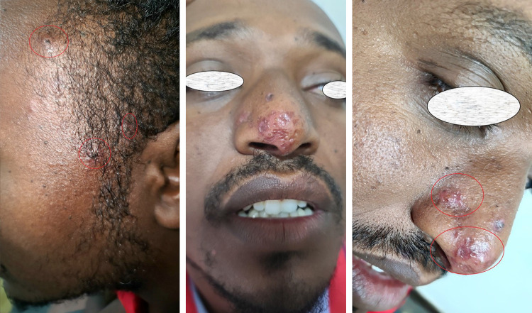
The first case was that of a 29-year-old man who presented with recurrent episodes of slightly pruritic erythematous papules on the anterior hairline and nose for more than 1 year. These lesions appeared as crops, became centrally indented, and were then covered with a centrally adherent eschar that resolved, leaving varioliform scars (Figure 1). Lesions were particularly frequent in the nose and hair-lines. No lesions were observed on the scalp or trunk. The lesions were more symptomatic during the summer.
Figure 1.
Crusted umbilicated papules on the anterior hair line and varioliform scarring over nose.
He visited another medical center for this complaint and was prescribed a prednisolone tablet, which improved but recurred after a few months.
Cutaneous examination revealed multiple reddish-brown, umbilicated papules with central necrosis covered with hemorrhagic crust, over the anterior hair-line and nose, and a varioliform scar over the tip of the nose (Figure 1). General physical and systemic examinations were normal.
On complete blood count, the total WBC count is 9.2 * 103, with 47.7% lymphocytes and 44.1% granulocytes, with a normal range of the other cell counts. C-reactive protein levels and routine blood chemistry results were within normal limits. No microorganism was discovered on AFB and Gram stain from the pustules. Tests for hepatitis B, syphilis, and HIV antibodies yielded negative results.
The patient was prescribed topical mometasone furoate cream and doxycycline at a daily oral dose of 100 mg, and significant improvements in papular and papulopustular lesions were observed after 8 weeks. He was placed on topical clindamycin gel and consulted in place of laser ablation for the varioliform scar after ensuring remission of the disease.
Case 2
The second case is a 58-year-old male patient with an 8-year history of multiple recurrent papules and pustules on the hair-line and central face. The patient complained of slight itching and burning as new lesions develop.
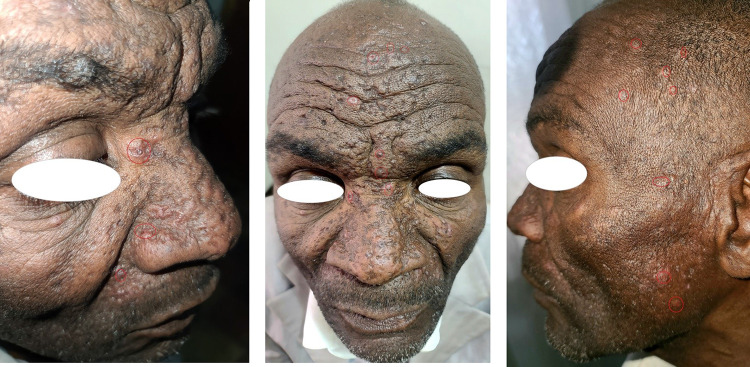
The number of lesions increased during the summer. With the resolution of the papules, varioliform scars appeared, and the face developed a marked cribriform appearance. He mostly complains about the scars it leaves behind (Figure 2). The patient had no other known medical illness.
Figure 2.
Multiple crusted umbilicated papules on the face and anterior hair line and varioliform scarring over the central face, and a single seborrheic keratosis papule over the right eye brow.
For this illness, he visited another medical facility and was prescribed topical betamethasone ointment for 2 weeks and advised to avoid excessive sun exposure, and his condition improved. After a couple of weeks, the lesion gradually returned, and, despite following the advice, it became recurrent.
Physical examination revealed reddish-brown, umbilicated papules with central necrosis, covered with round, adherent hemorrhagic crusts over the central face and hair-line, and extensive, depressed, varioliform scars distributed mainly on the mid-face and frontal scalp (Figure 2).
On Complete blood cell count, total WBC count was 7.5*109/L, with 36% lymphocytes and 56% granulocytes, Hgb of 13.9g/dl and serum creatinine is 1.1mg/dl. The erythrocyte sedimentation rate, liver function test, fasting blood glucose level, and C-reactive protein level were within the normal range. Tests for Hepatitis B and C, syphilis, and HIV antibodies were negative. Microbiological examination of the pustules revealed the presence of gram-positive cocci in groups.
The umbilicated papulopustular lesions and classic “varioliformis” scars led to a diagnosis of acne necrotica. Treatment was initiated with a topical clindamycin lotion, mometasone furoate cream, and doxycycline 100 mg PO once a day. After 2 months of treatment, the patient did not return for a follow-up appointment; therefore, he was contacted by phone and claimed that he was doing much better, but was too far away to come in person.
Discussion
Acne necrotica is a mysterious disorder that is unknown to many dermatologists, and usually not mentioned in recent texts. Acne necrotica was initially described by Bazin in 1851, which is a misnomer as it is not a variant of acne and bears only a superficial resemblance to an acneiform eruption,4 then acne frontalis seu varioliformis by Hebra, acne frontalis necrotica by Boeck, and the accepted term necrotizing lymphocytic folliculitis was first coined by Kossard et al in 1987.5
The disease onset is insidious with the appearance of red-brown papules on the nose, forehead, and frontal part of the scalp; however, in severe cases, the anterior chest and inter-scapular areas can also be affected. Initially red-brown follicular papules, often exudative, soon become pustule.4 As the pustule enlarges, its center becomes sunken and a dry scab develops, which is often hemorrhagic and gradually remodels, creating focal areas of necrosis over weeks, eventually leaving depressed varioliform scars.6 Some soreness and mild itching may be associated with the development of reddish-brown papules.
Although acne necrotica is usually localized on the face, with a peculiar distribution along the hairline, Shruti et al7 reported a case in which the entire scalp was covered with varioliform scars, giving it a remarkable “scar-studded appearance”.
Most notable is the fact that it is chronic over the course of several decades and always leaves ugly scars at the site of the disease. In some cases, emotional disturbances are associated with this condition.6
Necrotizing lymphocytic folliculitis is a rare and poorly understood chronic scarring follicular dermatosis characterized by necrotizing inflammation of follicles close to the scalp margins, resulting in multiple small round varioliform scars.6 It has been postulated that, acne necrotica varioliformis represents an acquired sensitivity to staphylococcal toxins, with subsequent formation of necrotic plugs and scarring.8 Furthermore, it was hypothesized that acne necrotica lesions begin as lymphocytic folliculitis, which were triggered by Cutibacterium acnes.7
The role of environmental and genetic factors has not yet been clearly established. Mechanical manipulation of pre-existing lesions, such as rubbing and scratching, may aggravate the disease, but not a cause.7 The association between acne necrotica and phenylbutazone treatment was reported in one patient,8 and herpes simplex virus was identified in a few cases of acne necrotica lesions.4
In 1928, Sabouraud recognized a milder variant, acne necrotica “miliaris.” Unlike the classic variant, it leaves no scars, is confined to the scalp, and is characterized by extremely itchy vesiculopustular lesions. Recently, micropapular and disseminated head and neck variants7 have also been described. This form of acne necrotica certainly needs to be distinguished from the cicatricial varioliform variant; however, it remains unclear whether this condition represents a minor variant of the same disease process or a different entity.9
The differential diagnosis is extensive and includes bacterial folliculitis, gram-negative folliculitis, acne vulgaris, tinea capitis (especially indolent Trichophyton tonsurans infection), eczema herpeticum, folliculitis decalvans, and eosinophilic pustular folliculitis. Other possible differential diagnoses include papulonecrotic tuberculosis, tertiary syphilis, and repetitive excoriations.
In scalp folliculitis, the lesions are typically distributed over the scalp and are smaller, usually more numerous, non-scarring, and itchy. Inflammatory and sometimes pustular lesions of acne necrotica on the face and upper aspects of the trunk can mimic acne vulgaris. However, acne necrotica begins in the third decade of life or later and often affects the scalp and lacks comedones, features that distinguish it from acne vulgaris. Compared to rosacea, the frequent involvement of the scalp and extra-facial sites and the lack of vascular symptoms in AN help distinguish between the two.
Lesions of lupus miliaris disseminatus faciei also require differentiation. These patients can also develop yellow-brown papules with a central adherent crust but are mainly concentrated around the eyes and cheeks, sparing the scalp, and do not heal with varioliform scars.
In most cases, histopathology helps confirm diagnosis and rule out other differential diagnoses. On histopathology, early lesions showed a marked perivascular and perifollicular lymphocytic infiltrate extending to the mid-dermis, associated with prominent sub-epidermal edema and exocytosis of lymphocytes into the external root sheath of centrally placed follicles and perifollicular epidermis. Necrotic keratinocytes were found at all levels of the epidermis and formed apoptotic bodies. Late lesions showed confluent necrosis of the central follicles, adjacent perifollicular epidermis, and the dermis. The central core of necrotic tissue showed only some hair fragments within the surface debris, indicating the presence of previous follicles.3
Other laboratory tests, such as aerobic culture, invariably grow Staphylococcus epidermidis or Staphylococcus aureus. Gram staining can detect numerous intracellular and extracellular gram-positive pleomorphic organisms, which is consistent with Cutibacterium acnes.6
Treatment is unrewarding, and usually needs to be continued for weeks or months. The clinical course is variable, with some cases resolving spontaneously, and others being treatment-resistant and requiring prolonged treatment for months, with frequent recurrence of lesions.2
If S. aureus is found, based on susceptibility testing, the appropriate antibiotic must be started, along with treatment of possible concomitant nasal carriage.6 Stritzler et al10 reported two cases of AN that achieved remission with 600,000 units of penicillin procaine administered once a week.
If S. aureus is not detected, tetracycline and erythromycin are the appropriate treatment options, possibly because of their anti-inflammatory effects. Anti-inflammatory treatment with topical or intralesional corticosteroids is considered a second-line treatment, which is also helpful in relieving pruritus. As a last resort, Isotretinoin has been recommended for the treatment of recalcitrant acne necrotica cases.6 Pitney et al5 reported significant improvements with Doxycycline, Erythromycin, and Isotretinoin in severe cases.
Lesion recurrence is common with decreasing doses or discontinuation of treatment. Once control is achieved, antibacterial or antiseptic lotions may substitute systemic antibiotics or isotretinoin as prophylaxis to prevent possible relapse.
Doxepin can be considered for patients who excoriate and manipulate lesions,11 and excision of larger scarred areas has been recommended.12
Conclusion
Acne necrotica (varioliformis) is a distinctive form of lymphocytic folliculitis with cutaneous involvement. Initially, only the superficial parts of the hair follicle are affected, and early and appropriate interventions can enable hair follicles to regenerate and promote new hair growth.
This refractory condition remains a challenge for every physician,4 and the resulting scars are a major concern for patients with remission. Acne necrotica is a disorder that requires reminiscing after it has been neglected for several decades.
Funding Statement
No funding was received.
Abbreviation
AN, Acne necrotica.
Ethics and Consent
Ethical approval was not required.
Written informed consent for publication of their details including patient photograph was obtained from the patients.
Disclosure
The author reports no conflict of interest in this work.
References
- 1.Ross EK, Shapiro J. Acne necrotica. In: Whiting D, Blume-Peytavi U, Tosti A, Trüeb RM, editors. Hair Growth and Disorders. Berlin, Heidelberg: SpringerVerlag; 2008:216–218. [Google Scholar]
- 2.Nikolić M, Perić J, Škiljević D. Acne Necrotica (Varioliformis) case report Serbian. J Dermatol Venereol. 2019;11(3):94–97. [Google Scholar]
- 3.Kossard S, Collins A, McCrossin I. Necrotizing lymphocytic folliculitis: the early lesion of acne necrotica (varioliformis). J Am Acad Dermatol. 1987;16:1007–1014. doi: 10.1016/S0190-9622(87)80408-5 [DOI] [PubMed] [Google Scholar]
- 4.Plewig G, Melnik B, Chen WC, Plewig G, Melnik B, Chen WC. Acne necrotica (necrotizing lymphocytic folliculitis). In: Plewig and Kligman’s Acne and Rosacea. 4th ed. Springer International Publishing; 2019:318–319. [Google Scholar]
- 5.Pitney LK, O’Brien B, Pitney MJ. Acne necrotica (necrotizing lymphocytic folliculitis): an enigmatic and underrecognised dermatosis. Australas J Dermatol. 2018;59:53–58. doi: 10.1111/ajd.12592 [DOI] [PubMed] [Google Scholar]
- 6.Griffiths CEM, Barker J, Bleiker T. Robert Chalmers, daniel Creamer. Acne necrotica (necrotizing lymphocytic folliculitis). In: Rook’s Textbook of Dermatology. 9th ed. John Wiley & Sons, Ltd; 2019:93.4–93.5. [Google Scholar]
- 7.Shruti S, Surabhi D, Varsha Gowda VM, Neha Dhankar Rajesh L, Pathi Kamal A, Aggarwal K. Story of a Scalp Studded with (Crateriform) Scars. Skin Appendage Disord. 2021;7:515–519. doi: 10.1159/000517239 [DOI] [PMC free article] [PubMed] [Google Scholar]
- 8.Hunter GA. Acne necrotica due to phenylbutazone. Br Med J. 1959;1(5114):113. doi: 10.1136/bmj.1.5114.113 [DOI] [PMC free article] [PubMed] [Google Scholar]
- 9.Ross EK, Tan E, Shapiro J. Update on primary cicatricial alopecias. J Am Acad Dermatol. 2005;53(1):1–37. doi: 10.1016/j.jaad.2004.06.015 [DOI] [PubMed] [Google Scholar]
- 10.Strizler C, Friedman R, Loveman AB. Acne necrotica. AMA Arch Dermatol Syph. 1951;64:464–469. doi: 10.1001/archderm.1951.01570100081013 [DOI] [PubMed] [Google Scholar]
- 11.Fisher DA. Acne necroticans (varioliformis) and staphylococcal aureus. J Am Acad Dermatol. 1988;1:1136–1138. doi: 10.1016/S0190-9622(88)80019-7 [DOI] [PubMed] [Google Scholar]
- 12.Sewon K, Masayuki A, Bruckner AL, et al. Acne necrotica (necrotizing lymphocytic folliculitis). In: Fitzpatrick’s Dermatology. 9th ed. McGraw-Hill Education; 2019:1531. [Google Scholar]


