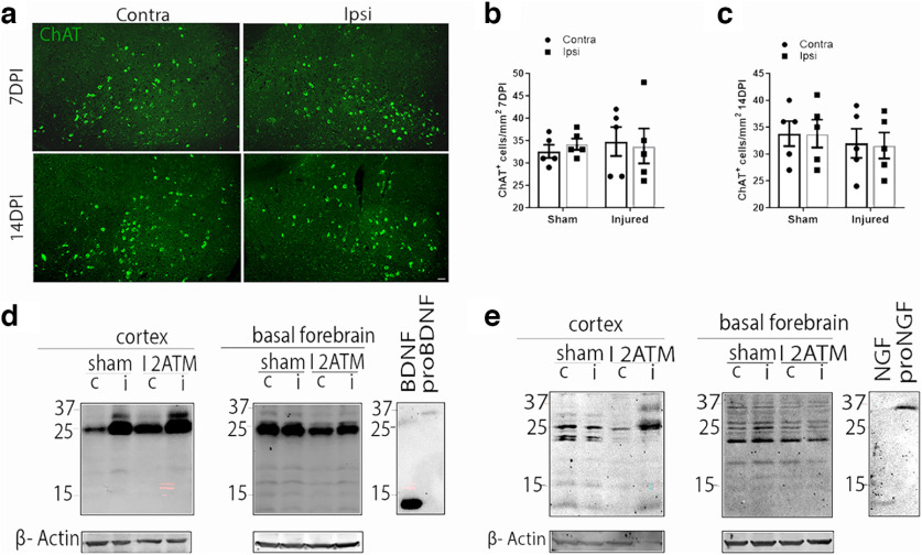
Figure 4.
The absence of p75NTR abrogates the retrograde loss of projecting basal forebrain neurons after cortical FPI. a, Coronal brain sections of the basal forebrain from injured p75NTR KO mice show immunostaining for ChAT (green) at 7DPI and 14DPI. Scale bar, 50 μm. b, Quantification of ChAT+ BFCNs in the ipsilateral versus contralateral side of the basal forebrain 7DPI in sham and injured p75NTR KO mice. Statistical analysis was performed using two-way ANOVA, Sidak’s multiple-comparisons tests. c, Quantification of ChAT+ BFCNs in the ipsilateral versus contralateral side of the basal forebrain 14DPI in sham and injured p75NTR KO mice. Statistical analysis was performed using two-way ANOVA, Sidak’s multiple comparisons tests. d, Cortex and basal forebrain tissue lysates obtained from 3DPI sham and TB1 p75NTR KO mice were probed for proBDNF (32 kDa) by Western blot; n = 3 sham and 3 injured brains. e, Cortex and basal forebrain tissue lysates harvested from 3DPI sham and TB1 p75NTR KO mice were probed for proNGF (37 kDa) by Western blot; n = 3 sham and 3 injured brains.

