Abstract
Proteins are not static but are flexible molecules that can adopt many different conformations. The HIV-1 protease is an important target for the development of therapies to treat AIDS, due to its critical role in the viral life cycle. We investigated several dynamics studies on the HIV-1 protease families to illustrate the significance of examining the dynamic behaviors and molecular motions for an entire understanding of their dynamics-structure–function relationships. Using computer simulations and principal component analysis approaches, the dynamics data obtained revealed that: (i) The flap regions are the most obvious sign of the evolution of conformational dynamics in HIV-1 protease; (ii) There are dynamic structural regions in some proteins that contribute to the biological function and allostery of proteins via appropriate flexibility. These regions are a clear sign of the evolution of conformational dynamics of proteins, which we call dynamozones. The flap regions are one of the most important dynamozones members that are critical for HIV-1 protease function. Due to the existence of other members of dynamozones in different proteins, we propose to consider dynamozones as a footprint of the evolution of the conformational dynamics of proteins.
Subject terms: Biochemistry, Biophysics
Introduction
Proteins are biomolecules that are regarded as the machinery of life. They are intrinsically dynamic, and their conformational variability is essential to their biological functions1,2. The function of a protein besides the structure also relies on its dynamics. Protein flexibility is necessary for biological function, ligand binding, and protein–protein or protein-nucleic acid interactions. A quantitative description of protein dynamics is essential for understanding living systems at a molecular level and probably also for the mechanisms leading to protein malfunction3,4.
Protein flexibility refers to the protein structure's internal dynamics, which is beneficial in the structural and functional aspects of proteins5. Plasticity and Conformational mobility represent key intrinsic features of proteins through evolution. Internal mobility eases the evolution of proteins to adopt conformational flexibility and thus provides the opportunity to develop novel functions. In addition, conformational flexibility allows proteins to better cope with harmful mutations which can lead to loss of function or altered function that result in disease5,6. Despite substantial proof that suggests that protein dynamics are under evolutionary selection, little is known about the molecular basis of the evolution of protein dynamics or how they affect function. An interesting case in the context of the dynamic-function relationship is that structural dynamics play a significant role in protein promiscuity, which almost means the ability of proteins to carry out several more or less related molecular works.
Indeed, every protein has the potential to accept many various conformations in the native state, so, many proteins are capable to perform several functions7,8. The structural diversity linked to protein flexibility constitutes a basis of protein evolvability6. In some cases, the protein exhibits one well-defined primary function together with several low-level “promiscuous activities.” Moonlighting proteins are other cases that can effectively carry out several relevant functions or even different duties linked to various molecular surfaces or active sites. Thus, it appears rational to attend that a particular trait of dynamics in functionally related protein regions (e.g., conformational variety, active-site “flexibility”, or “deformability”, results in the ability to stabilize different substrates, transition states of leaving groups) may be linked to many cases of protein promiscuity9,10.
Protein conformational dynamics play an important role in evolution, normal physiology, and pathophysiology. The evolution of proteins involves mutations that may lead to proteins adopting new functions and, in rare cases, new folds. Indeed, mutations of proteins can alter their conformations, dynamics, and stability, and thereby play critical roles in evolution and diseases. At a molecular level, protein evolution is dominated by neutral or nearly neutral mutations that have little effect on function11. Nonetheless, our comprehension of how proteins and species evolve is still elementary. There is a lack of detailed understanding of how proteins have evolved5,6.
One of the important questions about the evolution of proteins that should be further investigated is: How do conformational dynamics evolve as proteins evolve? The results of some studies show that the dynamics and the evolution of proteins share similarities12,13. Tang et al.14 found that there is a correspondence between the dynamics and evolution of protein structures. Their results show that the evolutionary mechanism of the proteins obtains both dynamical flexibility and evolutionary structural variation. Studying the evolution of protein conformational dynamics would not be possible without the use of computational and biophysical methodologies, that allow structural dynamics to be dissected in different protein variants. Much work has been done on the evolution of protein structure, but the role of protein dynamics in evolution has received attention in recent years.
Molecular dynamics (MD) simulations are a powerful tool to investigate the dynamic behavior of proteins in an aqueous solution and deepen our understanding of the relationship between protein structure and function3. The global molecular motions of the proteins can be obtained by applying the combination of MD simulation and essential dynamics (ED) analysis technique15.
HIV-1 protease (human immunodeficiency virus type 1 protease), an aspartyl protease, is responsible for the generation of structural proteins and viral enzymes critical to HIV viral maturation and infectivity. Thus, HIV-1 protease is a major drug target in the battle against HIV-1 infection, where the inactivation of the HIV-1 protease causes the production of immature, noninfectious viral particles16,17. There are about 750 experimentally determined available structures of this enzyme and this wide structural knowledge allows a study of a large number of conformations of protease complexes. The HIV-1 protease is one of the best-characterized cases of protein molecular evolution18. The HIV-1 protease is a homodimer with two identical monomers (chain A and chain B) each consisting of 99 amino acids. Each monomer has one α-helix (usually residues from 86 to 90) and nine β-sheets in the secondary structure. The residues of HIV-1 protease are numbered 1–99 and 100–198 (or 1′–99′) for chains A and B, respectively.
HIV-1 protease is a consisting of six structural segments (Fig. 1A): interface (residues 1–5/100–104, 95–99/194–198), fulcrum (residues 11–22/110–121), active site (residues 23–30/122–129), flap (residues 43–58/142–157), flap elbow (residues 35–42/134–141), and cantilever (residues 59–75/158–174).
Figure 1.
(A) Superposition of the crystal structures of the eleven selected HIV-1 proteases. Important regions of the HIV-1 proteases are labeled. (B) 3D structure of HIV-1 Protease (PDB code: 1HVI) showing the positions of mutated residues and also ligand position (light blue ellipse). Rendered using PyMOL.
The active site of the enzyme is formed at the dimer interface with each monomer a conserved catalytic triad (Asp25-Thr26-Gly27). It’s gated by two extended β-hairpin loops known as flaps. Two Asp25 residues (one from each monomer or chain) act as the catalytic residues and the conserved active site residues forms a symmetrical and highly hydrogen-bonded arrangement19,20.
Investigating protein flexibility may be important for the study of processes associated with conformational changes and state transitions21. Structural and dynamical studies of the HIV-1 protease normally focused on its more flexible region, the flaps, since they control the entrance/stabilization of ligands in the active site22,23. There is a large variety in the flap conformations in the unbound state, fluctuating between the closed, semi-open, and wide-open conformations24,25. In the closed/semi-open state, the catalytic site is shielded with two flaps and thus limits the entry of most of the ligands. The semi-open conformation is the dominant state in the ligand-free HIV-1 protease24–26. The flexibility of the flap is needed to facilitate the substrate access to and product release from the active site of the enzyme by an open and close mechanism27. The binding of protease substrate to the active site can be controlled by limiting the movement of the flap, thereby inhibiting HIV-1 protease activity24,28.
In order to indicate the relationship between the dynamics of proteins and their structure properties, we examine several dynamics studies. This study includes the investigations of the molecular motions and dynamic behaviors of the HIV-1 protease family in relation to their structure using computer simulation techniques. We adopted here the unbound form of the proteases of HIV-1 to investigate their dynamics-structure–function relationships. We focus mainly on the following facets: (i) dynamic behavior and collective motions of the HIV-1 protease family; (ii) the effect of point mutations on the molecular motions and stability of the HIV-1 protease family; (iii) correlation of some dynamic structural regions with the evolution of conformational dynamics in the HIV-1 proteases family.
The cross-correlation analysis and principal component analysis (PCA) were also performed to probe the difference in internal dynamics and conformational changes of the selected proteins induced by mutation. It is evident from the correlation map that almost in all proteases the flaps and flap elbow motions are highly correlated.
Results and discussion
Sequence analysis
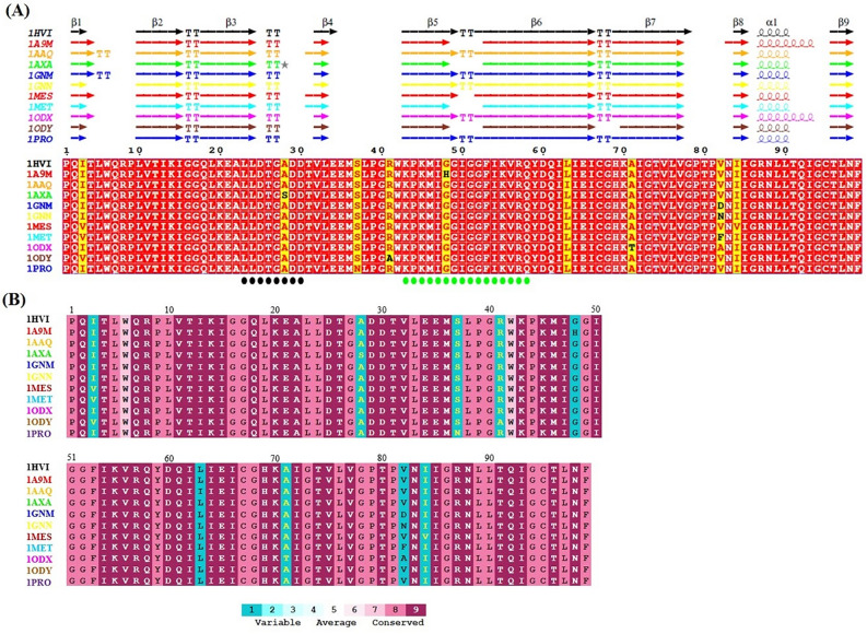
Proteins with sequence identity > 30% typically belong to the same family and have similar conformation and function. Such clear homologues are probably to have separated from a common ancestor and their sequences may show conserved differences between species of organisms. The simultaneous comparison of sequence and structure information is of significance to detect biological specificities in a group of proteins29,30. Multiple sequence alignment was performed using T-COFFEE and rendered by ESPript 3.0 using default parameters for residue similarity calculations, where boxed residues represent identical (red box, white character) and similar (Yellow box, red character) amino acid conservation (Fig. 2A). Residues boxed in red indicate strict conservation, while residues boxed in yellow indicate greater than or equal to 80% identity across the 11 homologs. Interestingly, the α-helix structure in proteases of the 1A9M and 1ODX is somewhat longer.
Figure 2.
(A) Sequence alignment produced by T-COFFEE and the figure was prepared with ESPript 3.0. Identical residues are written in white bold characters and boxed in red whereas similar residues are in red bold characters and boxed in yellow. Secondary structure elements related to each protein are indicated at the top of sequence blocks (α, α-helix; β, β-strand; TT, turn). Residues forming the active site (23–30) and the flap region (43–58) are labelled with a black circle and a green circle under the sequence alignment, respectively. (B) The Evolutionary Conservation prediction analysis of amino acid residues of all the selected proteins by Consurf server. The amino acids are colored according to their conservation grades and conservation levels. A grade of 1 indicates rapidly evolving sites, which are color-coded in turquoise; 5 shows sites that are evolving at an average rate, which are colored white; and 9 shows evolutionarily conserved sites, which are color-coded in maroon.
The structural and functional significance of a residue in protein structure is substantial for its evolutionary conservation. The importance of a given residue in conserving the structure and function of a protein can be inferred from the degree of conservation of the residue in a multiple sequence alignment of the protein and its homologues. The ConSurf31 is a bioinformatics tool for calculating the conservation pattern of a protein, which quantifies the degree of conservation at each aligned position. This program first identifies conserved positions using multiple sequence alignment, then calculates the evolutionary conservation rate using the empirical Bayesian method and provides the evolutionary conservation profiles of the structure or the sequence of the protein. ConSurf identifies functional regions in proteins, taking into account by considering the evolutionary relationships among their sequence homologs. ConSurf score ranges from 1 to 9, with 1 denoting rapidly evolving (variable) sites, 5 depicting sites that are evolving at an average rate, and 9 representing slowly evolving (evolutionary conserved) sites. The degree of conservation of the amino acid sites among the eleven homologues with similar sequences was estimated (Fig. 2B). Importantly, the information from the sequence logo of the proteins indicates that sequences are highly conserved in different proteins. As expected, the ConSurf analysis has revealed, that most of the amino acids in all the selected proteins are highly conserved. Similarly, this analysis indicated that the functional regions of all proteins are highly conserved.
Root mean square deviation (RMSD)
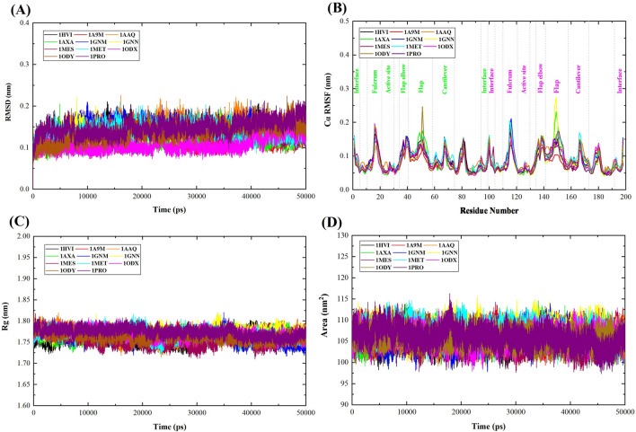
To obtain information about the conformational stability and assess the reliability of MD simulation, the RMSD of the backbone atoms of all the selected proteins was calculated. The value of RMSD has a negative correlation with the stability of the backbone atoms. The larger the value of RMSD, the more unstable the backbone atoms are32. Figure 3A shows the plot of RMSD for 1HVI (native protein) and ten proteins mutated. Initially, in the first 5000 ps, the RMSD was raised due to the "relaxation" of the proteins in the water environment, which is commonly observed in all MD simulation types. It is observed that all the proteins reach equilibrium after ~ 5 ns and present a steady behavior throughout the triplicates and thus suitable for exploring the dynamics of selected proteins.
Figure 3.
Plot illustrating (A) RMSD, (B) RMSF, (C) Rg, and (D) SASA for WT, and mutant proteins. The symbol coding scheme is as follows: wild-type protein (black color), mutant proteins 1A9M (red color), 1AAQ (orange color), 1AXA (green color), 1GNM (blue color), 1GNN (yellow color), 1MES (maroon color), 1MET (cyan color), 1ODX (magenta color), 1ODY (brown color), and 1PRO (violet color).
The RMSD average values for 1HVI, 1A9M, 1AAQ, 1AXA, 1GNM, 1GNN, 1MES, 1MET, 1ODX, 1ODY and 1PRO were found to be 0.146, 0.121, 0.146, 0.115, 0.144, 0.133, 0.133, 0.138, 0.107, 0.135 and 0.148 nm, respectively. 1PRO showed a higher RMSD value as compared to other proteins, whereas 1ODX showed the least value, which confirmed its greater stability than other proteins. The RMSD of wild and mutated proteins did not fluctuate convincingly, and all RMSD average values were less than 0.2 nm, thus, the equilibrium of all MD simulations is reliable. RMSD plots of these proteins showed that they displayed relatively similar stability. The replicates of each protein had slight variations in the RMSD values. Minor deviations in RMSD suggest the stable conformation of all proteins during this time period.
The plateau of RMSD values, observed at all simulations, is approximately similar between the wild-type and mutant proteins, indicating that all structures fluctuate around a stable average conformation. So, it is reasonable to evaluate its local fluctuations33.
Root mean square fluctuation (RMSF)
RMSF was calculated to study better the effect of amino acid mutations on the conformational flexibility of WT and mutant HIV-1 protease variants. The high value of RMSF shows the flexible region, while the low value of RMSF denotes limited movements during MD simulation. A fluctuation value of less than 2 Å is acceptable for a small protein34.
The comparison of the fluctuations between WT and mutant structures evidenced that the presence of the mutation resulted in no significant local flexibility alterations (Fig. 3B). The variation in the RMSF values suggested that the fluctuating behaviors were almost similar in the wild-type and mutant proteins except at the flap and fulcrum regions. The RMSF average values for mutant proteins were 0.0762, 0.0904, 0.0817, 0.0887, 0.0804, 0.0886, 0.0919, 0.0816, 0.0848, 0.0797 nm, for 1A9M, 1AAQ, 1AXA, 1GNM, 1GNN, 1MES, 1MET, 1ODX, 1ODY, and 1PRO, respectively, while the RMSF value for WT is 0.0864 nm (Table 1). According to the fluctuation score, we ranked the collected values as follows: 1MET > 1AAQ > 1GNM > 1MES > 1HVI (WT) > 1ODY > 1AXA > 1ODX > 1GNN > 1PRO > 1A9M. Therefore, 1MET showed larger fluctuations as compared to other proteins, whereas 1A9M showed the least fluctuations. In proteins 1A9M and 1ODX, the number of helix pitches in α-helix has increased, as a result, these two proteins have become more stable and show fewer fluctuations.
Table 1.
The calculated parameters for all the protein obtained after 50 ns MD simulations.
| Proteins | Average backbone RMSD (nm) | Average Cα-RMSF (nm) | Average Rg-protein (nm) | Average SASA (nm2) |
|---|---|---|---|---|
| 1HVI (WT) | 0.146 | 0.0864 | 1.759 | 105.677 |
| 1A9M | 0.121 | 0.0762 | 1.771 | 106.222 |
| 1AAQ | 0.146 | 0.0904 | 1.766 | 106.254 |
| 1AXA | 0.115 | 0.0817 | 1.767 | 105.925 |
| 1GNM | 0.144 | 0.0887 | 1.761 | 105.518 |
| 1GNN | 0.133 | 0.0804 | 1.772 | 107.363 |
| 1MES | 0.133 | 0.0886 | 1.754 | 105.314 |
| 1MET | 0.138 | 0.0919 | 1.768 | 107.140 |
| 1ODX | 0.107 | 0.0816 | 1.769 | 105.898 |
| 1ODY | 0.135 | 0.0848 | 1.762 | 105.717 |
| 1PRO | 0.148 | 0.0797 | 1.773 | 106.057 |
The RMSF plot showed that residual fluctuations are present in all proteins in several regions of the structure of the proteins. In addition to the N- and C-terminal residues, the regions around 17(116), 41(140), 52 (151), 67(166), and 81(180) show the biggest dynamic fluctuations. Residues 1–37 and 59–99 in each monomer are defined as the core region, while residues 43–58 constitute the flap region. It is worth noting that for all the proteins, changes observed in one monomer are almost always present also in the other. In the WT and mutant HIV-1 protease variants, there are two very stable regions in both monomers, one in the active site (residues 23–30/122–129) and the other in the α-helix formed by residues 86 to 90. As mentioned, the residues near the catalytic D25/D124 present a high degree of rigidity in all proteins, which is expected, as the catalytic function of these residues probably needs a well-defined stable three-dimensional structure.
Due to the fact that the conformational dynamics of the flap region of the protease are vital for the catalytic activity, our analysis for fluctuation focused on the flap region. Interestingly, after the check of the flap region, which includes residues 43–58, we noticed higher fluctuations in the 1ODY, 1GNN, and 1AXA proteins compared to the WT; while other areas exhibited similar behavior. The handedness feature of the flaps was also visible in the RMSF values in which one of the flaps has more fluctuation than the other35. The RMSF results are consistent with that of the RMSD.
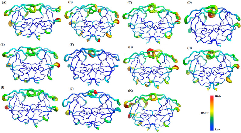
To visualize conformational fluctuations in selected proteins, we used a "sausage" plot to show the range of observed motions during simulation trajectories (Fig. 4). The thickness of the sausage plot is proportional to B-factor values and shows the extent of protein chain motion. The thinner segments denote the most stable regions of the protein, while thicker segments represent the more mobile regions. In most of the selected proteases, the highest mobility is found in the flaps and flap elbow regions, which should have significant functional implications because these regions are near the active site.
Figure 4.
Sausage representation for the selected proteases [(A) 1HVI; (B) 1A9M; (C) 1AAQ; (D) 1AXA; (E) 1GNM; (F) 1GNN; (G) 1MET; (H) 1MES; (I) 1ODX; (J) 1ODY; (K) 1PRO]. The thickness of the sausage indicates the mobility of the region. The color scale goes from blue (low RMSF values—poorly flexible regions) to red (high RMSF values—very flexible regions). The figures were prepared using the PyMOL software.
Radius of gyration (Rg)
The Rg calculated from the MD trajectory indicates the compactness or rigidity of a protein system during the simulation. Higher Rg values indicate less compactness of protein structure, while lower Rg values indicate more stability and compactness36. We performed Rg analysis to observe the conformational alterations and dynamic stability of the wild-type and mutant structures. To understand the changes in Rg with time, a plot was constructed (Fig. 3C). The average values of Rg calculated for each protein can be found in Table 1. The Rg plots of all the protein systems show fluctuations ranging lesser than 2 Å, which shows that the protein systems are stable. The average Rg values for 1HVI, 1A9M, 1AAQ, 1AXA, 1GNM, 1GNN, 1MES, 1MET, 1ODX, 1ODY, and 1PRO were found to be 1.759, 1.771, 1.766, 1.767, 1.761, 1.772, 1.754, 1.768, 1.769, 1.762 and 1.773 nm, respectively (Table 1). According to the fluctuation score, we ranked the collected values as follows: 1PRO > 1GNN > 1A9M > 1ODX > 1MET > 1AXA > 1AAQ > 1ODY > 1GNM > 1HVI (WT) > 1MES. The Rg plot suggested that the 1MES has tight packing than other proteins. Protein 1PRO showed a larger radius of gyration than other proteins, indicating that 1PRO, is less tightly packed. During the simulation, the WT and mutant proteins showed almost a similar pattern in terms of Rg values, indicating there were no important changes in the overall structure and folding of the protein after the mutation. In all of the proteins, the Rg results are in good agreement with that of RMSD and RMSF.
Solvent-accessible surface area (SASA)
The SASA analysis is used to measure the degree to which an amino acid is exposed to its environment. A higher SASA value denotes a diffused protein structure, while a lower SASA value represents a compact structure. An increase or decrease in SASA value represents a change in the structural conformation of the protein37. The SASA values of the WT and ten mutated proteins were analyzed for predicting how the mutations affect the structure of the native protein. The SASA values calculated for the WT and ten mutated proteins with time are shown in Fig. 3D, and average SASA values are depicted in Table 1.
The rank of collected average SASA values are listed as: 1GNN (107.363 nm2) > 1MET (107.140 nm2) > 1AAQ (106.254 nm2) > 1A9M (106.222 nm2) > 1PRO (106.057 nm2) > 1AXA (105.925 nm2) > 1ODX (105.898 nm2) > 1ODY (105.717 nm2) > 1HVI (105.677 nm2) > 1GNM (105.518 nm2) > 1MES (105.314 nm2). According to the Rg analysis, no significant difference was found between the wild and mutated protein, and a similar effect was also observed in the case of the SASA profile. Thus, the SASA results are also in settlement with the RMSD, RMSF, and Rg results.
Principal component analysis (PCA)
To gain deeper insight into the large-scale collective motions associated with conformation in the selected proteins, we performed PCA for analyzing the dominant protein conformational patterns in a principal components (PCs) phase space during 50 ns the MD simulations. In fact, we investigated the conformational behavior of the Cα atoms of the proteins by projecting them along the direction of the first three eigenvectors (PC1, PC2, and PC3).
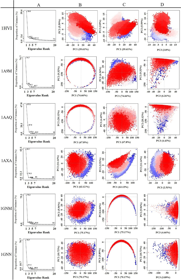
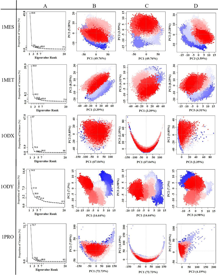
Figures 5 and 6 exhibit the first three PCs for selected proteins extracted from respective 50 ns MD simulation trajectories in the form of cluster groups. The 2D principal component plot between eigenvectors 1, 2, and 3 was drawn to compare acceptable conjoined motions. This 2D plot indicates the variations in the ensemble distribution for each conformation during the simulation interval, where each dot represents one conformation of the trajectory at a time t. The uninterrupted color representation (from blue to white to red) indicates the presence of substantial periodic bounces between conformers during MD simulations. There were three conformational states in all proteins, including the unstable conformational states (blue dots), the intermediate states (white dots), and the stable conformational states (red dots). These observations supported the compact and cluster motions for all selected proteins in their respective trajectory. Convincingly, dynamic motions of clusters in each extracted PC for the respective protein structure suggested the induction of collective fluctuation by point mutations as a function of the 50 ns MD simulation interval.
Figure 5.
Principal component analysis of the test proteins. This picture shows the proportion of variance (scree) plot (A), the projection of PC2 versus PC1 (B), PC3 versus PC1 (C), and PC2 versus PC3 (D) of the WT, 1A9M, 1AAQ, 1AXA, 1GNM and 1GNN, proteins during the simulation period.
Figure 6.
Principal component analysis of the test proteins. This picture shows the proportion of variance (scree) plot (A), the projection of PC2 versus PC1 (B), PC3 versus PC1 (C), and PC2 versus PC3 (D) of the 1MES, 1MET, 1ODX, 1ODY, and 1PRO proteins during the simulation period.
Principal components or eigenvectors are ranked according to decreasing eigenvalues, which directly correspond to their contribution to the overall conformational variance. Figures 5A and 6A show the scree plot of eigenvalues for the first 20 PC, which indicates the percentage of the total variance (mean-square fluctuation) captured by each PC based on their eigenvalue rank. In this diagram, tags on each point indicate the cumulative sum of variance accounted that by a specific eigenvector and its previous eigenvectors. Indeed, cumulative variance is shown as a function of the number of PCs. This Figure shows the first few eigenvalues at the beginning are associated with larger concerted motions but decline rapidly and attain more localized fluctuations.
Percent of the variance and cumulative variance for each of the proteins during the 50 ns of MD simulations are presented in Table 2. According to Table 2, PCA showed that the top 20 PCs could capture 73.3%, 94.7%, 96.4%, 88.7%, 93.3%, 92.7%, 77.2%, 73.8%, 89.7%, 57.9% and 92.2% of total variance during the 50 ns of MD simulations in 1HVI, 1A9M, 1AAQ, 1AXA, 1GNM, 1GNN, 1MES, 1MET, 1ODX, 1ODY and 1PRO proteins, respectively. From this result, also it was observed that the first three PCs were 49.0%, 90.2%, 93.2%, 76.7%, 85.7%, 85.3%, 58.6%, 52.4%, 79.2%, 26.9% and 84.0% of the total variance in the motion observed in the trajectories of 1HVI, 1A9M, 1AAQ, 1AXA, 1GNM, 1GNN, 1MES, 1MET, 1ODX, 1ODY and 1PRO, respectively. According to Table 2, PCA suggests that the properties of the motions described by the first three PCs are clearly different for all the proteins. The scree plot of all proteins indicates that the elbow point is located at the third PC, meaning the first three PCs appear to be significant (Figs. 5 and 6). As shown in Figs. 5A and 6A, after the third PC, there are no momentous variations in the eigen fraction till 20 eigenvalues, indicating a state of convergence in the respective proteins. These observations suggested that significant flexibility was produced in all proteins during the initial phase of 50 ns MD simulation which eventually diminished to attain a stable system. Moreover, a steady decrease in the amplitude of an eigen fraction further indicates an additional localized fluctuation in the protein structure to attain a favorable conformation. Comparing all proteins, the highest and lowest PC1 magnitude is 75.1% and 14.6% for 1GNM and 1ODY, respectively. Except for 1ODY and 1MET, the magnitude of PC1 in other proteins is significantly increased, which might correlate with the increased flap movement in the mutant proteins. The mutation in the 1MET protein did not alter the PC1 contribution significantly, but in the 1ODY protein, a lesser PC1 contribution was observed. These observations validated the result of higher flexibility of the mutated proteins compared to the native protein.
Table 2.
The eigenvalue, percent of the variance and cumulative variance for three principal components of each of the proteins during the 50 ns of MD simulations.
| Protein | Principle component (PC) | Eigenvalue | Variance (%) | Cumulative variance (%) |
|---|---|---|---|---|
| 1HVI (WT) | PC1 | 265.012 | 39.611 | 39.611 |
| PC2 | 37.407 | 5.591 | 45.203 | |
| PC3 | 25.440 | 3.802 | 49.005 | |
| 1A9M | PC1 | 2704.054 | 74.664 | 74.664 |
| PC2 | 337.502 | 9.319 | 83.983 | |
| PC3 | 226.812 | 6.263 | 90.246 | |
| 1AAQ | PC1 | 4278.441 | 74.335 | 74.335 |
| PC2 | 831.677 | 14.450 | 88.784 | |
| PC3 | 255.046 | 4.431 | 93.216 | |
| 1AXA | PC1 | 996.035 | 63.120 | 63.120 |
| PC2 | 175.818 | 11.142 | 74.261 | |
| PC3 | 39.660 | 2.513 | 76.775 | |
| 1GNM | PC1 | 2080.934 | 75.166 | 75.166 |
| PC2 | 164.879 | 5.956 | 81.121 | |
| PC3 | 128.989 | 4.659 | 85.781 | |
| 1GNN | PC1 | 1780.544 | 70.707 | 70.707 |
| PC2 | 269.991 | 10.722 | 81.428 | |
| PC3 | 98.061 | 3.894 | 85.322 | |
| 1MES | PC1 | 413.644 | 49.757 | 49.757 |
| PC2 | 43.914 | 5.282 | 55.040 | |
| PC3 | 29.853 | 3.591 | 58.631 | |
| 1MET | PC1 | 297.357 | 39.901 | 39.901 |
| PC2 | 61.484 | 8.250 | 48.151 | |
| PC3 | 32.091 | 4.306 | 52.457 | |
| 1ODX | PC1 | 1104.702 | 67.038 | 67.038 |
| PC2 | 162.356 | 9.852 | 76.890 | |
| PC3 | 38.706 | 2.349 | 79.239 | |
| 1ODY | PC1 | 50.291 | 14.642 | 14.642 |
| PC2 | 25.083 | 7.303 | 21.946 | |
| PC3 | 17.118 | 4.984 | 26.929 | |
| 1PRO | PC1 | 1766.330 | 72.730 | 72.730 |
| PC2 | 172.274 | 7.094 | 79.824 | |
| PC3 | 101.688 | 4.187 | 84.011 |
Except for 1ODY, the PC1 accounts for more than one-third of the total variance and strongly overcomes the total variance showing the global dynamics38. Interestingly, all the selected proteins, except 1ODY, showed a sharp increase in the percentage of variance corresponding to the first three PCs and covered more than > 50% of the total proportion of variance of atom positional fluctuations in each simulated protein. After that, the individual component contributions fall below 3%. In fact, these first three PCs account for a large proportion of the overall protein conformation and capture the most significant dominant motions, in other words, the fluctuations of the highest amplitude that are generally biologically relevant motions. These results showed that point mutations caused significant changes in the conformational motions of the selected proteins. Therefore, PCA dots images generated from the first three eigenvectors are used to observe the conformational transitions of these proteins39,40.
On a 2D principal component plot, the larger the cumulative variance on the two considered principal components, the more significant the distance between the points. It means that diverse conformations will have diverged while similar conformations will have grouped on the 2D PCA plot38. Comparing the 2D scatter plots of all proteins, it could be seen that the conformational states of the mutated proteins compared with wild protein had changed significantly. As shown in Figs. 5 and 6, except 1ODY, the PC2 versus PC1 plots for all proteins clearly indicated the conformers visit a large conformational space.
Evaluation of internal motions through the first three principal components shows that PC1 and PC2 are prominently related. As shown in Figs. 5 and 6, the PC2 versus PC1 plots obtained from the MD trajectories are almost varied for all proteins, which display differences in motion across the two first principal components. These observations clearly indicated the differences in protein motion and the conformational landscape between the proteins. This indicates that mutations in these proteins have caused conformational changes. However, the presence of overlap between blue and red colors indicates that the protein does revisit the same state during the simulation, even though it undergoes conformational changes. Also is an indication of the quality of sampling, which illustrates the simulation time is sufficient. As shown in PC2 versus PC1 plots, in the case of comparing proteins with each other, the 1ODY protein has a smaller phase space and lower performance flexibility than other proteins. In the 1ODY protein, the contribution of PC1 and PC2 to the variance is 14.6% and 7.3%, respectively, while other PCs contributed no more than 5.0%.
It can be found from Figs. 5 and 6 that in PC2 versus PC1 diagrams, the points of all proteins are almost evenly distributed near the midline, while in PC2 versus PC3 diagrams, the points of proteins are differently distributed. The closer the point distribution in Figs. 5 and 6 indicates that the protein system is more stable, so the 1ODX and 1AXA proteins are in a more stable state. In another word, 1ODX and 1AXA proteins exhibited the most favorable converged conformations and limited variation against other proteins during MD simulation; suggesting considerable stability as noted from the respective RSMD and RMSF values.
In some PC2 versus PC1 and PC3 versus PC1 diagrams, the projection of the points of proteins reveals a semicircle or U-shape pattern. This type of pattern has been attributed to random diffusion of motion in proteins, allowing only to inform on more accessible degrees-of-freedom for thermal motion along our studied time scale41.
Point mutations increase overall collective motions in selected proteins because these mutations effectively increase the Cα movements of proteins. In fact, these mutations have increased the flexibility of proteins. Except for 1ODY, the conformational space covered in other proteins proved to be broader than that of 1HVI. These results mean that after point mutation, the dynamics of the mutated proteins change, which may be required for proper protein function. Also, these fluctuations registered in each protein may be regarded as a requirement for the stability of the relevant protein during MD simulation as a function of time41. The PCA results are consistent with the RMSD, RMSF, Rg, and SASA results.
Porcupine plot
The main motions of protein residues can be better observed and analyzed by displaying eigenvectors as porcupine plots42. Porcupine plots are drawn using the custom-made program PyMOL43 to visualize the movements of the first three PCs obtained from the principal component analysis. It corresponds to an outline of protein motion, suggesting what part of the protein moves in concert and in which direction. In fact, the first and last eigenvectors from any PC were generated using the PyMOL tool and presented as a porcupine plot. The extent and direction of the most dominant motions of all proteins were visualized through porcupine plots using the ‘modevectors.py’ script (written by Sean M. Law) in PyMOL version 1.7. The plot shows a cone for each Cα atom reflecting the direction of its motion, where the length of the cone indicates the motion amplitude and the size of the cone specifies the number of such Cα atoms. The linear interpolations between the first and last eigenvectors are shown with the color transition from blue to red to highlight the conformational differences between them.
The porcupine plots showing the motions of all selected proteins, along the directions of PC1, PC2, and PC3 are presented in Fig. 7. The cones in black represent the direction of the concerted motion, and the length of the cones represents the extent of the motion. Based on the porcupine plots, it is evident that point mutation increases the overall movements of all mutant proteins as compared to the wild-type protein. All proteins showed anti-symmetric movement patterns in the flap, flap elbow, fulcrum, and cantilever regions. In all proteins, the flap and flap elbow regions experienced more flexibility compared to the different segments of the protease, as indicated by the length of the vectors. Thus, it is obvious that in all proteases the flaps and flap elbow motions are highly correlated. Flexible regions of the proteins are not simply the result of loose packing or instability but have been evolutionarily selected44. Considering that the conformational flexibility of the flap region is necessary for the activity of the HIV-1 protease, it can be concluded that the flap regions are one of the most important signs of the evolution of conformational dynamics in HIV-1 protease.
Figure 7.
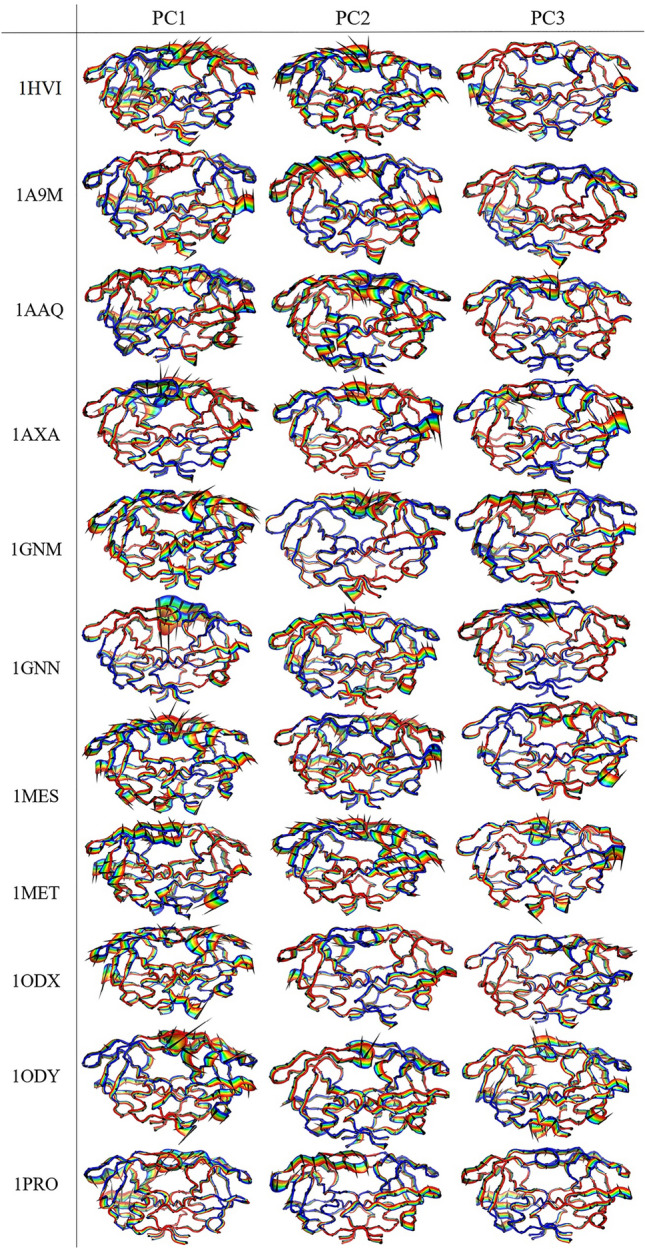
Porcupine plots corresponding to PC1, PC2, and PC3 obtained from PCA on the MD trajectories of the wild-type protein and the mutant proteins. The cones in black represent the direction of the concerted motion, and the length of the cones represents the magnitude of the movement.
Dynamical cross-correlated map (DCCM) analysis
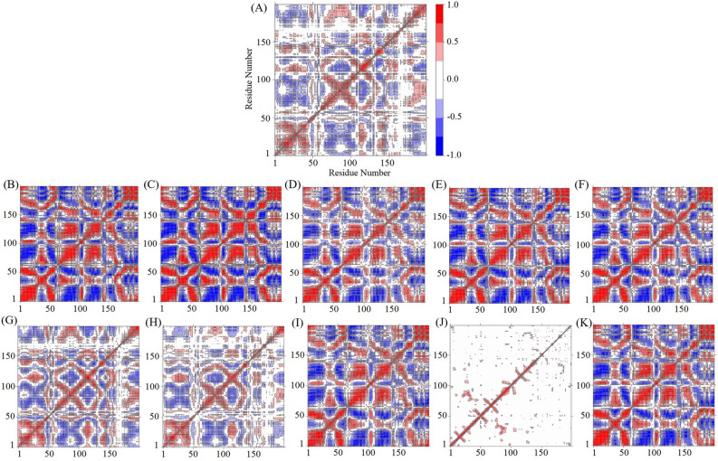
To investigate further the effects of point mutation on the conformational dynamics, DCCM analysis was used for Cα atom fluctuations during the 50-ns MD simulation for analyzing the correlation motions of each protein. The cross-correlation analysis could show the relationships between residues and between various regions by quantifying their relative motions. The correlation was normalized and varied from − 1.0 and + 1.0 (from dark blue to white to red) (Fig. 8). The positive correlation (red regions) represented residues moved in the same direction and the negative correlation (blue regions) meant residues moved in the opposite direction45. The deeper color indicated a stronger positive correlation or negative correlation. The white regions ranging from − 0.25 to 0.25 were considered as a low correction. A diagonal point represents the Cα atom of the same residue along both axes; therefore, the diagonal elements show the maximum correlation.
Figure 8.
Comparison of cross-correlation matrices of wild-type and mutant proteins. Cross correlation matrix of C-alpha atoms during 50 ns simulation for wild-type protein (A) and mutant proteins of 1A9M (B), 1AAQ (C), 1AXA (D), 1GNM (E), 1GNN (F), 1MES (G), 1MET (H), 1ODX (I), 1ODY (J), and 1PRO (K). The range of motion is shown by different colors in the panel. Red color denotes positive correlation whiles blue color denotes anti-correlation.
The cross-correlation matrix of the C-alpha displacement indicated complex correlated and anti-correlated motions in the wild-type protein and all mutant proteins except 1ODY. The diagonal parts show obvious correlated movements. In the cross-correlation matrix, the Ci,j elements of the matrix were found to be symmetrical about the diagonal. Interestingly, in all mutant proteins, correlated and anti-correlated motions increase. The only exception is the 1ODY mutant in which correlated and anti-correlated motions highly decrease. For the mutated proteins, we found deeper shades of red and blue, distinguishing regions of high correlation and anti-correlation that corroborate the fact that mostly global motions are seen for these proteins. In most mutant proteins, except 1ODY, almost all residues have concerted motions and they move in an anti-correlated fashion with the rest of the structure (Fig. 8). The point mutation has significantly changed the direction and the cooperativity of motion in 1ODY.
It can also be observed that the motion of atoms in all mutant proteins as compared to the wild-type protein is more compact, while the 1ODY mutated protein represented a very deformed form of the backbone atomic motion. The 1MES and 1MET proteins have approximately the same correlation and anti-correlation motions as the 1HVI protein, however, in these two mutant proteins, the intensity of the correlated and anti-correlation motions is slightly higher than that of the 1HVI protein. Thus, our results show that variations in sequence can lead to changes in dynamics without altering structure.
As seen from principal components and the dynamic cross-correlation maps, the point mutations change the major motions of the proteins and may improve the dynamic behavior of proteins. These results indicate that point mutations may have a pronounced effect on the conformational flexibility of proteins.
The conformational dynamics in the flaps of HIV-1 protease plays a critical role in the mechanism of substrate or inhibitor binding. Opening of the flaps is essential for the entrance of substrate into the binding cleft and flaps in the closed conformation sturdy the substrate for catalysis. The protease dimer is in dynamic equilibrium between the closed conformation and different open conformational states. These conformational changes are highly associated with the flexibility of the flaps of the protease46. Molecular dynamics studies show that mutations in the flap regions may affect the dynamics of the flap and thus the binding of protease inhibitors47. The results revealed that the mutations caused increased movement in the flap, and flap elbow regions. The presence of a region with such conformational dynamic diversity in the protein that also provides the necessary flexibility to perform the proper function of the protein indicates that the protein has also evolved dynamically in addition to structural evolution. Indeed, since the flexible regions of proteins are selected evolutionarily, it can be inferred that the flap regions are the most significant sign of the evolution of conformational dynamics in the HIV-1 protease.
Previous studies show that dynamic structural regions exist in various proteins whose conformational flexibility is essential for the function and allostery of proteins (Table 3). Thus, it is time to group these dynamic structural regions and choose a specific and unique name for these areas. Our suggestion for naming these regions is dynamozones. Dynamozones are dynamic structural regions in some proteins that contribute to the biological function and allostery of proteins via their convenient flexibility. These regions provide the flexibility needed for proteins to suitable function, and their presence is completely essential for protein function. One of the characteristics of these regions in most proteins is the presence of the amino acid glycine in their structure. Dynamozones are a subset of three main groups: loops, hinges, and linkers. One of the clear signs of the evolution of conformational dynamics of proteins is the presence of dynamozones in proteins because these dynamic regions have evolved in such a way that they workable the function and allostery of proteins. These regions via different mechanisms such as performing opening-closing movements for ligand binding to the active site, appropriate motions of the loops for placement of the catalytic residues in the suitable position for catalysis, switching from an inactive "out" conformation to an active "in" conformation to create the catalytically active form of the enzyme, proper motions of linkers in proteins to accept compact and extended conformations, movements of hinges to connect antibodies to antigens, etc. help various proteins to perform their proper function. Thus, we can say that dynamozones are a footmark of the evolution of the conformational dynamics of proteins. The details of some of the know dynamozone members are listed in Table 3.
Table 3.
List of some known members of Dynamozone.
| Structure | Dynamozones | Proteins | Role or Function | References |
|---|---|---|---|---|
| Loop | Flap |
• HIV protease • Plasmepsins • Beta-secretase • Cathepsin • Pepsins |
These regions control the entrance and stabilization of ligands in the active site | 67–70 |
| Loop | Loop (Residues 166–176) | • Triosephosphate isomerase | In the ligand-bound state, the loop moves for ∼7 Å as a rigid lid toward the active site and accepts a “closed” conformation. These motions of the rigid lid close to the active site are essential for the catalytic mechanism of the enzyme | 71–73 |
| Loop | Loop |
• Enolases • Aldolases |
Movements of the loop permit the catalytic residues to be oriented in a suitable position for catalysis | 74–76 |
| Loop | WPD loop | • Protein tyrosine phosphatases (PTPs) | This loop closes over the active site upon binding of the substrate, and loop closure permits the correct orientation of catalytic residues around the ligand | 77,78 |
| Loop | Met20 loop (Active site loop ( | • Dihydrofolate reductase (DHFR) | This loop acts as a lid that closes on the cofactor, thereby allowing DHFR to adopt occluded and closed conformations | 79–81 |
| Loop | Helical loop | • Lipases | This loop is important for the enzyme function, acting as a lid to open or close the hydrophobic active site | 82,83 |
| Loop | Long loop | • β1,4-galactosyl transferase | A displacement of more than 20 Å this long loop in protein provides binding sites for various ligands | 84 |
| Loop | Omega loop | • Cdc34-like E2 enzymes | This loop can act as a lid that regulates the accessibility of the catalytic site and disturbs the charging activity of ubiquitin until a conformational change toward an open state is promoted by phosphorylation | 85–87 |
| Linker | Flexible linker | • Calmodulin (CaM) | CaM has two globular domains connected by a short and flexible linker that permits the protein to accept a wide variety of extended and compact conformations | 88–90 |
|
Hinge Loop Linker |
• Hinge region • P-loop • hydrophobic “spines” • A-loop • αC helix • DFG motif • αB helix |
• Protein kinases (PKs) • Tyrosin kinase • Src protein kinase |
Hinge region: The hinge motion is necessary for the opening and closure of the kinase catalytic domain (CD) P-loop (β1-β2 loop or G-loop or Gly-rich loop): This flexible loop is very important for the coordination of ATP phosphates Hydrophobic “spines”: Two hydrophobic “spines” link the two lobes of protein kinase and dynamically connect all the elements important for catalysis A-loop (Activation loop): In the inactive state of the enzyme, the A-loop is folded onto itself, and its opening is required to create the catalytically active form αC helix: This helix in the “in” active conformation forms a hydrogen bond with the β3 strand for creating the catalytically active form of the enzyme DFG motif: This motif in the active site switches from an inactive (DFG-out) conformation to an active (DFG-in) conformation, which is necessary to create the catalytically active form of the enzyme αB helix: This helix creates a cavity, the so-called PIF pocket, which is very important for allosteric regulation of the protein kinases belonging to the AGC family |
91–96 |
| Hinge | Hinge region | • Lactoferrin | The hinge motions permit the formation of the complete iron-binding site in the closed states of lactoferrin | 97–100 |
| Hinge | Hinge region | • Immunoglobulins | The hinge region is connecting the Fab (Fragment antigen binding) region to the Fc (Fragment crystallizable) region | 101,102 |
Conclusions
Protein dynamics, structure, and function are highly correlated. In the case of the HIV-1 protease, the dynamics of conformational changes are very critical for enzyme function. In the present study, we have explored the effect of mutations on the dynamic behavior of the WT and mutant HIV-1 proteases variants using a combination of MD simulations, cross-correlation analysis, and PCA. Sequence similarities between proteins were strikingly similar and confirmed the assumption that proteins with greater sequence similarity behave similarly. The results indicate that mutations not only produce important effects on the correlated motions and flexibility of HIV-1 protease but also increase the stability of HIV-1 protease during the simulation period. These positively selected mutations introduce significant flexibility in important regions such as the flap and flap elbow. Further, the parameters such as RMSF, DCCM, porcupine plot, and the PCA analysis revealed increased fluctuation/motion in the mutated proteases in comparison to the wild-type structure. Our main results were as follows: (1) The flap regions are the most evident indication of the evolution of conformational dynamics in HIV-1 protease and are an excellent case for investigating the evolution of conformational dynamics; (2) Dynamozones are dynamic structural regions in some proteins that donate to the biological function and allostery of proteins through their proper flexibility; (3) Because of the presence of other members of dynamozones in various proteins, we offer to consider dynamozones as an indication of the evolution of the conformational dynamics of proteins. The flap regions are one of the most significant dynamozone members that are crucial for HIV-1 protease function.
Computational methods
Protein structures preparation
A set of eleven HIV-1 protease proteins with experimentally determined structures chosen from the Protein Data Bank (PDB), were used in this study48. The selected proteins' structures have been determined using X-ray crystallography, and their resolution and R-factor are less than 3.0 and 0.19, respectively. Molecular dynamics simulation approaches pave the way for an in-depth analysis of the effects of mutations on protein structure and dynamics. In the present study, mutations G48H, L63I, A28S, V82D, V82N, (I3V; I84V), (I3V; V82F), (A71T; V82A), (I3V; R41A) and S37N, which are related to structures 1A9M, 1AAQ, 1AXA, 1GNM, 1GNN, 1MES, 1MET, 1ODX, 1ODY and 1PRO, respectively, were analyzed to investigate the effect of mutations on the conformational dynamics of HIV-1 protease using molecular dynamics simulations (Fig. 1B). The PDB code: 1HVI was also selected as a wild-type protein. We selected a suitable mutation from each of the different parts of the HIV-1 protease to study the dynamic behavior of the proteases. The ligands, ions and water molecules were removed from the protein structures. The details about the selected HIV-1 protease variants are listed in Table 4.
Table 4.
Overview of the amino acid changes in the HIV-1 protease families.
| Simulated proteins (PDB ID) | Residue position | Amino acid change | Property change |
|---|---|---|---|
| 1HVI (WT) | |||
| 1A9M | 48 | G → H | Hydrophilic/+ * |
| 1AAQ | 63 | L → I | Hydrophobic/Hydrophobic |
| 1AXA | 28 | A → S | Hydrophobic/Hydrophilic |
| 1GNM | 82 | V → D | Hydrophobic/−** |
| 1GNN | 82 | V → N | Hydrophobic/Hydrophilic |
| 1MES | 3 | I → V | Hydrophobic/Hydrophobic |
| 84 | I → V | Hydrophobic/Hydrophobic | |
| 1MET | 3 | I → V | Hydrophobic/Hydrophobic |
| 82 | V → F | Hydrophobic/Hydrophobic | |
| 1ODX | 71 | A → T | Hydrophobic/Hydrophilic |
| 82 | V → A | Hydrophobic/Hydrophobic | |
| 1ODY | 3 | I → V | Hydrophobic/Hydrophobic |
| 41 | R → A | + /Hydrophobic | |
| 1PRO | 37 | S → N | Hydrophilic/Hydrophilic |
* + Positively charged amino acids; **− negatively charged amino acids.
Molecular dynamics simulation
In this study, all of the MD simulations were performed using the GROMACS 2016.349. The force field parameters were assigned according to the Amber99SB*-ILDN force field50. Protonation states were assigned to each structure using PDB2PQR51 through ProPKa52 at pH 7.0. The TIP3P water model53 was used to solvate the system, which was generated as a cubic box like area with a side of 1 nm such that the protein is covered appropriately with water molecules. All proteins were electrostatically neutralized by adding chlorine ions around the molecules. Each protein was minimized by the steepest descent algorithm up to a maximum of 50,000 steps and a convergence tolerance of 10 kJ mol−1 nm−1. The energy-minimized structure of the native protein and ten mutant proteins were used as the starting points for the MD simulations. Equilibration of the proteins was conducted in two phases NVT and NPT ensembles each for 100 ps. Particle-Mesh-Ewald (PME) method54 with a 1.0 nm cutoff was used to calculate the electrostatic interactions. During the MD simulation, the LINCS algorithm55 was used to constrain all the bonds. The temperature was kept constant (300 K) and pressure was maintained at 1 bar using the V-rescale thermostat56 and the Parrinello-Rahman barostat57, respectively. Following the equilibrium methods, MD simulations were performed for each of the native and mutant proteins with 3 repetitions for 50 ns.
Analysis of trajectory files
The resulting trajectory files of the simulations were studied employing different parameters existing in GROMACS utilities. From the refined trajectories, various parameters, such as root mean square deviation (RMSD), root mean square fluctuation (RMSF), the radius of gyration (Rg), and solvent-accessible surface area (SASA) for all proteins were calculated. The sequence alignments were created with T-Coffee58 and ESPript59. PCA was carried out using the Bio3D package60 and used to reveal the changes in the motion patterns of the protein systems.
To exclude the possibility of stochasticity and to confirm the first simulation performed, all 11 selected structures were simulated with different initial velocities with three repeats for 50 ns.
PCA analysis which is explained in detail below is one of the main analyzes performed for this research. To confirm that our simulation time is sufficient to investigate the overall dynamic behavior of the protein, we performed the cosine content analysis, which is part of the PCA method. As a result of this analysis, we can determine whether the protein exhibits random diffusion dynamics during the simulation. A cosine content close to 1 indicates random motion in the protein and non-convergence of the simulation time in the selected time interval, so it cannot be considered for PCA analysis. It has been reported that the cosine content close to 0.2 and sometimes up to 0.5 indicates the non-random diffusion dynamics of the protein during the simulation time and is reliable for further analysis, such as the free energy landscape (FEL)61,62.
In this study, we reported the replicates that had cosine content values of the first two eigenvectors (PC1 and PC2) close to 0. Based on chosen principal components (PC1 and PC2), we generated the FEL to calculate Gibbs's free energy value for selected structures. The cosine content values, Gibbs’s free energy values, and RMSD of the simulated structures are reported in the supplementary data file (Figures S1-S11, Table S1).
Principal component analysis
A principal component analysis was performed to investigate conformational flexibility and the collective motions of the selected proteins using the Bio3D package installed in the R program. This method is based on diagonalization of the covariance matrix of atomic fluctuations to obtain orthogonal eigenvectors and the corresponding eigenvalues. The eigenvectors are the principal components that represent the directions of the coordinated motions of atoms. The eigenvalues indicate the magnitude of the motions along the movement direction63. The ensemble formula used to obtain a covariance matrix with elements Cij for coordinates i and j is given as:
| 1 |
where xi and xj are the mass-weighted Cartesian coordinates of the ith and jth Cα atoms, N is the number of the Cα atoms considered, and and represent the time average over all the configurations obtained in MD simulation. In this study, by the Bio3D package, Cα atoms from 50,000 frames obtained through 50 ns trajectory were superimposed on the initial pose to minimize the root mean square variations between the equivalent residues using fit.xyz function64.
Dynamic cross-correlation map analysis
The cross-correlation analysis can provide information about the impact of mutations on protein dynamics by analyzing how atomic displacements were coupled65. The extent of correlative motion of two atoms (or two residues) can be denoted by the cross-correlation coefficient, Cij. It is defined by:
| 2 |
where i (j) means ith (jth) residue (or atom), Δxi and Δxj are the displacements from the mean position of i-th and j-th residues (or atoms), with respect to time, respectively, and N represents the number of Cα atoms. The angular brackets “〈 〉” illustrate the time average on the whole trajectory. The value of the cross-correlation coefficient is from − 1 to + 1. The positive value implies positively correlated movement (moving in the same direction), and the negative value implies anti-correlated movement (moving in the opposite direction). Higher values of the absolute value of Cij show two residues (or atoms) are more correlated (or anti-correlated)66. In this article, we calculated the cross-correlations for all Cα atomic fluctuations extracted from the MD trajectory by using the Bio3D packages of R.
Data availability
The datasets generated during and/or analysed during the current study are available from the corresponding author on reasonable request.
Supplementary Information
Acknowledgements
The authors would like to acknowledge the Department of biophysics of Tarbiat Modares University.
Author contributions
M.R. and M.T. conceived and designed the research, and M.R. and F.A.J. performed the simulations and prepared figures. M.R. Writing—original draft. M.T. supervised the project. All authors reviewed the manuscript.
Competing interests
The authors declare no competing interests.
Footnotes
Publisher's note
Springer Nature remains neutral with regard to jurisdictional claims in published maps and institutional affiliations.
Supplementary Information
The online version contains supplementary material available at 10.1038/s41598-023-40818-x.
References
- 1.Karplus M, McCammon JA. Molecular dynamics simulations of biomolecules. Nat. Struct. Biol. 2002;9:646–652. doi: 10.1038/nsb0902-646. [DOI] [PubMed] [Google Scholar]
- 2.Karplus M, Kuriyan J. Molecular dynamics and protein function. Proc. Natl. Acad. Sci. 2005;102:6679–6685. doi: 10.1073/pnas.0408930102. [DOI] [PMC free article] [PubMed] [Google Scholar]
- 3.Grant BJ, Gorfe AA, McCammon JA. Large conformational changes in proteins: Signaling and other functions. Curr. Opin. Struct. Biol. 2010;20:142–147. doi: 10.1016/j.sbi.2009.12.004. [DOI] [PMC free article] [PubMed] [Google Scholar]
- 4.Wlodawer A. Rational approach to AIDS drug design through structural biology. Annu. Rev. Med. 2002;53:595–614. doi: 10.1146/annurev.med.53.052901.131947. [DOI] [PubMed] [Google Scholar]
- 5.Schweizer L, Mueller L. Biased Signaling in Physiology, Pharmacology and Therapeutics. Elsevier; 2014. [Google Scholar]
- 6.Tokuriki N, Tawfik DS. Protein dynamism and evolvability. Science. 2009;324:203–207. doi: 10.1126/science.1169375. [DOI] [PubMed] [Google Scholar]
- 7.Dill KA, Chan HS. From Levinthal to pathways to funnels. Nat. Struct. Biol. 1997;4:10–19. doi: 10.1038/nsb0197-10. [DOI] [PubMed] [Google Scholar]
- 8.James LC, Tawfik DS. Conformational diversity and protein evolution–a 60-year-old hypothesis revisited. Trends Biochem. Sci. 2003;28:361–368. doi: 10.1016/S0968-0004(03)00135-X. [DOI] [PubMed] [Google Scholar]
- 9.Khersonsky O, Roodveldt C, Tawfik DS. Enzyme promiscuity: Evolutionary and mechanistic aspects. Curr. Opin. Chem. Biol. 2006;10:498–508. doi: 10.1016/j.cbpa.2006.08.011. [DOI] [PubMed] [Google Scholar]
- 10.Fornili A, Pandini A, Lu H-C, Fraternali F. Specialized dynamical properties of promiscuous residues revealed by simulated conformational ensembles. J. Chem. Theory Comput. 2013;9:5127–5147. doi: 10.1021/ct400486p. [DOI] [PMC free article] [PubMed] [Google Scholar]
- 11.Kimura M, Ohta T. On some principles governing molecular evolution. Proc. Natl. Acad. Sci. 1974;71:2848–2852. doi: 10.1073/pnas.71.7.2848. [DOI] [PMC free article] [PubMed] [Google Scholar]
- 12.Marsh JA, Teichmann SA. Parallel dynamics and evolution: Protein conformational fluctuations and assembly reflect evolutionary changes in sequence and structure. BioEssays. 2014;36:209–218. doi: 10.1002/bies.201300134. [DOI] [PubMed] [Google Scholar]
- 13.Campitelli P, Modi T, Kumar S, Ozkan SB. The role of conformational dynamics and allostery in modulating protein evolution. Annu. Rev. Biophys. 2020;49:267–288. doi: 10.1146/annurev-biophys-052118-115517. [DOI] [PubMed] [Google Scholar]
- 14.Tang Q-Y, Kaneko K. Dynamics-evolution correspondence in protein structures. Phys. Rev. Lett. 2021;127:098103. doi: 10.1103/PhysRevLett.127.098103. [DOI] [PubMed] [Google Scholar]
- 15.Amadei A, Linssen AB, Berendsen HJ. Essential dynamics of proteins. Proteins Struct. Function Bioinf. 1993;17:412–425. doi: 10.1002/prot.340170408. [DOI] [PubMed] [Google Scholar]
- 16.Wlodawer A, Vondrasek J. Inhibitors of HIV-1 protease: A major success of structure-assisted drug design. Annu. Rev. Biophys. Biomol. Struct. 1998;27:249–284. doi: 10.1146/annurev.biophys.27.1.249. [DOI] [PubMed] [Google Scholar]
- 17.Navia MA, et al. Three-dimensional structure of aspartyl protease from human immunodeficiency virus HIV-1. Nature. 1989;337:615–620. doi: 10.1038/337615a0. [DOI] [PubMed] [Google Scholar]
- 18.Tomasselli AG, Heinrikson RL. Targeting the HIV-protease in AIDS therapy: A current clinical perspective. Biochim. Biophys. Acta BBA Protein Struct. Mol. Enzymol. 2000;1477:189–214. doi: 10.1016/s0167-4838(99)00273-3. [DOI] [PubMed] [Google Scholar]
- 19.Perryman AL, Lin JH, McCammon JA. HIV-1 protease molecular dynamics of a wild-type and of the V82F/I84V mutant: Possible contributions to drug resistance and a potential new target site for drugs. Protein Sci. 2004;13:1108–1123. doi: 10.1110/ps.03468904. [DOI] [PMC free article] [PubMed] [Google Scholar]
- 20.Harte W, Jr, et al. Domain communication in the dynamical structure of human immunodeficiency virus 1 protease. Proc. Natl. Acad. Sci. 1990;87:8864–8868. doi: 10.1073/pnas.87.22.8864. [DOI] [PMC free article] [PubMed] [Google Scholar]
- 21.Cui Q, Bahar I. Normal Mode Analysis: Theory and Applications to Biological and Chemical Systems. CRC Press; 2005. [Google Scholar]
- 22.Batista PR, et al. Free energy profiles along consensus normal modes provide insight into HIV-1 protease flap opening. J. Chem. Theory Comput. 2011;7:2348–2352. doi: 10.1021/ct200237u. [DOI] [PubMed] [Google Scholar]
- 23.Batista PR, et al. Consensus modes, a robust description of protein collective motions from multiple-minima normal mode analysis—application to the HIV-1 protease. Phys. Chem. Chem. Phys. 2010;12:2850–2859. doi: 10.1039/b919148h. [DOI] [PubMed] [Google Scholar]
- 24.Tóth G, Borics A. Closing of the flaps of HIV-1 protease induced by substrate binding: A model of a flap closing mechanism in retroviral aspartic proteases. Biochemistry. 2006;45:6606–6614. doi: 10.1021/bi060188k. [DOI] [PubMed] [Google Scholar]
- 25.Tóth G, Borics A. Flap opening mechanism of HIV-1 protease. J. Mol. Graph. Model. 2006;24:465–474. doi: 10.1016/j.jmgm.2005.08.008. [DOI] [PubMed] [Google Scholar]
- 26.Sadiq SK, Noé F, De Fabritiis G. Kinetic characterization of the critical step in HIV-1 protease maturation. Proc. Natl. Acad. Sci. 2012;109:20449–20454. doi: 10.1073/pnas.1210983109. [DOI] [PMC free article] [PubMed] [Google Scholar]
- 27.Nicholson LK, et al. Flexibility and function in HIV-1 protease. Nat. Struct. Biol. 1995;2:274–280. doi: 10.1038/nsb0495-274. [DOI] [PubMed] [Google Scholar]
- 28.Ding F, Layten M, Simmerling C. Solution structure of HIV-1 protease flaps probed by comparison of molecular dynamics simulation ensembles and EPR experiments. J. Am. Chem. Soc. 2008;130:7184–7185. doi: 10.1021/ja800893d. [DOI] [PMC free article] [PubMed] [Google Scholar]
- 29.Lesk AM, Chothia C. How different amino acid sequences determine similar protein structures: The structure and evolutionary dynamics of the globins. J. Mol. Biol. 1980;136:225–270. doi: 10.1016/0022-2836(80)90373-3. [DOI] [PubMed] [Google Scholar]
- 30.Chothia C, Lesk AM. The relation between the divergence of sequence and structure in proteins. EMBO J. 1986;5:823–826. doi: 10.1002/j.1460-2075.1986.tb04288.x. [DOI] [PMC free article] [PubMed] [Google Scholar]
- 31.Landau M, et al. ConSurf 2005: The projection of evolutionary conservation scores of residues on protein structures. Nucleic Acids Res. 2005;33:W299–W302. doi: 10.1093/nar/gki370. [DOI] [PMC free article] [PubMed] [Google Scholar]
- 32.Kuzmanic A, Zagrovic B. Determination of ensemble-average pairwise root mean-square deviation from experimental B-factors. Biophys. J. 2010;98:861–871. doi: 10.1016/j.bpj.2009.11.011. [DOI] [PMC free article] [PubMed] [Google Scholar]
- 33.Knapp B, Frantal S, Cibena M, Schreiner W, Bauer P. Is an intuitive convergence definition of molecular dynamics simulations solely based on the root mean square deviation possible? J. Comput. Biol. 2011;18:997–1005. doi: 10.1089/cmb.2010.0237. [DOI] [PMC free article] [PubMed] [Google Scholar]
- 34.Kumar K, Anbarasu A, Ramaiah S. Molecular docking and molecular dynamics studies on β-lactamases and penicillin binding proteins. Mol. Biosyst. 2014;10:891–900. doi: 10.1039/c3mb70537d. [DOI] [PubMed] [Google Scholar]
- 35.Hornak V, Okur A, Rizzo RC, Simmerling C. HIV-1 protease flaps spontaneously open and reclose in molecular dynamics simulations. Proc. Natl. Acad. Sci. 2006;103:915–920. doi: 10.1073/pnas.0508452103. [DOI] [PMC free article] [PubMed] [Google Scholar]
- 36.Khan SA, Zia K, Ashraf S, Uddin R, Ul-Haq Z. Identification of chymotrypsin-like protease inhibitors of SARS-CoV-2 via integrated computational approach. J. Biomol. Struct. Dyn. 2021;39:2607–2616. doi: 10.1080/07391102.2020.1751298. [DOI] [PubMed] [Google Scholar]
- 37.Mazola Y, et al. A comparative molecular dynamics study of thermophilic and mesophilic β-fructosidase enzymes. J. Mol. Model. 2015;21:1–11. doi: 10.1007/s00894-015-2772-4. [DOI] [PubMed] [Google Scholar]
- 38.Hayward S, De Groot BL. Normal modes and essential dynamics. Mol. Model. Proteins. 2008;443:89–106. doi: 10.1007/978-1-59745-177-2_5. [DOI] [PubMed] [Google Scholar]
- 39.Maisuradze GG, Liwo A, Scheraga HA. Principal component analysis for protein folding dynamics. J. Mol. Biol. 2009;385:312–329. doi: 10.1016/j.jmb.2008.10.018. [DOI] [PMC free article] [PubMed] [Google Scholar]
- 40.Chen J. Drug resistance mechanisms of three mutations V32I, I47V and V82I in HIV-1 protease toward inhibitors probed by molecular dynamics simulations and binding free energy predictions. RSC Adv. 2016;6:58573–58585. [Google Scholar]
- 41.Hess B. Similarities between principal components of protein dynamics and random diffusion. Phys. Rev. E. 2000;62:8438. doi: 10.1103/physreve.62.8438. [DOI] [PubMed] [Google Scholar]
- 42.Haider S, Parkinson GN, Neidle S. Molecular dynamics and principal components analysis of human telomeric quadruplex multimers. Biophys. J. 2008;95:296–311. doi: 10.1529/biophysj.107.120501. [DOI] [PMC free article] [PubMed] [Google Scholar]
- 43.DeLano WL. The PyMOL Molecular Graphics System Version, 1.8. Schrödinger; 2002. [Google Scholar]
- 44.Sinha N, Smith-Gill SJ. Protein structure to function via dynamics. Protein Pept. Lett. 2002;9:367–377. doi: 10.2174/0929866023408508. [DOI] [PubMed] [Google Scholar]
- 45.Yuan Y, Zhan W, Yi H, Zhao Y, Song S. Molecular dynamics simulations study for the effect of cations hydration on the surface tension of the electrolyte solutions. Colloids Surf. A. 2018;539:80–84. [Google Scholar]
- 46.Freedberg DI, et al. Rapid structural fluctuations of the free HIV protease flaps in solution: Relationship to crystal structures and comparison with predictions of dynamics calculations. Protein Sci. 2002;11:221–232. doi: 10.1110/ps.33202. [DOI] [PMC free article] [PubMed] [Google Scholar]
- 47.Meher BR, Wang Y. Interaction of I50V mutant and I50L/A71V double mutant HIV-protease with inhibitor TMC114 (darunavir): Molecular dynamics simulation and binding free energy studies. J. Phys. Chem. B. 2012;116:1884–1900. doi: 10.1021/jp2074804. [DOI] [PMC free article] [PubMed] [Google Scholar]
- 48.Berman HM, et al. The protein data bank. Nucleic Acids Res. 2000;28:235–242. doi: 10.1093/nar/28.1.235. [DOI] [PMC free article] [PubMed] [Google Scholar]
- 49.Abraham MJ, et al. GROMACS: High performance molecular simulations through multi-level parallelism from laptops to supercomputers. SoftwareX. 2015;1:19–25. [Google Scholar]
- 50.Hornak V, et al. Comparison of multiple Amber force fields and development of improved protein backbone parameters. Proteins Struct. Funct. Bioinf. 2006;65:712–725. doi: 10.1002/prot.21123. [DOI] [PMC free article] [PubMed] [Google Scholar]
- 51.Dolinsky TJ, et al. PDB2PQR: Expanding and upgrading automated preparation of biomolecular structures for molecular simulations. Nucleic Acids Res. 2007;35:W522–W525. doi: 10.1093/nar/gkm276. [DOI] [PMC free article] [PubMed] [Google Scholar]
- 52.Li H, Robertson AD, Jensen JH. Very fast empirical prediction and rationalization of protein pKa values. Protein Struct. Funct. Bioinf. 2005;61:704–721. doi: 10.1002/prot.20660. [DOI] [PubMed] [Google Scholar]
- 53.Jorgensen WL, Chandrasekhar J, Madura JD, Impey RW, Klein ML. Comparison of simple potential functions for simulating liquid water. J. Chem. Phys. 1983;79:926–935. [Google Scholar]
- 54.Darden T, York D, Pedersen L. Particle mesh Ewald: An N⋅ log (N) method for Ewald sums in large systems. J. Chem. Phys. 1993;98:10089–10092. [Google Scholar]
- 55.Hess B, Bekker H, Berendsen HJ, Fraaije JG. LINCS: A linear constraint solver for molecular simulations. J. Comput. Chem. 1997;18:1463–1472. [Google Scholar]
- 56.Bussi G, Donadio D, Parrinello M. Canonical sampling through velocity rescaling. J. Chem. Phys. 2007;126:014101. doi: 10.1063/1.2408420. [DOI] [PubMed] [Google Scholar]
- 57.Parrinello M, Rahman A. Polymorphic transitions in single crystals: A new molecular dynamics method. J. Appl. Phys. 1981;52:7182–7190. [Google Scholar]
- 58.Notredame C, Higgins DG, Heringa J. T-Coffee: A novel method for fast and accurate multiple sequence alignment. J. Mol. Biol. 2000;302:205–217. doi: 10.1006/jmbi.2000.4042. [DOI] [PubMed] [Google Scholar]
- 59.Robert X, Gouet P. Deciphering key features in protein structures with the new ENDscript server. Nucleic Acids Res. 2014;42:W320–W324. doi: 10.1093/nar/gku316. [DOI] [PMC free article] [PubMed] [Google Scholar]
- 60.Grant BJ, Rodrigues AP, ElSawy KM, McCammon JA, Caves LS. Bio3d: An R package for the comparative analysis of protein structures. Bioinformatics. 2006;22:2695–2696. doi: 10.1093/bioinformatics/btl461. [DOI] [PubMed] [Google Scholar]
- 61.Topno NS, Kannan M, Krishna R. Interacting mechanism of ID3 HLH domain towards E2A/E12 transcription factor–An Insight through molecular dynamics and docking approach. Biochem. Biophys. Rep. 2016;5:180–190. doi: 10.1016/j.bbrep.2015.12.002. [DOI] [PMC free article] [PubMed] [Google Scholar]
- 62.Pandey B, et al. Alanine mutation of the catalytic sites of Pantothenate Synthetase causes distinct conformational changes in the ATP binding region. Sci. Rep. 2018;8:903. doi: 10.1038/s41598-017-19075-2. [DOI] [PMC free article] [PubMed] [Google Scholar]
- 63.Ichiye T, Karplus M. Collective motions in proteins: A covariance analysis of atomic fluctuations in molecular dynamics and normal mode simulations. Proteins Struct. Funct. Bioinf. 1991;11:205–217. doi: 10.1002/prot.340110305. [DOI] [PubMed] [Google Scholar]
- 64.Gedeon PC, Thomas JR, Madura JD. Accelerated molecular dynamics and protein conformational change: A theoretical and practical guide using a membrane embedded model neurotransmitter transporter. Mol. Model. Proteins. 2015;1215:253–287. doi: 10.1007/978-1-4939-1465-4_12. [DOI] [PMC free article] [PubMed] [Google Scholar]
- 65.Wan H, Hu J-P, Tian X-H, Chang S. Molecular dynamics simulations of wild type and mutants of human complement receptor 2 complexed with C3d. Phys. Chem. Chem. Phys. 2013;15:1241–1251. doi: 10.1039/c2cp41388d. [DOI] [PubMed] [Google Scholar]
- 66.Chillemi G, et al. Thr729 in human topoisomerase I modulates anti-cancer drug resistance by altering protein domain communications as suggested by molecular dynamics simulations. Nucleic Acids Res. 2008;36:5645–5651. doi: 10.1093/nar/gkn558. [DOI] [PMC free article] [PubMed] [Google Scholar]
- 67.Louis JM, Ishima R, Torchia DA, Weber IT. HIV-1 protease: Structure, dynamics, and inhibition. Adv. Pharmacol. 2007;55:261–298. doi: 10.1016/S1054-3589(07)55008-8. [DOI] [PubMed] [Google Scholar]
- 68.Lee J, Goodey NM. Catalytic contributions from remote regions of enzyme structure. Chem. Rev. 2011;111:7595–7624. doi: 10.1021/cr100042n. [DOI] [PubMed] [Google Scholar]
- 69.Karubiu W, Bhakat S, McGillewie L, Soliman ME. Flap dynamics of plasmepsin proteases: Insight into proposed parameters and molecular dynamics. Mol. Biosyst. 2015;11:1061–1066. doi: 10.1039/c4mb00631c. [DOI] [PubMed] [Google Scholar]
- 70.Kumalo HM, Bhakat S, Soliman ME. Investigation of flap flexibility of β-secretase using molecular dynamic simulations. J. Biomol. Struct. Dyn. 2016;34:1008–1019. doi: 10.1080/07391102.2015.1064831. [DOI] [PubMed] [Google Scholar]
- 71.Joseph D, Petsko GA, Karplus M. Anatomy of a conformational change: Hinged" lid" motion of the triosephosphate isomerase loop. Science. 1990;249:1425–1428. doi: 10.1126/science.2402636. [DOI] [PubMed] [Google Scholar]
- 72.Wade RC, Davis ME, Luty BA, Madura JD, McCammon JA. Gating of the active site of triose phosphate isomerase: Brownian dynamics simulations of flexible peptide loops in the enzyme. Biophys. J. 1993;64:9–15. doi: 10.1016/S0006-3495(93)81335-3. [DOI] [PMC free article] [PubMed] [Google Scholar]
- 73.Wang Y, Berlow RB, Loria JP. Role of loop− loop interactions in coordinating motions and enzymatic function in triosephosphate isomerase. Biochemistry. 2009;48:4548–4556. doi: 10.1021/bi9002887. [DOI] [PMC free article] [PubMed] [Google Scholar]
- 74.Zgiby S, Plater A, Bates M, Thomson G, Berry A. A functional role for a flexible loop containing Glu182 in the class II fructose-1, 6-bisphosphate aldolase from Escherichia coli. J. Mol. Biol. 2002;315:131–140. doi: 10.1006/jmbi.2001.5237. [DOI] [PubMed] [Google Scholar]
- 75.Pegan SD, et al. Active site loop dynamics of a class IIa fructose 1, 6-bisphosphate aldolase from Mycobacterium tuberculosis. Biochemistry. 2013;52:912–925. doi: 10.1021/bi300928u. [DOI] [PMC free article] [PubMed] [Google Scholar]
- 76.Hakobyan D, Nazaryan K. Molecular dynamics study of interaction and substrate channeling between neuron-specific enolase and B-type phosphoglycerate mutase. Proteins Struct. Funct. Bioinf. 2010;78:1691–1704. doi: 10.1002/prot.22686. [DOI] [PubMed] [Google Scholar]
- 77.Kamerlin SCL, Rucker R, Boresch S. A molecular dynamics study of WPD-loop flexibility in PTP1B. Biochem. Biophys. Res. Commun. 2007;356:1011–1016. doi: 10.1016/j.bbrc.2007.03.093. [DOI] [PubMed] [Google Scholar]
- 78.Hu X, Stebbins CE. Dynamics of the WPD loop of the Yersinia protein tyrosine phosphatase. Biophys. J. 2006;91:948–956. doi: 10.1529/biophysj.105.080259. [DOI] [PMC free article] [PubMed] [Google Scholar]
- 79.Francis K, Stojković V, Kohen A. Preservation of protein dynamics in dihydrofolate reductase evolution. J. Biol. Chem. 2013;288:35961–35968. doi: 10.1074/jbc.M113.507632. [DOI] [PMC free article] [PubMed] [Google Scholar]
- 80.Boehr DD, et al. A distal mutation perturbs dynamic amino acid networks in dihydrofolate reductase. Biochemistry. 2013;52:4605–4619. doi: 10.1021/bi400563c. [DOI] [PMC free article] [PubMed] [Google Scholar]
- 81.Tuttle LM, Dyson HJ, Wright PE. Side chain conformational averaging in human dihydrofolate reductase. Biochemistry. 2014;53:1134–1145. doi: 10.1021/bi4015314. [DOI] [PMC free article] [PubMed] [Google Scholar]
- 82.Jiang Y, Li L, Zhang H, Feng W, Tan T. Lid closure mechanism of Yarrowia lipolytica lipase in methanol investigated by molecular dynamics simulation. J. Chem. Inf. Model. 2014;54:2033–2041. doi: 10.1021/ci500163y. [DOI] [PubMed] [Google Scholar]
- 83.Rehm S, Trodler P, Pleiss J. Solvent-induced lid opening in lipases: A molecular dynamics study. Protein Sci. 2010;19:2122–2130. doi: 10.1002/pro.493. [DOI] [PMC free article] [PubMed] [Google Scholar]
- 84.Gunasekaran K, Ma B, Ramakrishnan B, Qasba PK, Nussinov R. Interdependence of backbone flexibility, residue conservation, and enzyme function: A case study on β1, 4-galactosyltransferase-I. Biochemistry. 2003;42:3674–3687. doi: 10.1021/bi034046r. [DOI] [PubMed] [Google Scholar]
- 85.Papaleo E, et al. Loop 7 of E2 enzymes: An ancestral conserved functional motif involved in the E2-mediated steps of the ubiquitination cascade. PLoS ONE. 2012;7:e40786. doi: 10.1371/journal.pone.0040786. [DOI] [PMC free article] [PubMed] [Google Scholar]
- 86.Papaleo E, et al. An acidic loop and cognate phosphorylation sites define a molecular switch that modulates ubiquitin charging activity in Cdc34-like enzymes. PLoS Comput. Biol. 2011;7:e1002056. doi: 10.1371/journal.pcbi.1002056. [DOI] [PMC free article] [PubMed] [Google Scholar]
- 87.Das R, et al. Allosteric activation of E2-RING finger-mediated ubiquitylation by a structurally defined specific E2-binding region of gp78. Mol. Cell. 2009;34:674–685. doi: 10.1016/j.molcel.2009.05.010. [DOI] [PMC free article] [PubMed] [Google Scholar]
- 88.Anthis NJ, Clore GM. The length of the calmodulin linker determines the extent of transient interdomain association and target affinity. J. Am. Chem. Soc. 2013;135:9648–9651. doi: 10.1021/ja4051422. [DOI] [PMC free article] [PubMed] [Google Scholar]
- 89.Krishnamurthy VM, Semetey V, Bracher PJ, Shen N, Whitesides GM. Dependence of effective molarity on linker length for an intramolecular protein− ligand system. J. Am. Chem. Soc. 2007;129:1312–1320. doi: 10.1021/ja066780e. [DOI] [PMC free article] [PubMed] [Google Scholar]
- 90.Shewmake TA, Solis FJ, Gillies RJ, Caplan MR. Effects of linker length and flexibility on multivalent targeting. Biomacromolecules. 2008;9:3057–3064. doi: 10.1021/bm800529b. [DOI] [PubMed] [Google Scholar]
- 91.Huse M, Kuriyan J. The conformational plasticity of protein kinases. Cell. 2002;109:275–282. doi: 10.1016/s0092-8674(02)00741-9. [DOI] [PubMed] [Google Scholar]
- 92.Cox S, Radzio-Andzelm E, Taylor SS. Domain movements in protein kinases. Curr. Opin. Struct. Biol. 1994;4:893–901. doi: 10.1016/0959-440x(94)90272-0. [DOI] [PubMed] [Google Scholar]
- 93.Dölker N, et al. The SH2 domain regulates c-Abl kinase activation by a cyclin-like mechanism and remodulation of the hinge motion. PLoS Comput. Biol. 2014;10:e1003863. doi: 10.1371/journal.pcbi.1003863. [DOI] [PMC free article] [PubMed] [Google Scholar]
- 94.Shan Y, et al. A conserved protonation-dependent switch controls drug binding in the Abl kinase. Proc. Natl. Acad. Sci. 2009;106:139–144. doi: 10.1073/pnas.0811223106. [DOI] [PMC free article] [PubMed] [Google Scholar]
- 95.Biondi RM, et al. Identification of a pocket in the PDK1 kinase domain that interacts with PIF and the C-terminal residues of PKA. EMBO J. 2000;19:979–988. doi: 10.1093/emboj/19.5.979. [DOI] [PMC free article] [PubMed] [Google Scholar]
- 96.Young MA, Gonfloni S, Superti-Furga G, Roux B, Kuriyan J. Dynamic coupling between the SH2 and SH3 domains of c-Src and Hck underlies their inactivation by C-terminal tyrosine phosphorylation. Cell. 2001;105:115–126. doi: 10.1016/s0092-8674(01)00301-4. [DOI] [PubMed] [Google Scholar]
- 97.Gerstein M, et al. Domain closure in lactoferrin: Two hinges produce a see-saw motion between alternative close-packed interfaces. J. Mol. Biol. 1993;234:357–372. doi: 10.1006/jmbi.1993.1592. [DOI] [PubMed] [Google Scholar]
- 98.Mizutani K, Mikami B, Hirose M. Domain closure mechanism in transferrins: new viewpoints about the hinge structure and motion as deduced from high resolution crystal structures of ovotransferrin N-lobe. J. Mol. Biol. 2001;309:937–947. doi: 10.1006/jmbi.2001.4719. [DOI] [PubMed] [Google Scholar]
- 99.Baker E, Baker H. Lactoferrin: Molecular structure, binding properties and dynamics of lactoferrin. Cell. Mol. Life Sci. 2005;62:2531–2539. doi: 10.1007/s00018-005-5368-9. [DOI] [PMC free article] [PubMed] [Google Scholar]
- 100.Nezlin R. Internal movements in immunoglobulin molecules. Adv. Immunol. 1990;48:1–40. doi: 10.1016/s0065-2776(08)60750-6. [DOI] [PubMed] [Google Scholar]
- 101.Kim H, Matsunaga C, Yoshino A, Kato K, Arata Y. Dynamical structure of the hinge region of immunoglobulin G as studied by 13C nuclear magnetic resonance spectroscopy. J. Mol. Biol. 1994;236:300–309. doi: 10.1006/jmbi.1994.1136. [DOI] [PubMed] [Google Scholar]
- 102.Price NE, Price NC, Kelly SM, McDonnell JM. The key role of protein flexibility in modulating IgE interactions. J. Biol. Chem. 2005;280:2324–2330. doi: 10.1074/jbc.M409458200. [DOI] [PubMed] [Google Scholar]
Associated Data
This section collects any data citations, data availability statements, or supplementary materials included in this article.
Supplementary Materials
Data Availability Statement
The datasets generated during and/or analysed during the current study are available from the corresponding author on reasonable request.


