Abstract
Macrophage polarization is an essential process involved in immune regulation. In response to different microenvironmental stimulation, macrophages polarize into cells with different phenotypes and functions, most typically M1 (pro-inflammatory) and M2 (anti-inflammatory) macrophages. Iron-based nanoparticles have been widely explored and reported to regulate macrophage polarization for various biomedical applications. However, the influence factors and modulation mechanisms behind are complicated and not clear. In this review, we systemically summarized different iron-based nanoparticles that regulate macrophage polarization and function and discussed the influence factors and mechanisms underlying the modulation process. This review aims to deepen the understanding of the modulation of macrophage polarization by iron-based nanoparticles and expects to provide evidence and guidance for subsequent design and application of iron-based nanoparticles with specific macrophage modulation functions.
Keywords: influence factors, iron-based nanoparticles, macrophage polarization, mechanism, modulation
Introduction
Macrophages are efficient effector cells of the innate immune response, secreting various molecules that regulate the inflammatory response, host defense, and immune homeostasis [1]. The diversified immune effect of macrophage is achieved by cell polarization, during which process cell subsets with different phenotypes are produced in response to microenvironmental stimulations. Currently, the most commonly studied macrophage polarization mode is the M1/M2 dichotomy, which means primary macrophages are polarized into the M1 phenotype with pro-inflammatory functions or the M2 phenotype with anti-inflammatory functions. The plasticity of macrophage polarization allows them to adapt to various physiological and pathological conditions. Therefore, understanding the regulation of macrophage polarization provides an important rationale for the development of immune therapeutic strategies for many diseases.
Iron-based nanoparticles are mainly divided into iron monomer, alloy, iron oxide and iron complex [2], which are widely used in industrial and medical fields because of their unique physicochemical properties. Among them, iron oxide nanoparticles (IONPs), including magnetite (Fe3O4), hematite (α-Fe2O3) and maghemite (γ-Fe2O3) nanoparticles, exhibit unique superparamagnetism and excellent biocompatibility and are widely used in the field of nanomedicine. Based on their superparamagnetic properties, iron oxide nanoparticles can be used in magnetic resonance imaging, separation of bio-molecules, magnetic hyperthermia, and magnetically targeted delivery of drugs [3], [4], [5].
Various iron-based nanoparticles have been shown to regulate the polarization and function of macrophages, thus holding the potential to be applied as an immune regulator for disease treatment. For example, the iron supplement ferumoxytol (2.73 mg Fe/mL) co-injected with cancer cells was found to promote the polarization of M1 macrophage in tumor microenvironment and inhibit the tumour growth and metastasis of subcutaneous adenocarcinoma in mice [6]. While a 34 nm-sized Prussian blue (PB) nanozyme (50 μg/mL or 500 μg/mL) applied to skin wounds could promote the anti-inflammatory phenotype of macrophages, relieving skin inflammation and accelerating wound healing and tissue regeneration [7]. The regulation of iron-based nanoparticles on macrophage polarization is a complex process, with significant variation under different conditions, which can be attributed to the interaction of multiple regulatory factors. This review starts with the characteristics and mechanism of macrophage polarization, summarizes the existing studies on the regulation of polarization by iron-based nanoparticles, and focuses on the material properties and possible mechanisms affecting iron-based nanoparticle-mediated macrophage polarization. This review may provide informative insight for further studies to develop novel immune modulation strategies using iron-based nanoparticles.
Polarization of macrophage
Origin of macrophage polarization
Tissue macrophages have a dual origin. Most tissue-resident macrophages are established before birth from yolk sacs or fetal precursors, which are independent of monocytes and capable of self-renewal [8]. While other adult-derived macrophages are terminally differentiated from circulating monocytes that arise from bone marrow and are released into the peripheral blood [9]. Embryonic macrophages mainly participate in tissue remodeling, whereas differentiated mononuclear macrophages and their ancestors constitute the mononuclear macrophage system, which acts as short-lived effector cells in tissues and assists in host defense [10]. Differentiated macrophages have plasticity, which can be further polarized into different phenotypes to play their roles. Polarization is a process in which macrophages respond functionally by producing different phenotypes to microenvironmental stimuli and signals in specific tissues [11]. In the 1960s, Mackness [12] reported an antimicrobial macrophage activation state known as classically activated macrophages (CAM, also known as M1). In 1992, the alternatively activated macrophages (AAM, also known as M2) were first proposed [13]. Subsequently, studies on the polarization of macrophages were deepened continuously, and it was found that the polarization of macrophages was varied, among which M1 macrophages and M2 macrophages were two extremely activated states [14].
Phenotypes and functions of macrophage polarization
Macrophages are polarized to M1 or M2 macrophages upon stimulation by the local cytokine milieu, inducing a pro-inflammatory response or promoting immune regulation and tissue remodelling.
M1 macrophages are induced by bacterial lipopolysaccharide (LPS) or other cytokines, such as granulocyte-macrophage colony-stimulating factor (GM-CSF), interferon-γ (IFN-γ) and tumor necrosis factor-α (TNF-α). The typical phenotype of M1 macrophages involves the secretion of high levels of pro-inflammatory cytokines (TNF-α, IL-1β, IL-6, IL-12, IL-23, IL-1α), high levels of Th1-recruiting chemokines (CXCL9, CXCL10, CXCL11), high expression of major histocompatibility complex class II (MHC II) and co-stimulatory molecules (CD40, CD80, CD86) and low levels of IL-10 [15]. Under physiological and pathological conditions, M1 macrophages mainly play their roles in bactericidal, tumor killing, tissue damaging, and hindering tissue regeneration and wound healing. For example, when infected by pathogens, M1 macrophages could activate the nicotinamide adenine dinucleotide phosphate (NADPH) oxidase system and produce reactive oxygen species (ROS) to facilitate their antibacterial function [16]. Moreover, tumor-infiltrating M1 macrophages function as tumor killers by secreting tumor growth inhibitors, including TNF and nitric oxide (NO) [17].
On the other hand, M2 macrophages are polarized by the stimulation of Th2 cytokines, such as IL4, IL10, and IL13, etc. [10, 18]. Phenotypically, M2 macrophages express high levels of anti-inflammatory molecules (TGFβ, IL-10), endocytic receptors (CD163, CD206, and CD301), and Th2 recruiting chemokines (CCL17, CCL18, CCL22, and CCL24) [19]. Functionally, M2 macrophages recruit Th2, basophils and eosinophils through the production of Th2-recruiting chemokines [20]. Besides, M2 macrophages scavenge debris and apoptotic cells with a strong phagocytosis capacity, and accelerate inflammation resolution, tissue regeneration and wound healing via anti-inflammatory molecules [10]. For instance, M2 macrophages effectively eliminate early apoptotic cells under the expression of Mer tyrosine kinase [21]. By Trem2 signaling, M2 macrophages promote epithelial proliferation, reduce mucosal damage, and accelerate wound healing [22]. In addition, M2 macrophages could promote angiogenesis and fibrosis by secreting vascular endothelial growth factor (VEGF) and epidermal growth factor (EGF) [23].
Regulatory factors of macrophage polarization
Macrophage polarization is a dynamic process that regulates the body in a balanced and integrated way [24]. Phenotypes of M1–M2 macrophage polarization can be reversed in vivo and in vitro. The M1/M2 axis balance of macrophages is the basis of maintaining body homeostasis. Once the balance is broken, chronic diseases and inflammation will be induced [25]. Therefore, understanding the regulatory factors affecting macrophage polarization is particularly crucial, which would provide an important rationale for developing disease treatment strategies.
Single regulatory factors
When macrophages encounter invading microorganisms or inflammatory microenvironment, their gene expression profiles change dramatically [26]. In this process, the direction of macrophage polarization can be regulated through the activation or inhibition of signaling pathways and downstream transcription factors. In addition, epigenetic mechanisms also mediate macrophage polarization by altering gene expression profiles [11]. Various single factor-mediated regulations at the transcription and post-transcription levels contribute to the dynamic and reversible polarization of macrophages.
Signaling molecules and transcription factors coordinate to regulate macrophage polarization. Different molecules have various effects on changing the polarization phenotype of macrophages. For example, alterations in AKT1/2, SHP-1/2, and TNF can regulate the polarization of macrophages. Different Akt kinase isoforms regulate macrophage polarization differentially, with Akt1 ablation promoting M1 phenotype and Akt2 knockout causing M2 phenotype [27]. By decreasing the production of TNF mRNA, which can block M2 gene expression in macrophages, the amount of M2 macrophages is enhanced [28]. In addition, the lack of STAT6, IRF4, JMJD3, PPARδ, and PPARγ blocks the M2 polarization phenotype and inhibits their anti-inflammatory effects. Studies have shown that PPARγ is required for alternatively activated macrophage maturation. Destruction of PPARγ impairs macrophage activation towards M2 [29]. Various genes associated with the mouse M2 macrophage phenotype are modulated by STAT6, such as arginase 1 (Arg1) and macrophage mannose-receptor 1 (Mrc1) [26]. Furthermore, the molecular switches of certain factors can completely reverse the polarization phenotype, such as RBP-J, Btk1, KLF4/6, let-7c, and DAB2 [30]. In particular, RBP-J inhibits the expression of the M2 macrophage characteristic genes and induces the activation of the M1 phenotype [31]. KLF6 promotes the M1 phenotype by cooperating with NF-κB and suppresses M2 targets by inhibiting PPAR-γ expression [32].
Epigenetics changes gene function by inducing or modifying the information encoded in DNA [33]. The epigenetic mechanisms involved in the modulation of macrophage polarization mainly include MicroRNA (miRNA), DNA methylation (DNAm), and histone modification. MiRNAs are short non-coding RNA molecules regulating gene expression at the post-transcriptional level. For instance, increased expression of miR-155 induces the M1 subtype with the secretion of TNF-α, whereas macrophages shift to M2 phenotype after miR-155 knockout [34, 35]. Besides, the abnormalities in DNAm patterns, especially the chemical modification of DNA cytosine residues, significantly affect the behavior of macrophages. Modification modes include DNA hypermethylation and DNA hypomethylation [36]. Various research proved that DNA hypermethylation was a determinant of macrophage polarization, leading to the development of inflammatory diseases [37]. Furthermore, the genes encoding enzymes catalyze post-translational modification of histones, which induce gene activation and gene silencing and are differentially expressed in M1/M2 macrophages [37]. For example, the promoters of pro-inflammatory cytokines TNF-α and IL-6 have a histone 3 lysine 36 (H3K36) dimethylation effect under the modification of specific methyltransferase SET, which inhibits NF-κB and ERK signaling and leads to decreased M1 polarization [38].
Systematic regulatory factors
There are significant metabolic differences between M1 and M2 macrophages, which make metabolic modulation a powerful factor in regulating the polarization of macrophages. In glycometabolism, M1 macrophages enhance the absorption of glucose, accelerating the aerobic glycolytic pathway and pentose phosphate pathway with the production of lactic acid and NADPH. At the same time, reactive oxygen species (ROS) and NO are produced intracellularly, which provide the cells with rapid energy and bactericidal activity which regulates [39, 40]. Under low oxygen levels, macrophages modulate polarization by changing the level of glucose metabolism at the site of inflammation. In this process, transcription factor HIF is the key mediator for macrophages to adapt to hypoxic conditions. HIF-1α regulates glycolysis through the NF-κB pathway, resulting in the production of pro-inflammatory cytokines and M1 phenotypes [41]. On the opposite, M2 macrophages up-regulate glucose metabolism to meet the energy demand. Apart from glycometabolism, M2 macrophages also obtain fuel through fatty acid oxidation, enhancing anti-inflammatory function [42]. In addition, the differential metabolism of arginine is one of the most accurate differentiators between M1 and M2 macrophages. Arginine is the common substrate of Arg1 and inducible nitric oxide synthase (iNOS). According to different activation states, different enzymes responsible for arginine metabolism induce different phenotypes [43]. In M1 macrophages, iNOS upregulation leads to the breakdown of arginine into citrulline and NO, which resists bacterial infection. On the contrary, in M2 macrophages, Arg1 is induced to produce polyamines and ornithine, which promote wound healing [44]. In addition, serine, glycine, and glutamine are also vital metabolic regulators of macrophage polarization [45]. As for iron metabolism, M1 macrophages express proteins connected with iron absorption and storage (ferritin, natural resistance-associated macrophage protein 1, and divalent metal transporter-1), restricting the utilization of iron in bacteria growth. While, M2 macrophages up-regulate molecules relevant to iron circulation and release (transferrin receptor, hemeoxygenase-1, and ferroportin), contributing to cell proliferation and wound healing [46].
In addition to metabolic modulation, physical factors also systematically regulate macrophage polarization. Changes in the physical microenvironment, including the structure, morphology, and stiffness of the extracellular matrix, affect the polarization phenotype of macrophages. Matrix stiffness affects cell adhesion, contraction, migration and differentiation by changing the elastic modulus of substrate and density of surface adhesion ligand and indirectly regulates cell polarization [47]. Furthermore, substrate pattern and surface roughness also affect cell phenotypes. Previous studies have reported that macrophages showed biphasic polarization in response to the size of substrate microgrooves [48], and the surface roughness synergically up-regulated the secretion of all inflammatory cytokines [49].
Iron-based nanoparticles modulate macrophage polarization
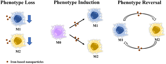
Numerous studies reported the role of iron-based nanoparticles in modulating macrophage polarization and function. Macrophage polarization is a dynamic and reversible process and involves the changes of a series of markers and signals. Macrophage polarization is not a dualistic model, while M1 and M2 represent the two extremes of the phenotype. By means of phenotypic loss, phenotypic induction, and phenotypic reversal, iron-based nanoparticles polarize macrophages toward either a pro- or anti-inflammatory phenotype (Figure 1).
Figure 1:
Iron-based nanoparticles modulate macrophage phenotype through phenotype loss, induction and reversal.
Polarization towards the pro-inflammatory phenotype
A variety of iron-based nanoparticles are found to promote a pro-inflammatory phenotype of macrophages by means of phenotype induction and phenotype reversal (Table 1). In most cases, IONPs promote the pro-inflammatory polarization of macrophages, while in rare cases PBNPs with specific coating materials [50], can also induce a M1-like phenotype. For example, the polyethylenimine-coated superparamagnetic iron oxide nanoparticles (SPION) induced M1 polarization dramatically, characterized by notable upregulation of typical M1-related genes such as CD80, IL-1β, TNF-α and so on [51]. Likewise, ferucarbotran and ferumoxytol, the two kinds of clinically used SPION, were reported to promote M1-like inflammatory response both in vitro and in vivo [52]. Furthermore, PBNPs coated with low molecular weight hyaluronic acid (LMWHA molecular weight [MW]< 5 kDa) have been shown to induce the pro-inflammatory phenotype and inhibit tumor growth [50, 53].
Table 1:
Iron-based nanoparticles inducing polarization towards pro-inflammatory phenotype.
| Polarization | Properties of nanoparticles | Experimental procedures | Marker changes in polarization | Ref. | |||||
|---|---|---|---|---|---|---|---|---|---|
| Iron-based nanoparticles | Core | Coating | Surface charge | Size (nm) | Cell types /Animal models | Methods | |||
| Phenotype Induction (M0 to M1) | PMA@Fe3O4 | Fe3O4 | PMA | – | 5 | RAW264.7 | 5 nmol/L, 24h | M1: TNF-α CD86 NF-κB ↑ M2: CD206 ↓ |
[63] |
| PEI-coated SPION | / | PEI | + | 139–228 | RAW264.7, THP-1, mouse peritoneal macrophages | 6.25–25μg/mL, 6h | M1: CD40 CD80 CD86 IL-1β TNF-α IL-6 IL-12 ↑ | [51] | |
| Resovist (ferucarbotran) | Fe3O4+γ-Fe2O3 | Dextran | + | / | RAW264.7, BMDM, Ana-1 | Fe 0–100μg/mL, 24h | M1: IL-1β IL-12p40/70 TNF-α IL-2 IL-10 ↑ | [52] | |
| Balb/c mice | Fe 25mg/kg | M1: IL-1β TNF-α ↑ | |||||||
| Feraheme (ferumoxytol) | γ-Fe2O3 | PSC | / | / | RAW264.7, BMDM, Ana-1 | Fe 0–100μg/mL | M1: IL-12p40/70 IL-4 IL-13 ↑ | [52] | |
| Balb/c mice | Fe 25mg/kg | M1: IL-1β TNF-α ↑ | |||||||
| PEG-Fns | Fns | PEG | / | 20 | RAW264.7 | Fe 320μmol/L, 12h | M1: TNF-α ↑ M2: IL-10 ↓ |
[64] | |
| Phenotype Reversal (M2 to M1) | Iron-dextran | / | Dextran | / | / | IL-4 treated BMDMs | 5 mg, 7d | M1: CD16/32 ↑ M2: CD206 ↓ |
[65] |
| Fe3O4-PLGA | Fe3O4 | PLGA | – | 283.8±24.1 | IL-4 treated RAW264.7 | 10 mg/mL | M1: TNF-α IL-1β iNOS ↑ M2: TGF-β IL-10 Arg1 ↓ |
[66] | |
| Fe3O4 | Fe3O4 | / | – | 453 | Lps treated BMDMs | 25 μg/mL, 24h | M2: IL-10 SOCS1 SOCS3 ↓ M1: TNF-α IL-12 ↑ |
[67] | |
| Fe3O4@D-SiO2 | Fe3O4 | Dendritic silica | – | 490x320 | IL-4 treated RAW264.7/BMDMs | 15 μg/mL, 24h | M1: CD80 CD86 CD64 ↑ M2: CD206 Arg-1 ↓ |
[68] | |
| CD206-Fe3O4-PLGA | Fe3O4 | PLGA | – | 294.3± 17.8 | Subcutaneous tumor-bearing mouse model | / | M1: CD86 ↓ | [66] | |
| PHNPs@DPA-S-S-BSA-MA@3-MA | Fe3O4 | / | – | 20 | MDA-MB-231 co-cultured RAW264.7/THP1 | 200 μg/mL, 24h | M1: CD86 iNOS IL-12 TNF-α ↑ M2: CD206 IL-10 Arg-1 ↓ |
[69] | |
| MDA-MB-231 tumor-bearing mouse model | 20 mg/kg | M1: CD86 iNOS IL-12 TNF-α ↑ | |||||||
| SPION | Fe3O4+γ-Fe2O3 | Carboxy-dextran | – | / | IL-4/IL-13 treated THP1 | Fe 20μg/mL, 24h/48h | M1: CD86 TNF-α ↑ | [70] | |
| Ferumoxytol | γ-Fe2O3 | PSC | / | / | MMTV-PyMT co-cultured RAW264.7 | Fe 2.73mg/mL, 6h | M1: ROS TNF-α CD86 ↑ M2: CD206 IL-10 ↓ |
[6] | |
| Mammary tumour model | Fe 2.73/8.37mg/mL | / | |||||||
| PEI-SPION | / | PEI | + | / | PMA treated THP1 | 4 μg/mL | M1: IL-1β IL-6 ↑ M2: IL-12 ↓ |
[71] | |
| Highly vascularized MDA-MB-231 xenograft tumors model | 1.2 mg/kg | M1: Mac3 iNOS ↑ M2: Arg-1 ↓ |
|||||||
| Fe@PDA-PEG | Fe3+ | mPEG-SH | / | 162 | IL-4 treated RAW264.7 | 24h | M1: iNOS IL-12 ↑ M2: IL-10 ↓ |
[72] | |
| Subcutaneous model of colon model | 30 mg/kg, 24h | M1: CD80 ↑ M2: CD206 ↓ |
|||||||
| PEG-Fns | Fns | PEG | / | 20 | SCC-7 co-cultured RAW264.7 | Fe 320μmol/L, 12h | M1: TNF-α ↑ M2: IL-10 ↓ |
[64] | |
| Subcutaneous tumor-bearing mouse model | Fe 800μmol/kg, 200 μL | M1: iNOS ↑ M2: CD163 ↓ |
|||||||
| LMWHA-MPB | PBNPs | LMWHA | – | 127 | IL-4 treated RAW264.7 | 50 μg/mL, 3h | M1: CD86 IL-12 ↑ M2: IL-10 ↓ |
[50] | |
| 4T1 tumor-bearing mouse model | 10 mg/kg | M1: CD86 ↑ M2: CD206 ↓ |
|||||||
| HA-PB/ICG | PBNPs | HA | – | 295 | IL-4 treated RAW264.7 | 1, 5, 20 and 50 μg/mL, 24h | M1: CD86 ↑ M2: CD206 ↓ |
[53] | |
| 4T1 tumor-bearing mouse model | 20 μg/mL, 8h | / | |||||||
| HION@Macs | / | HA | / | / | IL-4 treated RAW264.7 | 50 μg/mL, 24h | M1: CD80 iNOS ↑ M2: Arg-1 ↓ |
[73] | |
| 4T1 breast tumor bearing mouse model | 5 mg/kg, 24h | M1: TNF-α CD86 ↑ M2: CD206 ↓ |
|||||||
| Man-HMPB/HCQ | PBNPs | Mannose | – | 247 | IL-4 treated RAW264.7 | 10h | M1: CD86 IL-6 ↑ M2: CD206 IL-10 ↓ |
[57] | |
| 4T1 tumor model | 2.5 mg/kg | M1: CD86 IFN-γ TNF-α ↑ M2: CD206 IL-10 ↓ |
|||||||
| IMSN-PEG-TI | Iron manganese silicate nanoparticles | PEG | / | 122 | CT26 co-cultured mice abdominal macrophages | 100 μg/mL, 24h | M1: ly6C ↑ M2: CD206 ↓ |
[74] | |
| CT26-tumor-bearing mouse models | 10 mg/kg | M1: ly6C ↑ M2: CD206 ↓ |
|||||||
PMA, Amphiphilic polymer; PEI, Polyethyleneimine; PSC, Polyglucose sorbitol Carboxymethyl ether; Fns, Ferrihydrite nanoparticles; PEG, Polyethylene glycol; PLGA, Poly(lactic-co-glycolic) acid; PDA, polydiacetylene; mPEG-SH, thiol-terminated polyethylene glycol monomethyl ether; PHNPs@DPA-S-S-BSA-MA@3-MA, mannose (MA) modified porous hollow iron oxide nanoparticles (PHNPs) loaded with 3-methyladenine (3-MA) and blocked with bovine serum albumin (BSA); LMWHA-MPB, Mesoporous Prussian blue NPs with low molecular weight hyaluronic acid surface modification; HA-PB/ICG, Hyaluronic acid modified mesoporous Prussian blue nanoparticles loaded photosensitizer indocyanine green; HA, Hyaluronic acid; HION, Hyaluronic acid-decorated superparamagnetic iron oxide nanoparticles; Man-HMPB/HCQ, hollow mesoporous Prussian blue (HMPB) nanosystem with mannose decoration and hydroxychloroquine (HCQ) adsorption; IMSN-PEG-TI, TGF-β inhibitor (TI)-loaded PEGylated iron manganese silicate nanoparticles (IMSN); ROS, reactive oxygen species.
For diseases that are attributed to the lack of active pro-inflammatory M1 macrophages, reprogramming macrophages from immunosuppressive M2 to the killing mode of M1 could be an ideal strategy. It is widely reported that most of the tumor-associated macrophages (TAMs) in the tumor microenvironment are the typical M2 phenotype [54], [55], [56]. By tunning macrophages from M2-like TAMs to M1, iron-based nanoparticles can reverse the immunosuppressive status and promote effective tumour killing. For example, ferumoxytol inhibited tumor progression effectively by the promotion of M1 macrophages and inhibition of TAMs in mice [6]. Likewise, Hou et al. [57] designed and synthesized biological membrane-coated hollow mesoporous Prussian blue nanomaterials (PBNPs) for cancer therapy. This biomimetic nanosystem demonstrated dramatic effects on turning TAMs into M1 macrophages and suppressing tumor growth both in vitro and in vivo.
Polarization towards the anti-inflammatory phenotype
On the other hand, some iron-based nanoparticles are capable of inhibiting the inflammatory response and inducing M2 polarization, which has great potential to be applied for damaged tissue repair, wound healing, anti-inflammation, etc (Table 2). Iron-based nanoparticles can induce an anti-inflammatory phenotype of macrophage in three ways, loss of M1, phenotype induction from M0 to M2, and phenotype reversal from M1 to M2. Loss of M1 could be achieved by inhibiting pro-inflammatory cytokines release or reducing the numbers of M1 macrophages. For example, Park et al. reported that hyaluronan-coated PBNPs significantly suppressed both the function and population of M1 macrophage in an LPS-induced murine model, with great therapeutic effect for murine peritonitis [58]. Similarly, Chen et al. found that both 10 and 30 nm PEG-coated SPIONs inhibited the expression of LPS-induced pro-inflammatory factor, IL-6 and TNF-α, in a dose-dependent way [59]. However, in these models, whether the loss of M1 phenotype is associated with increased numbers and function of M2 macrophages remains unknown.
Table 2:
Iron-based nanoparticles inducing polarization towards anti-inflammatory phenotype.
| Polarization | Properties of nanoparticles | Experimental procedures | Marker changes in polarization | Ref. | |||||
|---|---|---|---|---|---|---|---|---|---|
| Iron-based nanoparticles | Core | Coating | Surface charge | Size, nm | Cell types/Animal models | Methods | |||
| Loss of M1 phenotype | Fe–cat NPs | Fe-cat complex | / | + | 40–150 | Lps treated RAW264.7 | 10–50 μg/mL | M1: TNF-α IL-1β IL-6 iNOS ↓ | [60] |
| Rat femur defect models | 20 μg/rat | M1: iNOS ↓ | |||||||
| MeLioN | Fe3O4 | Melittin | – | 202.4 | Intracranial arterial dolichoectasia model | 2.5 mg/kg | M1: CD86 TNF-α ↓ | [75] | |
| PBNPs | PBNPs | Hyaluronan | – | 180 | Lps treated RAW264.7 | 31.25 μg/mL, 24 h | M1: H2O2 CD86↓ | [58] | |
| PBNPs | PBNPs | PVP | – | 60 | Colitis mouse model | 40 mg/kg | M1: ROS TNF-α IL-6 IL-1β ↓ | [76] | |
| HPBZs | PBNPs | PVP | – | 78 | Lps treated RAW264.7 | / | M1: iNOS IL-1β NO ↓ | [77] | |
| Middle cerebral artery occlusion model | 40 μg/mL, 10 μL | M1: TNF-α IL-1β ↓ | |||||||
| Phenotype Induction (M0 to M2) | Fe–cat NPs | Fe-cat complex | / | + | 40–150 | RAW264.7 | 50 μg/mL | M2: Arg-1 CD163 CD206 ↑ | [60] |
| MHFPs | Fe3O4 | PAM | – | 185 | RAW264.7 | 40 μg/mL, alternating magnetic field (1.5 mT, 30 Hz, 2 h/day), 2 days | M1: CD32 iNOS↓ M2: Arg-1 CD206 IL-10↑ |
[78] | |
| Phenotype reversal (M1 to M2) | Prussian blue nanozyme | PBNPs | PVP | – | 67 | Lps treated RAW264.7 | 0–40 μg/mL, 24 h | M1: IL-6 TNF-α iNOS ROS CD16/32 ↓ M2: CD206 IL-10 Arg-1↑ |
[79] |
| Vascular balloon injury model | 6 weeks | M1: CD16/32 ↓ M2: CD206↑ |
|||||||
| Prussian blue nanozyme | PBNPs | / | – | 34 | Lps treated RAW 264.7 | 50 μg/mL, 24 h | M1: TNF-α IL-1β ROS ↓ M2: Arg-1↑ |
[7] | |
| Inflammation skin wound model | 50/500 μg, 14d | M1: TNF-α↓ | |||||||
| HMPB@curcumin@PDA | PBNPs | PDA | – | 188 | Lps treated RAW264.7 | 24 h | M1: ROS CD80 TNF-α IL-1β↓ M2: CD206 TGF-β IL-10 Arg-1↑ |
[80] | |
| Oral and maxillofacial inflammation model | 20 μg | M1: ROS TNF-α IL-1β ↓ | |||||||
| MPBzyme | PBNPs | NCM | – | 160.7 ± 6.08 | Glucose deprivation treated microglia | 10, 20, 40 μg/mL, 24 h | M1: CD16/32 iNOS TNF-α IL-6↓ M2: CD206 IL-10↑ |
[81] | |
| Transient middle cerebral artery occlusion model | 20 mg/kg, 24 h | M1: CD16/32 ↓ M2: CD206↑ |
|||||||
| HMPBzyme | Manganese prussian blue | BSA | – | 210 | Lps treated BMDMs | 80 μg/mL, 24 h | M1: IL-1β TNF-α HIF-1α ROS ↓ M2: Arg-1 IL-10↑ |
[61] | |
| Osteoarthritis model | 80 μg/mL, 24 h | M1: HIF-1α iNOS TNF-α IL-1β ROS ↓ | |||||||
| PBNPs | PBNPs | PVP | – | 80.2 | Lps treated RAW264.7 | 50 μg/mL | M1: TNF-α IL-1β↓ M2: IL-10 Arg-1↑ |
[62] | |
| Hepatic ischemia-reperfusion injury mouse model | 1 mg/kg, 24 h | M1: TNF-α IL-1β ↓ M2: IL-10↑ |
|||||||
Fe-cat NPs, Fe3+-(+)-catechin nanoparticles; MHFPs, Mesoporous hollow Fe3O4 nanoparticles; PAM, Polyacrylamide; PVP, Polyvinylpyrrolidone; HMPB, Hollow-structured manganese prussian blue; MPBzyme, Mesoporous Prussian blue nanozyme; NCM, Neutrophil-like cell membrane; HMPBzyme, Hollow-structured manganese prussian blue nanozyme; BSA, Bovine serum albumin; HPBZs, Hollow prussian blue nanozymes; MeLioN, Melittin-loaded L-arginine-coated iron oxide nanoparticle.
In addition to phenotypic loss, treatment of iron-based nanoparticles could directly induce the M2-like phenotype from either M0 or M1 phenotype. Liu et al. [60] developed self-assembled Fe3+-catechin nanoparticles (Fe-cat NPs) with a strong ability to facilitate M2 polarization from M0. By secreting anti-inflammatory cytokines, the Fe-cat NPs-treated macrophages contributed to efficient bone repair. For inflammation-related diseases characterized by massive accumulation of M1, the shift from M1 to M2 by iron-based nanoparticles could strongly diminish inflammation and reverses disease conditions. Fan et al. developed hollow-structured manganese prussian blue nanozyme with the ability to convert macrophages from M1 to M2, resulting in effective treatment of osteoarthritis [61]. In addition, Huang et al. also applied polyvinylpyrrolidone (PVP)-coated PBNPs to eliminate inflammation in the hepatic ischemia-reperfusion injury model, mainly by promoting M2 macrophages [62].
Influence factors of macrophage polarization by iron-based nanoparticles
As we summarized in Tables l and 2, different types of iron-based nanoparticles exert different influences on macrophage polarization. The modulation of iron-based nanoparticles on macrophage polarization is a complex process relying on the coordination of various factors [82], [83], [84], [85]. The inherent physicochemical properties of designed nanoparticles, such as composition, size, and surface characteristics, largely affect the interactions between nanoparticles and macrophages and are attributed to the differences in polarization (Figure 2).
Figure 2:
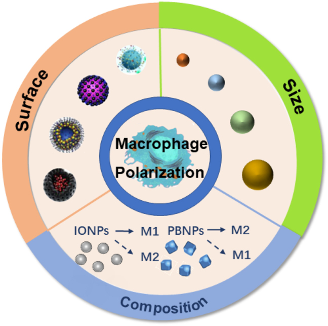
Internal influence factors of iron-based nanoparticles affecting macrophage polarization.
Composition
The different compositions and structures of iron-based nanoparticles notably affect their functions on macrophage polarization. In general, IONPs tend to induce a pro-inflammatory M1 phenotype, while PBNPs tend to induce an anti-inflammatory M2 phenotype. For example, one of the FDA-approved IONPs ferumoxytol strongly induced the M1 phenotype at a dose of 2.73 mg Fe/mL in vivo for effective inhibition of the growth and metastasis of mammary mouse tumor [6]. On the other hand, polyvinylpyrrolidone-coated PBNPs with an average size of 80 nm exhibited a notable induction of M2 phenotype in LPS-treated macrophage and was successfully applied for alleviating hepatic ischemia-reperfusion injury in mouse model [62]. However, the general principle is not always followed when other influencing factors become dominant, such as size, surface modifications, heterogeneous compositions, and external factors. Therefore, exceptions were found in special cases where PBNPs triggered M1 polarization and Fe3O4 nanoparticles induced M2 polarization. For instance, the PMA-coated mesoporous hollow Fe3O4 nanoparticles induced M0 to M2, which is largely affected by the addition of an alternating magnetic field [78]. In addition, the PB-based nanoparticles composed of hollow mesoporous Prussian blue, hydroxychloroquine (HCQ), and mannose decoration (Man-HMPB/HCQ) are reported to induce TAM to M1, probably due to the strong autophagy suppression effect of HCQ since inhibition of autophagy is known to contribute to the phenotypic reversal of TAMs [57].
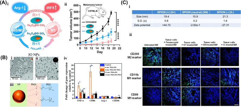
Furthermore, the differences in valence states of iron ions in nanoparticles also influence whether and how they induce macrophage polarizations. It was generally believed that magnetite IONPs (Fe3O4) are much more effective in inducing M1 polarization than hematite IONPs (Fe2O3) [68]. To compare the impact of states of iron (II and III) on macrophages polarization, Yu et al. synthesized the Fe2O3@D-SiO2 and Fe3O4@D-SiO2 nanoparticles with similar size (about 40 nm), parallel core–shell structures, identical ellipsoidal morphology, as well as the same surface modification by large-pore dendritic silica shell (D-SiO2). They found that only Fe3O4@D-SiO2 achieved significant M1 polarization from M0 or M2 macrophages, resulting in an excellent tumor suppression effect in the melanoma mouse model. Compared with Fe2O3@D-SiO2, Fe3O4@D-SiO2 induced much higher intracellular iron accumulation and thus triggered the interferon regulatory factor 5 (IRF-5) pathway, which is one of the key transcriptional factors that promoted the M1 polarization (Figure 3A). The reason for these different intracellular iron levels largely depends on their diversity in the dynamic process of iron endocytosis, intracellular degradation, and export rate from cells. In this model, Fe3O4@SiO2 exhibited a relatively higher uptake ability than Fe2O3@SiO2, which is probably due to the magnetism-induced aggregation [86].
Figure 3:
The effect of composition, size and surface of iron-based nanoparticles on the macrophage polarization. A. (i) Schematic diagram of the macrophage polarization effects of Fe2O3@SiO2 and Fe3O4@SiO2 with different the ratio (R) of intracellular iron accumulation. (ii) Antitumor properties of IONPs with different components in the melanoma tumor model. Reproduced with permission [68]. Copyright © 2019 American Chemical Society. B. TEM images (i, ii) and schematic diagram (iii) of nanoparticles with a polymer coating and surface PEGylation. (i. PMA/IO NPs; ii. PMA/IO-PEG NPs). (iv) Gene expression in RAW264.7 cells after IONPs treatment for 24 h. Reproduced with permission [63]. Copyright 2019, RSC Pub. C. (i) Characteristic of three differently charged SPION. (ii) Immunohistochemical staining of different treatment groups. The green area reflects the expression of CD206, CD11b and CD80. Reproduced with permission [90]. Copyright © 2020 Zhang, Cao, Liang, Tan, Luo, Xu and Saw.
Size
Iron-based nanoparticle-induced macrophage polarization is typically affected by the size properties of the materials. In particular, nanoparticles with similar composition but different sizes could exhibit different functions on macrophage polarization [87, 88]. For instance, Cheng et al. synthesized two groups of amphiphilic polymers (PMA)-modified Fe3O4 NPs and Au NPs with two different sizes, 4 and 14 nm, to investigate the influence of nanoparticles’ size on macrophage polarization [63]. They demonstrated that the 4 nm Fe3O4 NPs triggered M1 polarization more effectively than 14 nm Fe3O4 NPs, which might be due to the higher cellular uptake rate and intracellular accumulation of small nanoparticles. Meanwhile, the same trend was found with Au NPs (Figure 3B). Besides, Dalzon et al. compared the effects of two carboxymaltose-modified Fe2O3 nanoparticles with different sizes (20 and 100 nm) on the macrophages. They found that at a concentration of 1 mg/mL, the 100 nm Fe2O3 nanoparticles significantly inhibited the secretion of pro-inflammatory cytokines such as IL-6, TNF-α, and NO, as compared to smaller ones [89]. These results demonstrated that the size of iron-based nanoparticles indeed has an essential influence on the degree of macrophage polarization. However, whether and how the size factor contribute to the direction of macrophage polarization is unclear and requires further exploration in the future.
Surface
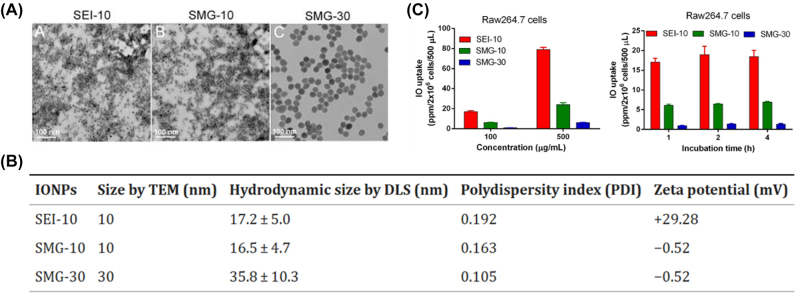
As another important characteristic of nanoparticles, the surface property is of great significance in influencing macrophage polarization, including surface charge, morphology, hydrophilicity, etc. In general, nanoparticles with a charged surface induce the polarization of M1 or M2 more easily than neutral ones. For example, Saw et al. explored the impact of the surface charge of nanoparticles on macrophage polarization by modifying ferumoxytol with different charges. They demonstrated that both positively and negatively charged SPIONs established a strong effect to repolarize macrophage from M2-like TAM to M1 and significantly suppressed tumor growth in the mouse model, while the neutral ones failed to induce polarization of macrophage [90]. It was presumed that the neutral surface limited the nanoparticles’ internalization by cells, and thus restrained the M1 polarization of ferumoxytol (Figure 3C). Consistently, a previous study has reported that PMA-coated IONPs significantly induced M1 polarization in RAW264.7, while further modifying IONPs with PEG outside the original layer dramatically reduced the effect of M1 polarization [63]. This is likely due to the limited uptake and endocytosis of PEGylation of nanoparticles by macrophages, resulting in less amount of particle accumulation and decreased activation of cellular signal and immune response [91]. The difference in cellular uptake of distinct surface modified-nanoparticles is supported by another research, since polyethyleneimine (PEI)-coated ultrasmall superparamagnetic IONPs induced much higher cellular uptake than PEG-modified IONPs (Figure 4) [87, 91].
Figure 4:
The effect of size and surface of iron-based nanoparticles on the cell uptake ability in macrophages. TEM images (A) and characteristics (B) of different IONPs. (C) Iron uptake in RAW264.7 cells measured by ICP-MS after co-culturing with different concentrations of IONPs for 1h or with the same concentration (100 μg/mL) for different incubation times. Reproduced with permission [87]. Copyright 2018, Springer Nature.
In addition, certain coating materials are known to have a strong macrophage modulating effect, therefore iron-based nanoparticles coated with them are inclined to gain the same effect as the coating material. For example, LMWHA can induce macrophage polarization from M2 to M1 phenotype [92, 93]. Therefore, iron-based nanoparticles coated with LMWHA could promote M1 polarization macrophages. Zhang’s group synthesized the LMWHA-modified mesoporous PBNPs (LMWHA-MPB) with an average size of 127 nm and further loaded them with photosensitizer indocyanine green (ICG) to form LMWHA-MPB/ICG. Both LMWHA-MPB and LMWHA-MPB/ICG promoted a M2 to M1 phenotype reversal in RAW264.7 cells [50, 53], which are opposite to most PBNPs reported by other studies [7, 62, 79], suggesting that the functional modifications on the surface of iron-based nanoparticles largely shift their effect on macrophage polarization.
Mechanisms of macrophage polarization by iron-based nanoparticles
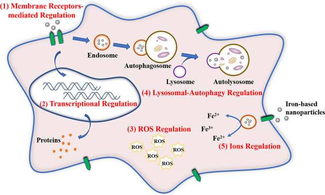
The interactions between macrophages and iron-based nanoparticles involve multiple procedures and diverse modulation pathways (Figure 5), including cellular signaling sensing and transduction, intracellular redox level rebalancing, lysosomal-autophagy modulation and iron metabolism alteration, etc. [84, 94].
Figure 5:
Modulation mechanisms of macrophage polarization by iron-based nanoparticles. ROS, reactive oxygen species.
Membrane receptors-mediated modulation
Surface receptors are the first-line sensors by which macrophages identify and respond to external signals. Various macrophage surface receptors including pattern recognition receptors (PRRs), scavenger receptors, complement receptors, cytokine receptors, etc., are responsible for phagocytosis and immunomodulation, mainly through the modulation of macrophage polarization. As important group members of PRRs, the Toll-like receptors (TLRs) family consists of several subtypes and is responsive to different danger signals. Among them, the activation of TLR-4 by lipopolysaccharide (LPS) strongly triggers M1 polarization to facilitate the killing of invaded bacteria, while blockage of TLR-4 limits its function effectively [95].
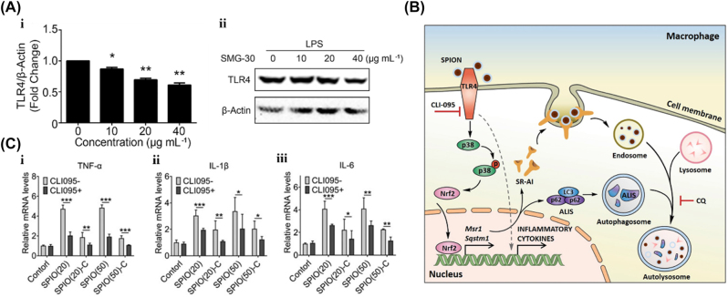
Importantly, iron-based nanoparticles are reported to interact with membrane receptors, such as TLR-4, to regulate macrophage polarization. For example, PEI-coated SPIONs initiated the TLR-4 cascade signaling pathway and promoted M1 polarization in both mouse and human-derived macrophages [51]. However, pre-treatment of RAW264.7 cells with the TLR-4 inhibitor CLI-095 strongly inhibited SPION-induced expression of M1 genes, suggesting that SPION-mediated M1 polarization is partly through inducing the TLR-4 pathway [51]. In addition, Ai et al. studied the interaction between two commercial SPIONs with macrophages, Resovist (ferucarbotran) and Feraheme (ferumoxytol). They found that both of the SPIONs interacted with TLR-4 and facilitated the M1-like phenotype through TLR-4-mediated p38-Nrf2 signaling pathway, with the secretion of pro-inflammatory cytokines like IL-12, TNF-α, and IL-1β (Figure 6B) [52]. Furthermore, inhibition of TLR-4 by CLI-095 greatly limited the expression of SPION-induced M1-like phenotype (Figure 6C) [96]. Taken together, these results demonstrated that TLR-4 acts as important membrane receptors for SPIONs to induce M1 polarization. On the other hand, blocking the expression or function of TLR-4 by iron-based nanoparticles is capable of limiting the LPS-induced M1 polarization. Chen et al. found that a 30 nm PEG-coated SPIONs decreased the transcription and expression of TLR-4 in a dose-dependent manner, which notably decreased the immune-activation function of LPS both in the mouse- and human-derived macrophages (Figure 6A) [97].
Figure 6:
The membrane receptor-mediated modulation effect by iron-based nanoparticles. A. (i) The relative mRNA level and (ii) expression of TLR4 in RAW264.7 cells stimulated by different concentrations of SMG-30 and LPS (1 μg/mL). β-actin was used as a control. Reproduced with permission [59]. Copyright 2020, John Wiley and Sons. B. Schematic diagram of the mechanism of TLR-4 and autophagy pathway activated by SPION. Reproduced with permission [52]. Copyright 2019, Elsevier. C. The relative mRNA levels of (i) TNF-α, (ii) IL-1β, and (iii) IL-6 in SPIO-treated RAW264.7 cells with or without CLI-095. Reproduced with permission. Copyright 2022, Oxford University Press.
Transcriptional modulation
Several key transcriptional factors in the modulation of macrophage polarization are found to be selectively alternated upon IONPs treatment. A group of transcriptional factors is responsible for M1 polarization, including signal transducer and activator of transcription-1 (STAT-1), IRF-5, nuclear factor-κB (NF-κB), activator protein-1 (AP-1), etc. On the contrary, activation of STAT-3, STAT-6, peroxisome proliferator-activated receptor-γ (PPAR-γ), etc., are reported to facilitate M2 polarization.
Upregulation of M1-related transcriptional factors directly results in M1 polarization and the secretion of pro-inflammatory cytokines. For example, the hyaluronic acid-decorated SPIONs has a notable effect on shifting macrophages from M2 to M1 phenotype mainly through the activation of NF-κB [73]. In addition, Fe3O4@D-SiO2 was found to promote the activation of IRF-5, thus inducing the polarization to M1 [68]. Similarly, Cheung et al. reported that IRF-5 was highly elevated by carboxymethylated IONPs, resulting in polarization from M2 to M1 [98]. Furthermore, bioinformatic gene analysis revealed that transcription factors like NF-κB and AP-1 were significantly upregulated by DMSA-coated Fe3O4 magnetic nanoparticles, and thus M1-like pro-inflammatory reactions were induced [99]. On the other hand, suppressing key transcriptional factors restricted the ability of macrophage polarization and their specific functions. For instance, hollow Prussian blue nanozymes were able to inhibit NF-κB and efficiently limited the M1 polarization induced by LPS, with down-regulated expression of inducible nitric oxide synthase (iNOS), cyclooxygenase-2 and IL-1β [77].
ROS modulation
During M1 macrophage-mediated anti-microbicidal process, reactive oxygen species (ROS) generate and act as the main weapon to destroy foreign pathogens. In addition, it is revealed that intracellular levels of ROS are involved in the modulation of macrophage polarization [100, 101]. In general, excessive intracellular ROS induces M1 polarization, mainly by activation of MAPK, STAT1 and NF-κB signaling, while ROS scavenging suppresses M1-mediated pro-inflammatory effects and leads to an M2 phenotype. During the differentiation process of macrophages from monocytes, the existence of ROS is essential for the subsequent M2 polarization, since the complete inhibition of ROS during differentiation strongly limited the later M2 or TAMs polarization [102]. As for the function of mitochondrial ROS on macrophage polarization, they could induce M1 or M2 polarization according to different reports, which are required for further studies to explain [103, 104].
It is well-known that IONPs can produce massive ROS by triggering Fenton reactions. Therefore, a variety of IONPs induces macrophage polarization through the modulation of ROS. For instance, Kim et al. demonstrated that the combination of dextran-coated IONPs with ascorbic acid was capable of eliciting vast ROS production and facilitating the pro-inflammatory phenotype of macrophages, which exhibited excellent bactericidal function against intracellular staphylococcus aureus [105]. In addition, Fan et al. found that alternating magnetic field further amplified ROS level in macrophages treated by ferrimagnetic vortex-domain iron oxide nanoring and graphene oxide (FVIOs-GO), and stimulated macrophage polarization from TAM to M1 [106]. Besides, the Prussian blue-modified ferritin nanoparticles (PB-Ft NPs) possessed ROS-inducing abilities by their peroxidase-like activity, particularly under 808 nm laser irradiation, and thus promoted M1 polarization. The enhanced peroxidase-like activity of PB-Ft NPs largely depends on the increased temperature under external laser irradiation [107].
In contrast, the elimination of excessive ROS by iron-based nanoparticles reverses the M1 phenotype and relieves the inflammatory condition. It is reported that a series of PBNPs exert ROS scavenging effects and suppress M1 polarization [7, 58, 79, 80, 108]. Due to their excellent anti-inflammatory ability, PBNPs have been widely studied for the treatment of peritonitis, maxillofacial infection, vascular restenosis and skin wound in different tissue. For example, Fan et al. reported that the hollow-structured manganese Prussian blue nanozyme (HMPBzymes) with intracellular ROS clearing activities induced M1 to M2 macrophage polarization under the hypoxia condition and suppressed inflammation in mice osteoarthritic model [61, 109].
Lysosomal-autophagy modulation
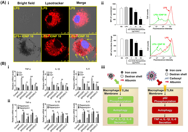
Nanoparticles’ internalization, transportation, and degradation greatly relied on the intracellular membrane structure-based organelle system such as endosomes, lysosomes and autophagosomes, which also acted as important regulatory pathways for macrophage polarization. The co-localization of nanoparticles with lysosomes sometimes causes lysosome dysfunctions with high-permeable membranes. Thus, the damaged lysosomes would release their containing enzymes like cathepsin-B and trigger NLRP3-mediated inflammation, which contributes to M1-like polarization (Figure 7A) [110]. For example, Song et al. demonstrated that PMA-coated IONPs caused M1 polarization mainly by inducing lysosomal damage. The same phenomenon was also observed in Au nanoparticles-treated macrophages [63]. Furthermore, the dysfunction of specific secretory lysosomes may block the transportation pathway of certain cytokines, thus inhibiting the function of macrophages. The commercially-used IONPs, Resovist, was reported to inhibit LPS-induced IL-1β generation by hindering the secretory lysosome-mediated excretion, and thus partially reduced the inflammatory phenotype in murine microglial cells [111].
Figure 7:
The Lysosomal-autophagy modulation effect by iron-based nanoparticles. A. After pre-treatment with IONPs (1–50 μg Fe/mL), (i) the morphology of lysosomes with Lysotracker (red) staining, (ii) lysosome acidity measured by lysosome sensor and (iii) cell permeability with acridine orange staining in microglia stimulated by LPS. IONPs treated cells had scattered dark brown dots. The arrows represent the colocalization of dark brown dots and lysosomes. Reproduced with permission [111]. Copyright 2013, Springer Nature. B. The relative mRNA levels of TNF-α, IL-1β, and IL-6 in SPIO-treated RAW264.7 cells (i) with autophagy inhibitor CQ and (ii) autophagy enhancer rapamycin, respectively. (iii) Schematic diagram of carboxyl-modified SPIONs inhibited macrophage autophagy and inflammatory cytokine secretion. Reproduced with permission [96].
As an essential intracellular process for energy recycles and stress handling, autophagy is commonly involved in the modulation of immune response including macrophage polarization. In general, the high level of autophagy induces macrophage polarization to the M1 phenotype. Ai’s group has conducted a series of studies to clarify the modulation effect of autophagy on polarization. They found that both Resovist (ferucarbotran) and Feraheme (ferumoxytol), the two clinically applied SPION, can trigger the autophagy-mediated pro-inflammatory response in vitro and in vivo. Importantly, suppression of autophagy by the autophagy inhibitor, chloroquine, dramatically inhibited the SPION-mediated secretion of inflammatory cytokines, strongly suggesting that autophagy acted as one of the important modulation factors for M1-like polarization (Figure 7B) [52, 96]. Recently, further studies demonstrated that compared with bare IONPs, IONPs with carboxyl modification on the surface induced a much lower level of autophagy in macrophages, leading to reduced immune activation [96].
Ion modulation
Most iron-based nanoparticles ultimately undergo degradation by acid organelles and release iron ions into cells. These released ions would further lead to a series of physiological changes and serve as an important regulator of macrophage polarization [112, 113]. Studies have proved that, after the treatment with IONPs, intracellular iron levels were increased in a dose- and time-dependent manner [114]. Theoretically, endocytosis of iron-based nanoparticles with higher iron leaching tends to contribute to a higher level of intracellular irons and trigger M1 polarization. Notably, iron chelate desferrioxamine (DFO) blocked the polarization effect of SPIONs in macrophages [70, 112]. In addition, Resovist-induced polarization from M2 to M1 was strongly limited by the pre-chelation of cellular iron in human-derived macrophages. Also, with the addition of DFO, carboxy-dextran-coated SPIONs failed to increase the expression of M1 makers, such as ferritin and cathepsin L in THP-1 [70]. These results strongly highlight the crucial roles of degraded iron ions in modulating macrophage polarization.
Summary and perspectives
Since the increasing studies on the design, synthesis, and biomedical applications of iron-based nanoparticles, their immune modulation effects have received much attention. After entering human bodies, most nanoparticles will be captured and metabolized by the reticuloendothelial system (RES), such as monocytes and macrophages. Upon uptake of iron-based nanoparticles, the polarization and function of macrophages can be modulated. In this review, we systemically summarized different iron-based nanoparticles that could regulate macrophage polarization and function and discussed the influence factors and mechanisms underlying the modulation process, with the expectation of providing evidence and guidance for subsequent design and application of iron-based nanoparticles with specific macrophage modulation functions.
In summary, the effect of iron-based nanoparticles on macrophage polarization is a systemic result influenced by the intrinsic physicochemical properties of nanoparticles including composition, size, surface, and so on. Besides, the addition of various external factors such as magnetic fields, laser irradiation, and temperature can trigger specific properties of iron-based nanoparticles and influence macrophage polarization in unconventional ways. Multiple mechanisms are involved in the modulation of macrophage polarization by iron-based nanoparticles, including membrane receptors interference, transcriptional modulation, ROS rebalancing, lysosomal-autophagy pathway, and iron ions release. Although some of the regularity has been revealed, there are still essential issues requiring further studies: (1) Among all the influencing factors and regulating mechanisms, which is dominant in macrophage polarization over others under specific conditions? (2) How do different factors and mechanisms coordinate with each other and determine the final direction of macrophage polarization? (3) Under different biological conditions, do the same iron-based nanoparticles present identical or different functions on macrophage polarization? (4) How to rationally design iron-based nanoparticles to achieve precise modulation and control of macrophage polarization? In the future, more in-depth research and studies considering the above issues are expected to gain a better understanding of iron-based nanoparticles’ modulation on macrophages and promote their better applications to biomedicine.
Footnotes
Research funding: This work was supported by the National Natural Science Foundation of China (51832001, 61821002), the Natural Science Foundation of Jiangsu Province (BK20220824, BK20222002), the National Key Research and Development Project (2021YFA1201403, 2021YFA0716304), and the Nanjing Science and technology development Foundation (202205066).
Author contributions: All authors have accepted responsibility for the entire content of this manuscript and approved its submission.
Competing interests: Authors state no conflict of interest.
Informed consent: Not applicable.
Ethical approval: Not applicable.
Contributor Information
Jingyi Sheng, Email: shengjingyi@hotmail.com.
Ning Gu, Email: guning@seu.edu.cn.
References
- 1.Laskin DL, Sunil VR, Gardner CR, Laskin JD. Macrophages and tissue injury: agents of defense or destruction? Annu Rev Pharmacol Toxicol. 2011;51:267. doi: 10.1146/annurev.pharmtox.010909.105812. [DOI] [PMC free article] [PubMed] [Google Scholar]
- 2.Mondal P, Anweshan A, Purkait MK. Green synthesis and environmental application of iron-based nanomaterials and nanocomposite: a review. Chemosphere. 2020;259:127509. doi: 10.1016/j.chemosphere.2020.127509. [DOI] [PubMed] [Google Scholar]
- 3.Chen O, Riedemann L, Etoc F, Herrmann H, Coppey M, Barch M, et al. Magneto-fluorescent core-shell supernanoparticles. Nat Commun. 2014;5:1–8. doi: 10.1038/ncomms6093. [DOI] [PMC free article] [PubMed] [Google Scholar]
- 4.Key J, Leary JF. Nanoparticles for multimodal in vivo imaging in nanomedicine. Int J Nanomed. 2014;9:711. doi: 10.2147/ijn.s53717. [DOI] [PMC free article] [PubMed] [Google Scholar]
- 5.Figuerola A, Di Corato R, Manna L, Pellegrino T. From iron oxide nanoparticles towards advanced iron-based inorganic materials designed for biomedical applications. Pharmacol Res. 2010;62:126–43. doi: 10.1016/j.phrs.2009.12.012. [DOI] [PubMed] [Google Scholar]
- 6.Zanganeh S, Hutter G, Spitler R, Lenkov O, Mahmoudi M, Shaw A, et al. Iron oxide nanoparticles inhibit tumour growth by inducing pro-inflammatory macrophage polarization in tumour tissues. Nat Nanotechnol. 2016;11:986–94. doi: 10.1038/nnano.2016.168. [DOI] [PMC free article] [PubMed] [Google Scholar]
- 7.Sahu A, Jeon J, Lee MS, Yang HS, Tae G. Antioxidant and anti-inflammatory activities of Prussian blue nanozyme promotes full-thickness skin wound healing. Mater Sci Eng C. 2021;119:111596. doi: 10.1016/j.msec.2020.111596. [DOI] [PubMed] [Google Scholar]
- 8.Yona S, Kim K-W, Wolf Y, Mildner A, Varol D, Breker M, et al. Fate mapping reveals origins and dynamics of monocytes and tissue macrophages under homeostasis. Immunity. 2013;38:79–91. doi: 10.1016/j.immuni.2013.05.008. [DOI] [PMC free article] [PubMed] [Google Scholar]
- 9.Epelman S, Lavine KJ, Randolph GJ. Origin and functions of tissue macrophages. Immunity. 2014;41:21–35. doi: 10.1016/j.immuni.2014.06.013. [DOI] [PMC free article] [PubMed] [Google Scholar]
- 10.Shapouri-Moghaddam A, Mohammadian S, Vazini H, Taghadosi M, Esmaeili SA, Mardani F, et al. Macrophage plasticity, polarization, and function in health and disease. J Cell Physiol. 2018;233:6425–40. doi: 10.1002/jcp.26429. [DOI] [PubMed] [Google Scholar]
- 11.Sica A, Mantovani A. Macrophage plasticity and polarization: in vivo veritas. J Clin Invest. 2012;122:787–95. doi: 10.1172/jci59643. [DOI] [PMC free article] [PubMed] [Google Scholar]
- 12.Mackaness G. The immunological basis of acquired cellular resistance. J Exp Med. 1964;120:105–20. doi: 10.1084/jem.120.1.105. [DOI] [PMC free article] [PubMed] [Google Scholar]
- 13.Stein M, Keshav S, Harris N, Gordon S. Interleukin 4 potently enhances murine macrophage mannose receptor activity: a marker of alternative immunologic macrophage activation. J Exp Med. 1992;176:287–92. doi: 10.1084/jem.176.1.287. [DOI] [PMC free article] [PubMed] [Google Scholar]
- 14.Mosser DM, Edwards JP. Exploring the full spectrum of macrophage activation. Nat Rev Immunol. 2008;8:958–69. doi: 10.1038/nri2448. [DOI] [PMC free article] [PubMed] [Google Scholar]
- 15.Porta C, Riboldi E, Ippolito A, Sica A. Molecular and epigenetic basis of macrophage polarized activation. Semin Immunol. 2015;27:237–48. doi: 10.1016/j.smim.2015.10.003. [DOI] [PubMed] [Google Scholar]
- 16.Bermudez S, Khayrullina G, Zhao Y, Byrnes KR. NADPH oxidase isoform expression is temporally regulated and may contribute to microglial/macrophage polarization after spinal cord injury. Mol Cell Neurosci. 2016;77:53–64. doi: 10.1016/j.mcn.2016.10.001. [DOI] [PMC free article] [PubMed] [Google Scholar]
- 17.Klimp A, De Vries E, Scherphof G, Daemen T. A potential role of macrophage activation in the treatment of cancer. Crit Rev Oncol-Hematol. 2002;44:143–61. doi: 10.1016/s1040-8428(01)00203-7. [DOI] [PubMed] [Google Scholar]
- 18.Kurowska-Stolarska M, Stolarski B, Kewin P, Murphy G, Corrigan CJ, Ying S, et al. IL-33 amplifies the polarization of alternatively activated macrophages that contribute to airway inflammation. J Immunol. 2009;183:6469–77. doi: 10.4049/jimmunol.0901575. [DOI] [PubMed] [Google Scholar]
- 19.Murray PJ, Allen JE, Biswas SK, Fisher EA, Gilroy DW, Goerdt S, et al. Macrophage activation and polarization: nomenclature and experimental guidelines. Immunity. 2014;41:14–20. doi: 10.1016/j.immuni.2014.06.008. [DOI] [PMC free article] [PubMed] [Google Scholar]
- 20.Mantovani A, Sica A, Sozzani S, Allavena P, Vecchi A, Locati M. The chemokine system in diverse forms of macrophage activation and polarization. Trends Immunol. 2004;25:677–86. doi: 10.1016/j.it.2004.09.015. [DOI] [PubMed] [Google Scholar]
- 21.Zizzo G, Hilliard BA, Monestier M, Cohen PL. Efficient clearance of early apoptotic cells by human macrophages requires M2c polarization and MerTK induction. J Immunol. 2012;189:3508–20. doi: 10.4049/jimmunol.1200662. [DOI] [PMC free article] [PubMed] [Google Scholar]
- 22.Seno H, Miyoshi H, Brown SL, Geske MJ, Colonna M, Stappenbeck TS. Efficient colonic mucosal wound repair requires Trem2 signaling. Proc Natl Acad Sci USA. 2009;106:256–61. doi: 10.1073/pnas.0803343106. [DOI] [PMC free article] [PubMed] [Google Scholar]
- 23.Funes SC, Rios M, Escobar-Vera J, Kalergis AM. Implications of macrophage polarization in autoimmunity. Immunology. 2018;154:186–95. doi: 10.1111/imm.12910. [DOI] [PMC free article] [PubMed] [Google Scholar]
- 24.Sica A, Larghi P, Mancino A, Rubino L, Porta C, Totaro MG, et al. Macrophage polarization in tumour progression. Semin Cancer Biol. 2008;18:349–55. doi: 10.1016/j.semcancer.2008.03.004. [DOI] [PubMed] [Google Scholar]
- 25.Tarique AA, Logan J, Thomas E, Holt PG, Sly PD, Fantino E. Phenotypic, functional, and plasticity features of classical and alternatively activated human macrophages. Am J Respir Cell Mol Biol. 2015;53:676–88. doi: 10.1165/rcmb.2015-0012oc. [DOI] [PubMed] [Google Scholar]
- 26.Lawrence T, Natoli G. Transcriptional regulation of macrophage polarization: enabling diversity with identity. Nat Rev Immunol. 2011;11:750–61. doi: 10.1038/nri3088. [DOI] [PubMed] [Google Scholar]
- 27.Arranz A, Doxaki C, Vergadi E, Martinez de la Torre Y, Vaporidi K, Lagoudaki ED, et al. Akt1 and Akt2 protein kinases differentially contribute to macrophage polarization. Proc Natl Acad Sci USA. 2012;109:9517–22. doi: 10.1073/pnas.1119038109. [DOI] [PMC free article] [PubMed] [Google Scholar]
- 28.Kratochvill F, Neale G, Haverkamp JM, Van de Velde L-A, Smith AM, Kawauchi D, et al. TNF counterbalances the emergence of M2 tumor macrophages. Cell Rep. 2015;12:1902–14. doi: 10.1016/j.celrep.2015.08.033. [DOI] [PMC free article] [PubMed] [Google Scholar]
- 29.Odegaard JI, Ricardo-Gonzalez RR, Goforth MH, Morel CR, Subramanian V, Mukundan L, et al. Macrophage-specific PPARγ controls alternative activation and improves insulin resistance. Nature. 2007;447:1116–20. doi: 10.1038/nature05894. [DOI] [PMC free article] [PubMed] [Google Scholar]
- 30.Murray PJ. Macrophage polarization. Annu Rev Physiol. 2017;79:541–66. doi: 10.1146/annurev-physiol-022516-034339. [DOI] [PubMed] [Google Scholar]
- 31.Xu H, Zhu J, Smith S, Foldi J, Zhao B, Chung AY, et al. Notch-RBP-J signaling regulates the transcription factor IRF8 to promote inflammatory macrophage polarization. Nat Immunol. 2012;13:642–50. doi: 10.1038/ni.2304. [DOI] [PMC free article] [PubMed] [Google Scholar]
- 32.Date D, Das R, Narla G, Simon DI, Jain MK, Mahabeleshwar GH. Kruppel-like transcription factor 6 regulates inflammatory macrophage polarization. J Biol Chem. 2014;289:10318–29. doi: 10.1074/jbc.m113.526749. [DOI] [PMC free article] [PubMed] [Google Scholar]
- 33.Natoli G. Maintaining cell identity through global control of genomic organization. Immunity. 2010;33:12–24. doi: 10.1016/j.immuni.2010.07.006. [DOI] [PubMed] [Google Scholar]
- 34.Bala S, Marcos M, Kodys K, Csak T, Catalano D, Mandrekar P, et al. Up-regulation of microRNA-155 in macrophages contributes to increased tumor necrosis factor α (TNFα) production via increased mRNA half-life in alcoholic liver disease. J Biol Chem. 2011;286:1436–44. doi: 10.1074/jbc.m110.145870. [DOI] [PMC free article] [PubMed] [Google Scholar]
- 35.Bala S, Csak T, Saha B, Zatsiorsky J, Kodys K, Catalano D, et al. The pro-inflammatory effects of miR-155 promote liver fibrosis and alcohol-induced steatohepatitis. J Hepatol. 2016;64:1378–87. doi: 10.1016/j.jhep.2016.01.035. [DOI] [PMC free article] [PubMed] [Google Scholar]
- 36.Yu J, Qiu Y, Yang J, Bian S, Chen G, Deng M, et al. DNMT1-PPARγ pathway in macrophages regulates chronic inflammation and atherosclerosis development in mice. Sci Rep. 2016;6:30053. doi: 10.1038/srep30053. [DOI] [PMC free article] [PubMed] [Google Scholar]
- 37.Zhou D, Yang K, Chen L, Zhang W, Xu Z, Zuo J, et al. Promising landscape for regulating macrophage polarization: epigenetic viewpoint. Oncotarget. 2017;8:57693. doi: 10.18632/oncotarget.17027. [DOI] [PMC free article] [PubMed] [Google Scholar]
- 38.Xu G, Liu G, Xiong S, Liu H, Chen X, Zheng B. The histone methyltransferase Smyd2 is a negative regulator of macrophage activation by suppressing interleukin 6 (IL-6) and tumor necrosis factor α (TNF-α) production. J Biol Chem. 2015;290:5414–23. doi: 10.1074/jbc.m114.610345. [DOI] [PMC free article] [PubMed] [Google Scholar]
- 39.Aktan F. iNOS-mediated nitric oxide production and its regulation. Life Sci. 2004;75:639–53. doi: 10.1016/j.lfs.2003.10.042. [DOI] [PubMed] [Google Scholar]
- 40.West AP, Brodsky IE, Rahner C, Woo DK, Erdjument-Bromage H, Tempst P, et al. TLR signalling augments macrophage bactericidal activity through mitochondrial ROS. Nature. 2011;472:476–80. doi: 10.1038/nature09973. [DOI] [PMC free article] [PubMed] [Google Scholar]
- 41.Takeda N, O’Dea EL, Doedens A, Kim J-W, Weidemann A, Stockmann C, et al. Differential activation and antagonistic function of HIF-α isoforms in macrophages are essential for NO homeostasis. Genes Dev. 2010;24:491–501. doi: 10.1101/gad.1881410. [DOI] [PMC free article] [PubMed] [Google Scholar]
- 42.Batista-Gonzalez A, Vidal R, Criollo A, Carreño LJ. New insights on the role of lipid metabolism in the metabolic reprogramming of macrophages. Front Immunol. 2020;10:2993. doi: 10.3389/fimmu.2019.02993. [DOI] [PMC free article] [PubMed] [Google Scholar]
- 43.Corraliza IM, Soler G, Eichmann K, Modolell M. Arginase induction by suppressors of nitric oxide synthesis (IL-4, IL-10 and PGE2) in murine bone-marrow-derived macrophages. Biochem Biophys Res Commun. 1995;206:667–73. doi: 10.1006/bbrc.1995.1094. [DOI] [PubMed] [Google Scholar]
- 44.Munder M, Eichmann K, Modolell M. Alternative metabolic states in murine macrophages reflected by the nitric oxide synthase/arginase balance: competitive regulation by CD4+ T cells correlates with Th1/Th2 phenotype. J Immunol. 1998;160:5347–54. doi: 10.4049/jimmunol.160.11.5347. [DOI] [PubMed] [Google Scholar]
- 45.Kieler M, Hofmann M, Schabbauer G. More than just protein building blocks: how amino acids and related metabolic pathways fuel macrophage polarization. FEBS J. 2021;288:3694–714. doi: 10.1111/febs.15715. [DOI] [PMC free article] [PubMed] [Google Scholar]
- 46.Recalcati S, Locati M, Cairo G. Systemic and cellular consequences of macrophage control of iron metabolism. Semin Immunol. 2012;24:393–8. doi: 10.1016/j.smim.2013.01.001. [DOI] [PubMed] [Google Scholar]
- 47.Irwin EF, Saha K, Rosenbluth M, Gamble L, Castner DG, Healy KE. Modulus-dependent macrophage adhesion and behavior. J Biomater Sci Polym Ed. 2008;19:1363–82. doi: 10.1163/156856208786052407. [DOI] [PubMed] [Google Scholar]
- 48.Wójciak-Stothard B, Madeja Z, Korohoda W, Curtis A, Wilkinson C. Activation of macrophage-like cells by multiple grooved substrata. Topographical control of cell behaviour. Cell Biol Int. 1995;19:485–90. doi: 10.1006/cbir.1995.1092. [DOI] [PubMed] [Google Scholar]
- 49.Refai AK, Textor M, Brunette DM, Waterfield JD. Effect of titanium surface topography on macrophage activation and secretion of proinflammatory cytokines and chemokines. J Biomed Mater Res A. 2004;70:194–205. doi: 10.1002/jbm.a.30075. [DOI] [PubMed] [Google Scholar]
- 50.Zhang H, Zhang X, Ren Y, Cao F, Hou L, Zhang Z. An in situ microenvironmental nano-regulator to inhibit the proliferation and metastasis of 4T1 tumor. Theranostics. 2019;9:3580. doi: 10.7150/thno.33141. [DOI] [PMC free article] [PubMed] [Google Scholar]
- 51.Mulens-Arias V, Rojas JM, Pérez-Yagüe S, Morales MP, Barber DF. Polyethylenimine-coated SPIONs trigger macrophage activation through TLR-4 signaling and ROS production and modulate podosome dynamics. Biomaterials. 2015;52:494–506. doi: 10.1016/j.biomaterials.2015.02.068. [DOI] [PubMed] [Google Scholar]
- 52.Jin R, Liu L, Zhu W, Li D, Yang L, Duan J, et al. Iron oxide nanoparticles promote macrophage autophagy and inflammatory response through activation of toll-like Receptor-4 signaling. Biomaterials. 2019;203:23–30. doi: 10.1016/j.biomaterials.2019.02.026. [DOI] [PubMed] [Google Scholar]
- 53.Zhang H, Pei Y, Zhang X, Zhu L, Hou L, Chang J, et al. Engineering of an intelligent cascade nanoreactor for sequential improvement of microenvironment and enhanced tumor phototherapy. Appl Mater Today. 2020;18:100494. doi: 10.1016/j.apmt.2019.100494. [DOI] [Google Scholar]
- 54.Lin X, Fang Y, Jin X, Zhang M, Shi K. Modulating repolarization of tumor-associated macrophages with targeted therapeutic nanoparticles as a potential strategy for cancer therapy. ACS Appl Bio Mater. 2021;4:5871–96. doi: 10.1021/acsabm.1c00461. [DOI] [PubMed] [Google Scholar]
- 55.Miao X, Leng X, Zhang Q. The current state of nanoparticle-induced macrophage polarization and reprogramming research. Int J Mol Sci. 2017;18:336. doi: 10.3390/ijms18020336. [DOI] [PMC free article] [PubMed] [Google Scholar]
- 56.Shi L, Gu H. Emerging nanoparticle strategies for modulating tumor-associated macrophage polarization. Biomolecules. 2021;11:1912. doi: 10.3390/biom11121912. [DOI] [PMC free article] [PubMed] [Google Scholar]
- 57.Hou L, Gong X, Yang J, Zhang H, Yang W, Chen X. Hybrid-membrane-decorated Prussian blue for effective cancer immunotherapy via tumor-associated macrophages polarization and hypoxia relief. Adv Mater. 2022;34:2200389. doi: 10.1002/adma.202200389. [DOI] [PubMed] [Google Scholar]
- 58.Mathew AP, Rajendrakumar SK, Mohapatra A, Vasukutty A, Revuri V, Mondal J, et al. Hyaluronan-coated Prussian blue nanoparticles relieve LPS-induced peritonitis by suppressing oxidative species generation in tissue-resident macrophages. Biomater Sci. 2022;10:1248–56. doi: 10.1039/d1bm01796a. [DOI] [PubMed] [Google Scholar]
- 59.Chen Y, Zeng Z, Ying H, Wu C, Chen S. Superparamagnetic iron oxide nanoparticles attenuate lipopolysaccharide-induced inflammatory responses through modulation of toll-like receptor 4 expression. J Appl Toxicol. 2020;40:1067–75. doi: 10.1002/jat.3967. [DOI] [PubMed] [Google Scholar]
- 60.Kong Y, Liu F, Ma B, Wang W, Li L, Xu X, et al. Intracellular pH-responsive iron-catechin nanoparticles with osteogenic/anti-adipogenic and immunomodulatory effects for efficient bone repair. Nano Res. 2022;15:1153–61. doi: 10.1007/s12274-021-3618-2. [DOI] [Google Scholar]
- 61.Xiong H, Zhao Y, Xu Q, Xie X, Wu J, Hu B, et al. Biodegradable hollow-structured nanozymes modulate phenotypic polarization of macrophages and relieve hypoxia for treatment of osteoarthritis. Small. 2022;18:2203240. doi: 10.1002/smll.202203240. [DOI] [PubMed] [Google Scholar]
- 62.Huang Y, Xu Q, Zhang J, Yin Y, Pan Y, Zheng Y, et al. Prussian blue scavenger Ameliorates hepatic ischemia-reperfusion injury by inhibiting inflammation and reducing oxidative stress. Front Immunol. 2022;13:891351. doi: 10.3389/fimmu.2022.891351. [DOI] [PMC free article] [PubMed] [Google Scholar]
- 63.Cheng J, Zhang Q, Fan S, Zhang A, Liu B, Hong Y, et al. The vacuolization of macrophages induced by large amounts of inorganic nanoparticle uptake to enhance the immune response. Nanoscale. 2019;11:22849–59. doi: 10.1039/c9nr08261a. [DOI] [PubMed] [Google Scholar]
- 64.Yang Y, Tian Q, Wu S, Li Y, Yang K, Yan Y, et al. Blue light-triggered Fe2+-release from monodispersed ferrihydrite nanoparticles for cancer iron therapy. Biomaterials. 2021;271:120739. doi: 10.1016/j.biomaterials.2021.120739. [DOI] [PubMed] [Google Scholar]
- 65.Kroner A, Greenhalgh AD, Zarruk JG, Dos Santos RP, Gaestel M, David S. TNF and increased intracellular iron alter macrophage polarization to a detrimental M1 phenotype in the injured spinal cord. Neuron. 2014;83:1098–116. doi: 10.1016/j.neuron.2014.07.027. [DOI] [PubMed] [Google Scholar]
- 66.Zhou Y, Que K-T, Tang H-M, Zhang P, Fu Q-M, Liu Z-J. Anti-CD206 antibody-conjugated Fe3O4-based PLGA nanoparticles selectively promote tumor-associated macrophages to polarize to the pro-inflammatory subtype. Oncol Lett. 2020;20:298. doi: 10.3892/ol.2020.12161. [DOI] [PMC free article] [PubMed] [Google Scholar]
- 67.Kodali V, Littke MH, Tilton SC, Teeguarden JG, Shi L, Frevert CW, et al. Dysregulation of macrophage activation profiles by engineered nanoparticles. ACS Nano. 2013;7:6997–7010. doi: 10.1021/nn402145t. [DOI] [PMC free article] [PubMed] [Google Scholar]
- 68.Gu Z, Liu T, Tang J, Yang Y, Song H, Tuong ZK, et al. Mechanism of iron oxide-induced macrophage activation: the impact of composition and the underlying signaling pathway. J Am Chem Soc. 2019;141:6122–6. doi: 10.1021/jacs.8b10904. [DOI] [PubMed] [Google Scholar]
- 69.Li K, Lu L, Xue C, Liu J, He Y, Zhou J, et al. Polarization of tumor-associated macrophage phenotype via porous hollow iron nanoparticles for tumor immunotherapy in vivo. Nanoscale. 2020;12:130–44. doi: 10.1039/c9nr06505a. [DOI] [PubMed] [Google Scholar]
- 70.Laskar A, Eilertsen J, Li W, Yuan X-M. SPION primes THP1 derived M2 macrophages towards M1-like macrophages. Biochem Biophys Res Commun. 2013;441:737–42. doi: 10.1016/j.bbrc.2013.10.115. [DOI] [PubMed] [Google Scholar]
- 71.Mulens-Arias V, Rojas JM, Sanz-Ortega L, Portilla Y, Pérez-Yagüe S, Barber DF. Polyethylenimine-coated superparamagnetic iron oxide nanoparticles impair in vitro and in vivo angiogenesis. Nanomedicine. 2019;21:102063. doi: 10.1016/j.nano.2019.102063. [DOI] [PubMed] [Google Scholar]
- 72.Rong L, Zhang Y, Li W-S, Su Z, Fadhil JI, Zhang C. Iron chelated melanin-like nanoparticles for tumor-associated macrophage repolarization and cancer therapy. Biomaterials. 2019;225:119515. doi: 10.1016/j.biomaterials.2019.119515. [DOI] [PubMed] [Google Scholar]
- 73.Li CX, Zhang Y, Dong X, Zhang L, Liu MD, Li B, et al. Artificially reprogrammed macrophages as tumor-tropic immunosuppression-resistant biologics to realize therapeutics production and immune activation. Adv Mater. 2019;31:1807211. doi: 10.1002/adma.201807211. [DOI] [PubMed] [Google Scholar]
- 74.Xu B, Cui Y, Wang W, Li S, Lyu C, Wang S, et al. Immunomodulation-enhanced nanozyme-based tumor catalytic therapy. Adv Mater. 2020;32:2003563. doi: 10.1002/adma.202003563. [DOI] [PubMed] [Google Scholar]
- 75.Vu HD, Huynh PT, Ryu J, Kang UR, Youn SW, Kim H, et al. Melittin-loaded iron oxide nanoparticles prevent intracranial arterial dolichoectasia development through inhibition of macrophage-mediated inflammation. Int J Biol Sci. 2021;17:3818. doi: 10.7150/ijbs.60588. [DOI] [PMC free article] [PubMed] [Google Scholar]
- 76.Zhao J, Cai X, Gao W, Zhang L, Zou D, Zheng Y, et al. Prussian blue nanozyme with multienzyme activity reduces colitis in mice. ACS Appl Mater Interfaces. 2018;10:26108–17. doi: 10.1021/acsami.8b10345. [DOI] [PubMed] [Google Scholar]
- 77.Zhang K, Tu M, Gao W, Cai X, Song F, Chen Z, et al. Hollow prussian blue nanozymes drive neuroprotection against ischemic stroke via attenuating oxidative stress, counteracting inflammation, and suppressing cell apoptosis. Nano Lett. 2019;19:2812–23. doi: 10.1021/acs.nanolett.8b04729. [DOI] [PubMed] [Google Scholar]
- 78.Guo W, Wu X, Wei W, Wang Y, Dai H. Mesoporous hollow Fe3O4 nanoparticles regulate the behavior of neuro-associated cells through induction of macrophage polarization in an alternating magnetic field. J Mater Chem B. 2022;10:5633–43. doi: 10.1039/d2tb00527a. [DOI] [PubMed] [Google Scholar]
- 79.Feng L, Dou C, Xia Y, Li B, Zhao M, El-Toni AM, et al. Enhancement of nanozyme permeation by endovascular interventional treatment to prevent vascular restenosis via macrophage polarization modulation. Adv Funct Mater. 2020;30:2006581. doi: 10.1002/adfm.202006581. [DOI] [Google Scholar]
- 80.Da J, Li Y, Zhang K, Ren J, Wang J, Liu X, et al. Functionalized prussian blue nanozyme as dual-responsive drug therapeutic nanoplatform against maxillofacial infection via macrophage polarization. Int J Nanomed. 2022;17:5851–68. doi: 10.2147/ijn.s385899. [DOI] [PMC free article] [PubMed] [Google Scholar]
- 81.Feng L, Dou C, Xia Y, Li B, Zhao M, Yu P, et al. Neutrophil-like cell-membrane-coated nanozyme therapy for ischemic brain damage and long-term neurological functional recovery. ACS Nano. 2021;15:2263–80. doi: 10.1021/acsnano.0c07973. [DOI] [PubMed] [Google Scholar]
- 82.Mulens-Arias V, Rojas JM, Barber DF. The use of iron oxide nanoparticles to reprogram macrophage responses and the immunological tumor microenvironment. Front Immunol. 2021;12:693709. doi: 10.3389/fimmu.2021.693709. [DOI] [PMC free article] [PubMed] [Google Scholar]
- 83.Dukhinova MS, Prilepskii AY, Shtil AA, Vinogradov VV. Metal oxide nanoparticles in therapeutic regulation of macrophage functions. Nanomaterials (Basel) 2019;9:1631. doi: 10.3390/nano9111631. [DOI] [PMC free article] [PubMed] [Google Scholar]
- 84.Reichel D, Tripathi M, Perez JM. Biological effects of nanoparticles on macrophage polarization in the tumor microenvironment. Nanotheranostics. 2019;3:66–88. doi: 10.7150/ntno.30052. [DOI] [PMC free article] [PubMed] [Google Scholar]
- 85.Ali LM, Pinol R, Villa-Bellosta R, Gabilondo L, Millan A, Palacio F, et al. Cell compatibility of a maghemite/polymer biomedical nanoplatform. Toxicol In Vitro. 2015;29:962–75. doi: 10.1016/j.tiv.2015.04.003. [DOI] [PubMed] [Google Scholar]
- 86.Krajewski M, Brzozka K, Tokarczyk M, Kowalski G, Lewinska S, Slawska-Waniewska A, et al. Impact of thermal oxidation on chemical composition and magnetic properties of iron nanoparticles. J Magn Magn Mater. 2018;458:346–54. doi: 10.1016/j.jmmm.2018.03.047. [DOI] [Google Scholar]
- 87.Feng Q, Liu Y, Huang J, Chen K, Huang J, Xiao K. Uptake, distribution, clearance, and toxicity of iron oxide nanoparticles with different sizes and coatings. Sci Rep. 2018;8:1–13. doi: 10.1038/s41598-018-19628-z. [DOI] [PMC free article] [PubMed] [Google Scholar]
- 88.Huang J, Bu L, Xie J, Chen K, Cheng Z, Li X, et al. Effects of nanoparticle size on cellular uptake and liver MRI with polyvinylpyrrolidone-coated iron oxide nanoparticles. ACS Nano. 2010;4:7151–60. doi: 10.1021/nn101643u. [DOI] [PMC free article] [PubMed] [Google Scholar]
- 89.Dalzon B, Torres A, Reymond S, Gallet B, Saint-Antonin F, Collin-Faure V, et al. Influences of nanoparticles characteristics on the cellular responses: the example of iron oxide and macrophages. Nanomaterials. 2020;10:266. doi: 10.3390/nano10020266. [DOI] [PMC free article] [PubMed] [Google Scholar]
- 90.Zhang W, Cao S, Liang S, Tan CH, Luo B, Xu X, et al. Differently charged super-paramagnetic iron oxide nanoparticles preferentially induced M1-like phenotype of macrophages. Front Bioeng Biotechnol. 2020;8:537. doi: 10.3389/fbioe.2020.00537. [DOI] [PMC free article] [PubMed] [Google Scholar]
- 91.Andrade RGD, Reis B, Costas B, Lima SAC, Reis S. Modulation of macrophages M1/M2 polarization using carbohydrate-functionalized polymeric nanoparticles. Polymers (Basel) 2020;13:88. doi: 10.3390/polym13010088. [DOI] [PMC free article] [PubMed] [Google Scholar]
- 92.Rayahin JE, Buhrman JS, Zhang Y, Koh TJ, Gemeinhart RA. High and low molecular weight hyaluronic acid differentially influence macrophage activation. ACS Biomater Sci Eng. 2015;1:481–93. doi: 10.1021/acsbiomaterials.5b00181. [DOI] [PMC free article] [PubMed] [Google Scholar]
- 93.Lyle DB, Breger JC, Baeva LF, Shallcross JC, Durfor CN, Wang NS, et al. Low molecular weight hyaluronic acid effects on murine macrophage nitric oxide production. J Biomed Mater Res A. 2010;94:893–904. doi: 10.1002/jbm.a.32760. [DOI] [PubMed] [Google Scholar]
- 94.Nascimento CS, Alves ÉAR, Melo CP, Corrêa-Oliveira R, Calzavara-Silva CE. Immunotherapy for cancer: effects of iron oxide nanoparticles on polarization of tumor-associated macrophages. Nanomedicine. 2021;16:2633–50. doi: 10.2217/nnm-2021-0255. [DOI] [PubMed] [Google Scholar]
- 95.Sameer AS, Nissar S. Toll-like receptors (TLRs): structure, functions, signaling, and role of their polymorphisms in colorectal cancer susceptibility. BioMed Res Int. 2021;2021:1157023. doi: 10.1155/2021/1157023. [DOI] [PMC free article] [PubMed] [Google Scholar]
- 96.Deng D, Fu S, Cai Z, Fu X, Jin R, Ai H. Surface carboxylation of iron oxide nanoparticles brings reduced macrophage inflammatory response through inhibiting macrophage autophagy. Regen Biomater. 2022;9:rbac018. doi: 10.1093/rb/rbac018. [DOI] [PMC free article] [PubMed] [Google Scholar]
- 97.Chen Y, Zeng Z, Ying H, Wu C, Chen S. Superparamagnetic iron oxide nanoparticles attenuate lipopolysaccharide-induced inflammatory responses through modulation of toll-like receptor 4 expression. J Appl Toxicol. 2020;40:1067–75. doi: 10.1002/jat.3967. [DOI] [PubMed] [Google Scholar]
- 98.Su Y, Yang F, Chen L, Cheung PCK. Mushroom carboxymethylated beta-d-Glucan functions as a macrophage-targeting carrier for iron oxide nanoparticles and an inducer of proinflammatory macrophage polarization for immunotherapy. J Agric Food Chem. 2022;70:7110–21. doi: 10.1021/acs.jafc.2c01710. [DOI] [PubMed] [Google Scholar]
- 99.Liu Y, Chen Z, Gu N, Wang J. Effects of DMSA-coated Fe3O4 magnetic nanoparticles on global gene expression of mouse macrophage RAW264.7 cells. Toxicol Lett. 2011;205:130–9. doi: 10.1016/j.toxlet.2011.05.1031. [DOI] [PubMed] [Google Scholar]
- 100.Lu Y, Rong J, Lai Y, Tao L, Yuan X, Shu X. The degree of Helicobacter pylori infection affects the state of macrophage polarization through crosstalk between ROS and HIF-1alpha. Oxid Med Cell Longev. 2020;2020:5281795. doi: 10.1155/2020/5281795. [DOI] [PMC free article] [PubMed] [Google Scholar]
- 101.He C, Carter AB. The metabolic prospective and redox regulation of macrophage polarization. J Clin Cell Immunol. 2015;6:371. doi: 10.4172/2155-9899.1000371. [DOI] [PMC free article] [PubMed] [Google Scholar]
- 102.Covarrubias A, Byles V, Horng T. ROS sets the stage for macrophage differentiation. Cell Res. 2013;23:984–5. doi: 10.1038/cr.2013.88. [DOI] [PMC free article] [PubMed] [Google Scholar]
- 103.Formentini L, Santacatterina F, Nunez de Arenas C, Stamatakis K, Lopez-Martinez D, Logan A, et al. Mitochondrial ROS production protects the intestine from inflammation through functional M2 macrophage polarization. Cell Rep. 2017;19:1202–13. doi: 10.1016/j.celrep.2017.04.036. [DOI] [PubMed] [Google Scholar]
- 104.Yuan Y, Chen Y, Peng T, Li L, Zhu W, Liu F, et al. Mitochondrial ROS-induced lysosomal dysfunction impairs autophagic flux and contributes to M1 macrophage polarization in a diabetic condition. Clin Sci (Lond) 2019;133:1759–77. doi: 10.1042/cs20190672. [DOI] [PubMed] [Google Scholar]
- 105.Yu B, Wang Z, Almutairi L, Huang S, Kim MH. Harnessing iron-oxide nanoparticles towards the improved bactericidal activity of macrophage against Staphylococcus aureus. Nanomedicine. 2020;24:102158. doi: 10.1016/j.nano.2020.102158. [DOI] [PMC free article] [PubMed] [Google Scholar]
- 106.Liu X, Yan B, Li Y, Ma X, Jiao W, Shi K, et al. Graphene oxide-grafted magnetic nanorings mediated magnetothermodynamic therapy favoring reactive oxygen species-related immune response for enhanced Antitumor efficacy. ACS Nano. 2020;14:1936–50. doi: 10.1021/acsnano.9b08320. [DOI] [PubMed] [Google Scholar]
- 107.Li H, Zhang W, Ding L, Li XW, Wu Y, Tang JH. Prussian blue-modified ferritin nanoparticles for effective tumor chemo-photothermal combination therapy via enhancing reactive oxygen species production. J Biomater Appl. 2019;33:1202–13. doi: 10.1177/0885328218825175. [DOI] [PubMed] [Google Scholar]
- 108.Zhou T, Yang X, Chen Z, Yang Y, Wang X, Cao X, et al. Prussian blue nanoparticles stabilize SOD1 from ubiquitination-proteasome degradation to rescue intervertebral disc degeneration. Adv Sci (Weinh) 2022;9:2105466. doi: 10.1002/advs.202105466. [DOI] [PMC free article] [PubMed] [Google Scholar]
- 109.Díaz-Bulnes P, Saiz ML, López-Larrea C, Rodríguez RM. Crosstalk between hypoxia and ER stress response: a key regulator of macrophage polarization. Front Immunol. 2020;10:2951. doi: 10.3389/fimmu.2019.02951. [DOI] [PMC free article] [PubMed] [Google Scholar]
- 110.Mirshafiee V, Sun B, Chang CH, Liao YP, Jiang W, Jiang J, et al. Toxicological profiling of metal oxide nanoparticles in liver context reveals pyroptosis in kupffer cells and macrophages versus apoptosis in hepatocytes. ACS Nano. 2018;12:3836–52. doi: 10.1021/acsnano.8b01086. [DOI] [PMC free article] [PubMed] [Google Scholar]
- 111.Wu H-Y, Chung M-C, Wang C-C, Huang C-H, Liang H-J, Jan T-R. Iron oxide nanoparticles suppress the production of IL-1beta via the secretory lysosomal pathway in murine microglial cells. Part Fibre Toxicol. 2013;10:1–11. doi: 10.1186/1743-8977-10-46. [DOI] [PMC free article] [PubMed] [Google Scholar]
- 112.Sindrilaru A, Peters T, Wieschalka S, Baican C, Baican A, Peter H, et al. An unrestrained proinflammatory M1 macrophage population induced by iron impairs wound healing in humans and mice. J Clin Invest. 2011;121:985–97. doi: 10.1172/jci44490. [DOI] [PMC free article] [PubMed] [Google Scholar]
- 113.Recalcati S, Locati M, Marini A, Santambrogio P, Zaninotto F, De Pizzol M, et al. Differential regulation of iron homeostasis during human macrophage polarized activation. Eur J Immunol. 2010;40:824–35. doi: 10.1002/eji.200939889. [DOI] [PubMed] [Google Scholar]
- 114.Rojas JM, Sanz-Ortega L, Mulens-Arias V, Gutierrez L, Perez-Yague S, Barber DF. Superparamagnetic iron oxide nanoparticle uptake alters M2 macrophage phenotype, iron metabolism, migration and invasion. Nanomedicine. 2016;12:1127–38. doi: 10.1016/j.nano.2015.11.020. [DOI] [PubMed] [Google Scholar]


