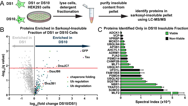
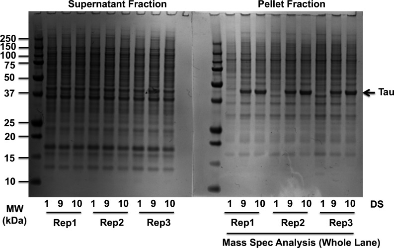
Figure 1. A proteomic approach to identify tau aggregate interactors.
(A) Tau aggregates were partially purified from DS1 and DS10 HEK293 cells expressing tauRD-YFP. Detergent fractionation enabled generation of a sarkosyl-insoluble fraction containing tau aggregates. Proteins were resolved by SDS-PAGE and then extracted from individual lanes for analysis by LC-MS/MS. (B) Volcano plot showing proteins enriched in the sarkosyl-insoluble fraction as a fold enrichment from cells expressing tauRD-YFP aggregates (DS10, dark blue dots) over cells expressing tauRD-YFP that does not form aggregates (DS1, gray dots). The red line indicates a false discovery rate of 1.5%. Gene ontology (GO) term enrichment analyses of biological processes is also shown for select GO terms: orange dots, chaperone-mediated protein folding (chaperone folding); green dots, regulation of ubiquitination (Ub regulation); teal dots, ubiquitin-dependent protein catabolic process (Ub degradation). (C) Spectral indices for a selection of the proteins identified only in the DS10-insoluble fraction. Viable knockouts are shown as green bars. Non-viable knockouts are shown as black bars. Error bars represent the SEM of three extracted protein SDS-PAGE gel bands.


