Improved strategies for ex vivo expansion of human hematopoietic stem cells (HSCs) are necessary to supply the large demand of cells required for certain clinical applications such as gene therapy/editing and cord blood (CB) transplantation. In a recent paper published in Nature, Sakurai et al. reported that the combination of three small molecules, namely a phosphoinositide 3-kinase activator peptide, a thrombopoietin receptor agonist and the pyrimidoindole derivative UM171, was sufficient to stimulate the expansion of umbilical CB HSCs, providing the first chemically defined cytokine-free expansion medium for human HSCs.
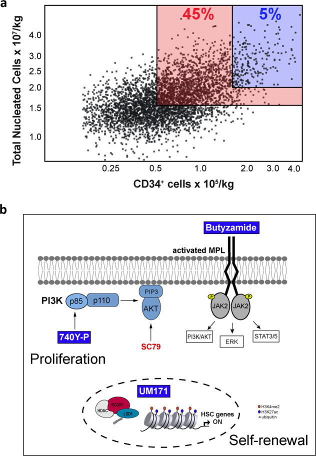
Sustenance of adult hematopoiesis is dependent on the multipotent hematopoietic stem cell (HSC) and relies on a strictly regulated balance between self-renewal and commitment to mature blood cells.1 The regenerative capacity of these cells makes HSC transplantation a lifesaving treatment for several hematologic malignancies and blood diseases.2 HSCs for transplantation are obtained either from their niche in the bone marrow or from circulating peripheral blood after mobilization.3 Another available source of HSCs is umbilical cord blood (CB) which offers many advantages including permissibility to HLA mismatches,4 low risk of chronic graft-versus-host disease5,6 and low risk of relapse of the underlying malignancy. However, the risk of transplantation-related mortality after a CB transplant is higher compared to other graft sources because of the low cell dose (10 times fewer cells) which delays neutrophil recovery causing more infections and a higher risk of graft failure.7 Thus, larger CB units are selected to circumvent the cell dose issue but they represent only 5% of the units in the banks.8 Consequently, physicians are selecting larger but poorly HLA-matched CBs as choice is quite limited. However, this practice is somewhat counterproductive as each HLA mismatch will increase transplant-related mortality by ∼5%–10%. Given these challenges, ex vivo expansion of long-term HSCs from small CB units represents an appealing solution. Indeed, the reported expansion protocol using the pyrimidoindole derivative UM171 significantly increases the therapeutic availability of CB units as physicians can now use 45% of the units in the banks as opposed to 5% as cell dose is no longer a limiting factor (Fig. 1a and ref. 9). However, a large proportion (∼55%) of the banks’ CB units remains unavailable for clinical use due to insufficient cell dose. Thus, the further development of enhanced strategies to expand HSCs would contribute to utilization of even very small CB units in the banks (Fig. 1a).
Fig. 1. Strategies to increase cord blood units availability for transplantation.

a Scatter plot of CB units cryopreserved in the Héma-Québec CB bank. Black dots represent individual CB units according to total nucleated cells (TNC) and CD34+ cell dose adjusted for a 70 kg patient. Blue square, percentage of CB units meeting minimal cell dose criteria for single CB transplantation; red square, percentage of CB units meeting minimal cell dose criteria for UM171-expanded single CB transplantation. The remaining 55% of CB units do not meet minimum criteria for therapeutic use; thus, improved expansion technologies would allow the use of these smaller CBs and likely improve the HLA match of the CBs selected for transplantation as number of available CBs would increase. b Representation of the molecular pathways activated by the chemical components of the 3a medium (740Y-P, butyzamide and UM171) described by Sakurai et al.14.
HSC expansion involves two main mechanisms: restricted differentiation which is secured by CoREST1 degradation under the control of UM17110 and sustained cell division which is provided by cytokines and other medium additives. Akin to the work described for embryonic stem cells,11 the authors have successfully replaced the growth factors which typically induced HSC proliferation with 2 molecules, an agonist of phosphoinositide 3-kinase (PI3K), 740Y-P, and a well-described thrombopoietin (THPO) analog, butyzamide.
This newly defined 2a (2 activators) medium was efficient in promoting short-term expansion (7 days) of CD34+CD38– cells from CB but ineffective for extended cultures (14 days) showing low numbers of CD34+ cells and predominance of megakaryocytic cells indicating ongoing differentiation. To overcome this differentiation, the authors supplemented the 2a “proliferative-biased” medium with two additional small molecules, best known for their anti-differentiation potential, UM17112 or StemRegenin-113 (3a medium, Fig. 1b) and reported a significant improvement of functional HSC counts in long-term cultures (10 or 30 days) only in the presence of UM171. This new 3a medium, which now included UM171 at a higher dose than used in the clinical trials (70 nM vs 35 nM), was further improved by the replacement of albumin with polyvinyl alcohol (PVA), and even further by the replacement of PVA with the polyvinyl caprolactam-polyvinyl acetate-polyethylene glycol (PCL-PVAc-PEG).14 Transcriptomic analyses of expanded cells revealed that phenotypically primitive CD34hiEPCR+ cells are enriched for HLF, a key stemness transcription factor. With these findings, Sakurai et al. suggest that these new “chemically-defined” conditions selectively expand functional HSPCs for up to 30 days with the possibility of additional clinical translation.
This approach raises the interest of the polymer-based culture system for HSC expansion and maintenance. Engineered hydrogels have been used to mimic the in vivo HSC niche as these materials provide a 3D scaffold that promotes cellular interactions. In addition, reduced levels of ROS were reported by the use of zwitterionic hydrogels, supporting a 73-fold expansion of functional HSCs.15 Moreover, the induced activation of the PI3K class I isoform through the mitogenic agonist 740Y-P peptide contributed to the CD34+ cell expansion. Nevertheless, activating the upstream PI3K pathway may affect a broad range of cellular processes. Importantly, the 740Y-P peptide is relatively poorly characterized and consequently, it remains unclear whether additional (non-PIK3) off-targets are at play here. The inability of an AKT (the major PI3K downstream target) activating molecule SC79 to reproduce the impact of 740Y-P underlines the complexity of the mechanism. Moreover, what is the role of THPO signaling in this context and how does one explain its synergy with PI3K activation by 740Y-P?
Finally, this study highlights the central role of CoREST1 degradation in preventing HSC differentiation and suggests that optimization of UM171 dosage is warranted. Although some work remains to fully characterize components of the PI3K involved in HSC proliferation, we remark on the important contribution of this work in offering additional strategies to allow the use of very small CB units in the banks that could benefit the so-called “donorless” patients but also to improve the HLA match of the CBs of those patients who have adequately sized CBs. Most excitingly, it will be interesting to test this strategy with HSCs from other sources to verify whether it is applicable in gene editing/therapy trials.
References
- 1.Till JE. Radiat. Res. 1961;14:213–222. doi: 10.2307/3570892. [DOI] [PubMed] [Google Scholar]
- 2.Chivu-Economescu M, Rubach M. Curr. Stem Cell Res. Ther. 2016;12:124–133. doi: 10.2174/1574888X10666151026114241. [DOI] [PubMed] [Google Scholar]
- 3.Tong J, et al. Blood Cells. 1994;20:351–362. [PubMed] [Google Scholar]
- 4.Zhu X, et al. Stem Cells Transl. Med. 2021;10(Suppl 2):S62–S74. doi: 10.1002/sctm.20-0495. [DOI] [PMC free article] [PubMed] [Google Scholar]
- 5.Lazaryan A, et al. Biol. Blood Marrow Transpl. 2016;22:134–140. doi: 10.1016/j.bbmt.2015.09.008. [DOI] [PMC free article] [PubMed] [Google Scholar]
- 6.Kok LMC, et al. Hum. Cell. 2020;33:243–251. doi: 10.1007/s13577-019-00297-7. [DOI] [PMC free article] [PubMed] [Google Scholar]
- 7.Eapen M, et al. Blood. 2014;123:133–140. doi: 10.1182/blood-2013-05-506253. [DOI] [PMC free article] [PubMed] [Google Scholar]
- 8.Barker JN, et al. Blood Adv. 2019;3:1267–1271. doi: 10.1182/bloodadvances.2018029157. [DOI] [PMC free article] [PubMed] [Google Scholar]
- 9.Dumont-Lagacé M, et al. Cell. Ther. 2022;28:1–5. [Google Scholar]
- 10.Chagraoui J, et al. Cell Stem Cell. 2021;28:48–62. doi: 10.1016/j.stem.2020.12.002. [DOI] [PubMed] [Google Scholar]
- 11.Kalkan T, et al. Development. 2017;144:1221–1234. doi: 10.1242/dev.142711. [DOI] [PMC free article] [PubMed] [Google Scholar]
- 12.Fares I, et al. Science. 2014;345:1509–1512. doi: 10.1126/science.1256337. [DOI] [PMC free article] [PubMed] [Google Scholar]
- 13.Boitano AEA, et al. Science. 2010;329:1345–1348. doi: 10.1126/science.1191536. [DOI] [PMC free article] [PubMed] [Google Scholar]
- 14.Sakurai M, et al. Nature. 2023;615:127–133. doi: 10.1038/s41586-023-05739-9. [DOI] [PubMed] [Google Scholar]
- 15.Bai T, et al. Nat. Med. 2019;25:1566–1575. doi: 10.1038/s41591-019-0601-5. [DOI] [PubMed] [Google Scholar]


