Abstract
Background
The microbiota of the upper respiratory tract is increasingly recognized as a gatekeeper of respiratory health. Despite this, the microbiota of healthy adults remains understudied. To address this gap, we investigated the composition of the nasopharyngeal and oropharyngeal microbiota of healthy adults, focusing on the effect of Streptococcus pneumoniae carriage, smoking habits, and contact with children.
Results
Differential abundance analysis indicated that the microbiota of the oropharynx was significantly different from that of the nasopharynx (P < 0.001) and highly discriminated by a balance between the classes Negativicutes and Bacilli (AUC of 0.979). Moreover, the oropharynx was associated with a more homogeneous microbiota across individuals, with just two vs. five clusters identified in the nasopharynx. We observed a shift in the nasopharyngeal microbiota of carriers vs. noncarriers with an increased relative abundance of Streptococcus, which summed up to 30% vs. 10% in noncarriers and was not mirrored in the oropharynx. The oropharyngeal microbiota of smokers had a lower diversity than the microbiota of nonsmokers, while no differences were observed in the nasopharyngeal microbiota. In particular, the microbiota of smokers, compared with nonsmokers, was enriched (on average 16-fold) in potential pathogenic taxa involved in periodontal diseases of the genera Bacillus and Burkholderia previously identified in metagenomic studies of cigarettes. The microbiota of adults with contact with children resembled the microbiota of children. Specifically, the nasopharyngeal microbiota of these adults had, on average, an eightfold increase in relative abundance in Streptococcus sp., Moraxella catarrhalis, and Haemophilus influenzae, pathobionts known to colonize the children’s upper respiratory tract, and a fourfold decrease in Staphylococcus aureus and Staphylococcus lugdunensis.
Conclusions
Our study showed that, in adults, the presence of S. pneumoniae in the nasopharynx is associated with a shift in the microbiota and dominance of the Streptococcus genus. Furthermore, we observed that smoking habits are associated with an increase in bacterial genera commonly linked to periodontal diseases. Interestingly, our research also revealed that adults who have regular contact with children have a microbiota enriched in pathobionts frequently carried by children. These findings collectively contribute to a deeper understanding of how various factors influence the upper respiratory tract microbiota in adults.
Video Abstract
Supplementary Information
The online version contains supplementary material available at 10.1186/s40168-023-01640-9.
Keywords: Microbiota, Nasopharynx, Oropharynx, Streptococcus pneumoniae, Healthy adults
Background
The microbiota of the human upper respiratory tract (URT) has an important role in human health since it modulates the colonization of commensal bacteria and provides colonization resistance against pathogens [1]. The URT comprises several structures, among which the nasopharynx and the oropharynx are distinctive, as they are the preferential niches of important human pathobionts, including Streptococcus pneumoniae (or pneumococcus). Pneumococcus is a gram-positive facultative anaerobe that is known to be the main cause of bacterial respiratory infections worldwide [2]. Risk groups for pneumococcal disease include young children, the elderly, and immunocompromised individuals of all ages [3]. Colonization is mostly asymptomatic and is very frequent in children under 5 years of age, in which it is often higher than 50% [4–6]. In contrast, in adults, it has been reported as being between 20 and 40% [7–9]. Two important factors that contribute to increased pneumococcal colonization and persistence in adults include contact with children and smoking [9–12]. To what extent the microbiota composition is a risk factor for pneumococcal colonization has not been explored. Nonetheless, to the best of our knowledge, while there are studies in children, the microbiota of the nasopharynx and oropharynx remains poorly characterized since the few microbiota studies of the upper respiratory tract in adults have been associated with disease status [13–16].
Understanding the factors that shape and characterize a healthy microbiota is a fundamental step in strategies aimed at promoting a healthy state, for example, through the use of live biotherapeuticals [17].
Here, we comprehensively analyzed the composition of the nasopharyngeal and oropharyngeal microbiota of immunocompetent healthy adults aged between 25 and 50 years old. The specific aims of our study were (i) to compare the microbiota of pneumococcal carriers vs. noncarriers in the nasopharynx and oropharynx and (ii) to understand how individual characteristics such as age, sex, contact with children, and smoking habits shape the microbiota of the nasopharynx and oropharynx.
Methods
Study population and study design
A case–control study was designed with the aim of evaluating potential differences between the nasopharyngeal and oropharyngeal bacterial microbiota of immunocompetent healthy adults colonized with S. pneumoniae (cases) and noncolonized individuals (controls).
The study was nested in a prospective 6-month longitudinal study that aimed to characterize the dynamics of S. pneumoniae colonization in healthy adults [9]. The original study was conducted between February 2015 and December 2016 and enrolled 87 immunocompetent adults aged between 25 and 50 years old living in the Lisbon metropolitan area, Portugal. Detailed information about sample collection and study design is described in the supplementary information (“Sample collection”) and in Almeida et al. [9]. Briefly, nasopharyngeal and oropharyngeal samples were collected using appropriate swabs and were immediately stored in STGG medium. All samples were kept at − 80 °C. The presence of pneumococci was screened by classical culture based methods and real-time PCR targeting the genes lytA and piaB [9].
The study was approved by the ethical committee of Instituto de Higiene e Medicina Tropical, Universidade Nova de Lisboa, and was registered at the National Commission of Data Protection (ref. 3803/2014). Signed informed consent was obtained from all participants; samples and questionnaires were processed anonymously.
In the current study, cases were defined as individuals who were found to carry pneumococci in the nasopharynx and/or oropharynx at a minimum of three time points at least 1 month apart from each other. Controls were defined as individuals who were sampled at least five times (1 month apart from each other) during the 6-month period and were never colonized with pneumococci. For both cases and controls, samples collected within 1 month of antibiotic use were excluded. For both cases and controls, three samples per individual were selected for analyses. The three samples were, as much as possible, distant in time and covered different seasons.
DNA extraction
Nasopharyngeal and oropharyngeal samples were maintained at − 80 °C in STGG. Samples were thawed on ice, and for each sample, 200 μL was pipetted and added to 200 μL of lysis buffer (MagNA Pure Compact Nucleic Acid Isolation Kit, Roche Diagnostics, GmbH). Samples were incubated at 37 °C for 20 min. DNA extraction was performed with the MagNA Pure Compact System (Roche) according to the manufacturer’s instructions. For every run, water samples (used as negative controls) were extracted in parallel. DNA was stored at − 20 °C. Water samples were used as technical negative controls and were processed in parallel with biological samples to control for potential contaminations arising during manipulation and testing.
Total bacterial load quantification
To prepare a standard curve for total bacterial load quantification, the method described by Bogaert et al. [18] was followed with some modifications. Representative strains of eight bacterial species that commonly colonize the upper respiratory tract were used: Corynebacterium accolens, Dolosigranulum pigrum, Haemophilus influenzae, Moraxella catarrhalis, Streptococcus mitis, Streptococcus oralis, S. pneumoniae, and Streptococcus pseudopneumoniae. First, serial dilutions of the frozen stocks of each strain were performed to quantify the bacterial load. Except for H. influenzae, which was cultured on chocolate agar, all other species were cultured on blood agar plates. Corynebacterium accolens cultures were incubated overnight at 37 °C in anaerobic conditions; Dolosigranulum pigrum cultures were incubated overnight at 37 °C in aerobic conditions; and the remaining bacterial species were incubated overnight at 37 °C in a 5% CO2 atmosphere. On the following day, CFU/mL was estimated for each frozen stock. Afterwards, a mixture containing 104 CFU/mL of each species was prepared. DNA extraction of the mixture was carried out as described above. DNA was quantified on a NanoDrop and used as a reference for total bacterial load quantification.
To evaluate the quality of the nasopharyngeal and oropharyngeal samples, the total bacterial load of each sample was quantified by qPCR using universal primers and probes that target the 16S rDNA gene [18]: 16Sfw-5′-CGA AAG CGT GGG GAG CAA A-3′, 16Srev-5′-GTT CGT ACT CCC CAG GCG G-3′, and FAM-ATTAGATACCCTGGTAGTCCA-MGB. qPCRs were performed in a final volume of 25 μL containing 12.5 μl of 1 × master mix (FastStart TaqMan® Probe Master, Roche), 1 μL of each primer (0.4 μM), 1 μL of probe (0.2 μM), 7 μL of H2O, and 2.5 μL of DNA. DNA amplification was performed in CFX96™ Real-Time System Amplification (Bio-Rad). The thermocycling conditions were 50 °C for 2 min and 95 °C for 10 min followed by 45 amplification cycles of 95 °C for 15 s and 60 °C for 1 min. In each 16S qPCR run, multiple negative controls (one per every 16 reactions) and serial dilutions (in duplicate) of the DNA extracted from the species mixture (100 to 10−5 ng/μL) were included. The latter was included to obtain a standard curve for each qPCR.
A standard curve was considered valid when the difference between Ct values of consecutive dilutions did not exceed three Ct values and the paired results obtained for a given dilution did not exceed 0.5 Ct. Samples with a bacterial load lower than the DNA extraction negative control were considered of low quality and, whenever possible, were replaced by other samples from the same individual following the inclusion and exclusion criteria described above.
16S rRNA gene amplicon sequencing
For all samples, the V4 region of 16S rRNA was amplified using forward (5′-GTGCCAGCMGCCGCGGTAA-3′) and reverse (5′-GGACTACHVGGGTWTCTAAT-3′) primers previously described [19]. PCR was conducted in a final volume of 25 μL containing 10 μL of 2 × master mix, 2.5 μL of primer barcode (2 μM), 2.5 μL of universal primer (2 μM), and 10 μL of DNA. The thermocycling conditions were 94 °C for 3 min, 35 amplification cycles of 94 °C for 1 min, 50 °C for 1 min, 72 °C for 105 s, and a final extension of 72 °C for 10 min. Each sample was run in triplicate. After that, triplicates were pooled and submitted to a next-generation sequencing platform for indexing and pair-end sequencing (2 × 250 bp) on a MiSeq platform. Amplification and sequencing were performed at the Genomics Unit of Instituto Gulbenkian da Ciência.
Bioinformatic processing
Divisive Amplicon Denoising Algorithm 2 (DADA2) [20] was used to denoise and taxonomically assign the 16S rRNA sequences following the authors’ online pipeline tutorial 1.16 (https://benjjneb.github.io/dada2/tutorial.html). DADA2 was run on R version 3.6.2 [21]. The parameters used in each step of the DADA2 workflow were those predefined and recommended in the pipeline except for the parameters that are data driven, specifically trimming and inference of error rates. Sequences were trimmed in the position in which the 25th percentile of the quality score was above 30 (see supplementary information and Fig. S1); error inference rates were calculated using the entire dataset and pooled sequences. In brief, reads were filtered and trimmed to remove sequencing errors, based on their quality scores (Phred scores) and on the identification of ambiguous bases in both forward and reverse reads. Subsequently, an estimation of the error rates (i.e., possible transitions or transversion point mutations), made by MiSeq platform, was performed. This step aimed to achieve the following: (i) infer amplicon sequence variants (ASVs) based on the estimated error rates mentioned earlier, (ii) dereplicate reads to obtain unique sequences, and (iii) remove singletons. Afterward, the forward and reverse reads were ready for merging. The final step involved identifying and removing ASVs that could potential be chimeric sequences originating from defective PCR amplification (for example, resulting from pairing of incomplete parental sequences). Taxonomy was assigned using the Silva v132 database as a reference [22].
Several additional approaches were used to remove potential contaminants by filtering ASVs and samples. First, ASVs attributed to Eukaryota and Archaea were excluded. Second, ASVs were filtered according to a frequency-based approach described by Davis et al. [23]. This approach is based on the observation that the probability of having contaminants is higher when the DNA concentration is lower. Briefly, for each ASV, a regression line was fitted to the number of reads as a function of DNA concentrations measured by 16S qPCR in each sample. If the number of reads of an ASV was observed to decrease linearly with increased DNA concentration, it was considered a contaminant and was excluded. Otherwise, it was kept in the analysis. ASVs were filtered according to their relative abundance and were kept if they were present in at least two samples, with a relative abundance within each sample higher than 0.1% [24]. Finally, samples that had fewer than 1000 reads after all ASV filters were excluded [14, 25].
For ASVs that were shown to be significantly different in the differential abundance analysis, NCBI BLAST searches were performed (using MegaBLAST) to identify the presumptive species. Species assignment was based exclusively on hits with 100%, as differences in one or more nucleotides result in assignment of different ASVs.
Statistical analysis
All analyses described below were performed in R version 3.6.2 (Boston, MA, USA).
Statistical analysis of the study population
The baseline characteristics of the study population and samples were stratified by the presence/absence of pneumococci. To compare characteristics between strata, the chi-square or Student’s t-test was used in conformity with the type of data. Bacterial DNA quantification was stratified by the presence/absence of pneumococci and by the anatomical site (oropharynx or nasopharynx). Bacterial DNA quantification, per stratum, was summarized by their geometric mean and respective standard deviation (SD). The Wilcoxon rank-sum test with continuity correction was used for multiple comparisons of groups two by two. The Benjamini–Hochberg procedure was used to control for the false discovery rate at the level of 0.05. Differences between strata were considered statistically significant if the adjusted P-value was < 0.05.
Statistical analysis of nasopharyngeal and oropharyngeal microbiota profiles
The statistical analysis of the microbiota was performed using compositional data analysis methods. In brief, for each sample, count reads were normalized using the centered log2-ratio (CLR) transformation [26]. This transformation allows us to account for the complex compositional data structure of metagenomic studies and to reduce the likelihood of spurious correlations. The microbiome package for the CLR transformation, which replaces ASV read counts with exact zero relative abundance with a pseudocount before calculating the logarithms, was used [27].
To identify homogeneous bacterial communities in the nasopharynx and oropharynx, a hierarchical clustering approach was employed. The samples were transformed using the CLR transformation, and the Euclidean distance between samples was used for clustering. The Ward’s minimum variance method [28] was used to agglomerate samples that share similar taxonomic profiles. To determine the optimal number of clusters, the gap statistics proposed by Tibshirani et al. [29] were used. For cross-validation, a random forest model classifier with 500 trees was utilized. The out-of-bag error, representing the percentage of misclassified samples, was estimated, and the confusion matrix was examined to assess the degree of cluster overlap. Each cluster was characterized by the two most abundant genera it contained.
To study associations between clusters and the pneumococcal carrier state, mixed general linear models with a logit link function were used. A model was fit to each microbiota profile using the microbiota profile comprising the higher number of samples as a reference. Individuals were introduced as a random variable to account for repeated measurements. In addition, models were adjusted for sociodemographic characteristics and environmental factors (individual’s age, gender, having contact with children, smoking habits, and season in which the sample was collected). Associations between variables and the clusters were calculated using odds ratios (ORs) and corresponding confidence intervals (CIs) at 95%. A CI that did not include 1 was considered statistically significant.
Microbiota α-diversity
The abundance-based diversity of the microbiota groups was estimated using Hill's first five numbers [30]. Hill’s numbers have a scaling parameter, known as the order of diversity (q), that modulates sensitivity toward more abundant or rare taxonomic units. The higher the order, the higher the importance attributed to abundant taxonomic units. The Hill numbers of orders 0, 1, and 2 are related to three popular diversity indexes known as richness, Shannon’s index, and Simpson’s index, respectively, with the advantage of having the replication principle (i.e., when doubling the number of taxonomic units in a system, the diversity is also doubled). Evenness was measured according to the steepness of the diversity profile from the Hill number of order 0 to the Hill number of order 1 (the higher the steepness, the lower the community evenness). The Hill numbers were calculated for each sample using the abundance-based estimates at the taxa level of genus and then by calculating the geometric mean for each group. Differences in Hill numbers between groups were calculated using the Mann‒Whitney test.
Differential abundance analysis of microbiota
To evaluate if there were significant differences between the microbiota of different groups, a permutational multivariate analysis of variance (PERMANOVA) [31] implemented on the Adonis algorithm of the R Vegan package was utilized. PERMANOVA was performed on the Euclidean distance matrix with CLR-transformed read counts. To ensure the reliability of the results, the assumption of variance homogeneity was checked. If a significant effect was found, a differential abundance analysis to determine which taxa were differentially abundant between groups of samples was performed. Since the data were very sparse, we made use of a zero-inflated Gaussian mixed model (ZIGMM) [32] implemented in R with metagenomeSeq [31]. The cumulative sum scaling method was used to normalize sequence counts based on the lower-quartile abundance of features. Data were also filtered to maintain a threshold of ASVs that were present in at least 75% of the samples, a step needed to avoid unreliable fold-change estimates [32]. Only ASVs with more than 1.5 log fold-change differences and an adjusted P-value ≤ 0.05 were considered [31]. Volcano plots of the log10 of statistical significance (P-value) vs. log2 of the magnitude of change (fold-change) were used to visualize the results.
Microbial signatures, that is, groups of microbial taxa that are predictive of a phenotype of interest, were further identified using the algorithm selbal developed in R [33]. Briefly, this algorithm takes the log ratio of the geometric mean of the taxa from two groups and tests for association with the response variable by fitting a logistic model. The model that maximizes the area under the receiver operating characteristic (AUC) curve is then selected.
Dynamics of microbiota carriage
The dynamics of individual nasopharyngeal and oropharyngeal clusters were represented by alluvial plots and stratified by pneumococcus carriage. The number of individuals who changed clusters was reported as proportions and compared using a chi-squared test.
Temporal changes in the nasopharyngeal and oropharyngeal microbiota were analyzed at the genus level by comparing the microbiota of each individual in consecutive samples (first and second, second and third). Differences between the composition of microbiota in consecutive samples were expressed as volatility (Aitchison distance), calculated using the Euclidean distance on the CLR transformed data [34]. The Wilcoxon rank-sum test was used to compare volatility values.
Results
Study population
The records of 87 individuals who were followed-up for 6 months were reviewed retrospectively. Fifty-nine individuals met the following criteria to be included in the current study: 12 pneumococcal carriers with at least three samples (collected 1 month apart from each other) positive for pneumococci and 47 pneumococcal noncarriers (negative for pneumococci on at least five occasions separated 1 month apart from each other). None of the individuals included had samples collected within 1 month of antibiotic use.
The baseline characteristics of the study population are summarized in Table 1. There were no significant differences between carriers and noncarriers when mean age, sex, smoking status, and antibiotic consumption in the previous six months were compared. Pneumococcal carriers were more likely to have regular contact with children than nonpneumococcal carriers (83.3% vs. 38.3%, P = 0.014) and to have received seasonal flu vaccination (33.2% vs. 7.4%, P = 0.015). Antibiotic consumption prior to sample collection occurred in a minority of samples (and always in a period exceeding 1 month from sample collection), with no significant differences between carriers and noncarriers (Table 1).
Table 1.
Baseline characteristics of the study population stratified by the presence/absence of pneumococci
| Characteristics | Carriers N = 12 | Noncarriers N = 47 | P-valuea |
|---|---|---|---|
| Mean age ± standard deviation (years) | 36.3 ± 5.7 | 37.5 ± 7.1 | 0.142b |
| Gender male, n (%) | 5 (41.7) | 22 (46.8) | 1.000 |
| Living with children 18 years old, n (%) | 10 (83.3) | 18 (38.3) | 0.014 |
| Smoker, n (%) | 5 (41.7) | 20 (42.6) | 1.000 |
| Chronic diseases, n (%) | 6 (50.0) | 12 (25.5) | 0.158 |
| Seasonal flu vaccination, n (%) | 4 (33.4) | 2 (7.4) | 0.015 |
| Vaccination with PCV13, n (%) | 4 (33.4) | 6 (12.8) | 0.189 |
| Antibiotic consumption 6 months before enrollment, n (%) | 3 (25) | 8 (17) | 0.679 |
Characteristics with a P-value less than 0.05 are highlighted in bold
aIndividual characteristics (with the exception of mean age) were compared with Pearson’s chi-squared test
bMean age between groups was compared with Student’s t-test
.
Samples analyzed
For each of the 59 individuals, paired samples collected from the oropharynx and the nasopharynx at three time points were analyzed, resulting in a total of 354 samples. Antibiotic consumption between visits occurred 8.3% of the time. Samples were collected throughout the year with no significant differences between pneumococcal carriers and noncarriers by sampling season (Table 2).
Table 2.
Characteristics of samples included in the study according to individuals’ pneumococcal carrier state
| Characteristics | Pneumococcal carriers' samples N = 72 | Pneumococcal noncarriers' samples N = 282 | P-valuea | |
|---|---|---|---|---|
| Antibiotic consumption between sampling, n (%) | 6 (8.3) | 20 (7.1) | 0.799 | |
| Sampling season, n (%) | Spring | 22 (30.6) | 80 (28.4) | 0.826 |
| Summer | 18 (25.0) | 78 (27.7) | 0.761 | |
| Autumn | 6 (8.3) | 50 (17.7) | 0.077 | |
| Winter | 26 (36.1) | 74 (26.2) | 0.130 |
aSample characteristics were compared with Pearson’s chi-squared test
Bacterial DNA quantification, processing of 16S rRNA gene data, and identification of clusters
The geometric mean of the total bacterial load based on 16S rRNA gene quantification in the nasopharynx (19.58 pg/μL) was significantly lower (P < 0.001) than the bacterial DNA quantity in the oropharynx (961.27 pg/μL) independent of the pneumococcal carrier state. In the nasopharynx, the geometric mean of the total bacterial load of pneumococcal carriers was higher than that of noncarriers (43.91 pg/μL vs. 15.89 pg/μL, P < 0.009). In the oropharynx, the corresponding numbers were 1237.57 pg/μL (carriers) and 901.23 pg/μL (noncarriers, P = 0.233) (Fig. 1).
Fig. 1.
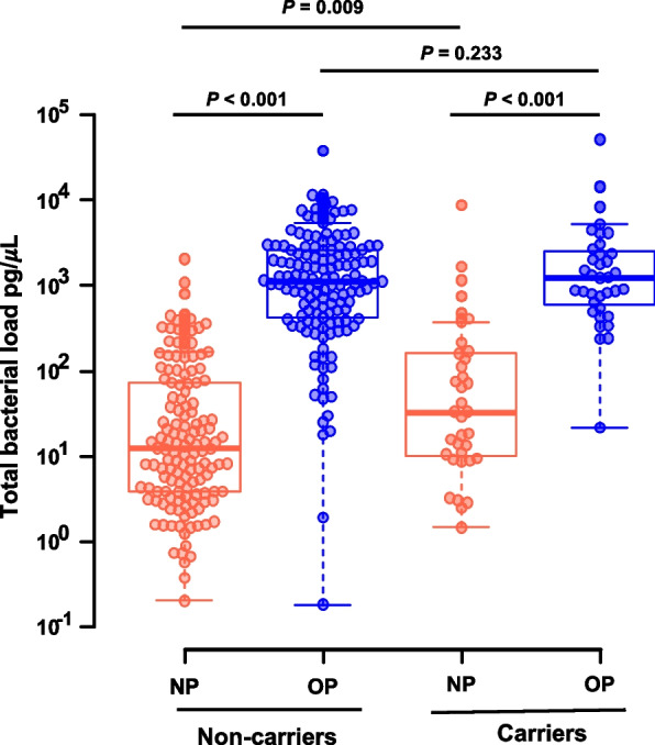
Total bacterial load of nasopharyngeal (NP) and oropharyngeal (OP) samples. The Wilcoxon rank-sum test with continuity correction and the Benjamini–Hochberg procedure were used to adjust for the false discovery rate
Processing of the raw metagenomic sequencing data was performed for all 354 samples as detailed in the supplementary information (“Processing of raw metagenomic sequencing data” and Fig. S1 therein).
A total of 9,027,200 reads were received. The average number of reads per sample was 22,071 (range 2 and 38,536), which were clustered in 14,669 ASVs. After removing sequences from Eukarya and Archaea, there were a total of 8,079,020 reads with a medium number of 24,652 reads per sample (range between 2 and 38,536) and 187 singletons. The SILVA database assigned 6,589 ASVs to bacteria. A total of 108 reads were unassigned, with the majority (105 reads) sampled from the nasopharynx.
To identify groups of samples that shared closer bacterial taxonomic profiles with each other, hierarchical clustering was performed. Two main groups were identified, and these, with few exceptions, segregated nasopharyngeal samples from oropharyngeal samples (Fig. 2).
Fig. 2.
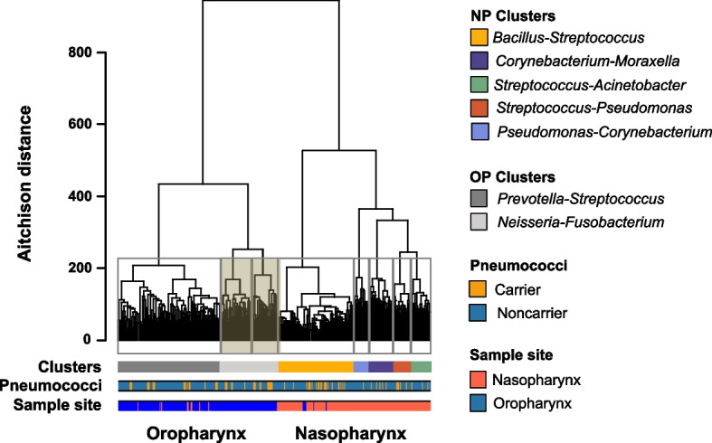
Oropharynx and nasopharynx microbiota profiles. Dendrogram showing the clusters identified by hierarchical cluster analysis performed on the Euclidean distance of the centered log-ratio transformed data (Aitchison distance). The gray lines surrounding clades represent the clusters identified by the Calinski and Harabasz index. Clusters inside the gray rectangle are the oropharyngeal clusters merged after cross-validation by random forest analysis. Bars below the dendrogram indicate clusters, samples in which pneumococcus was identified, and sampling site
A total of eight clusters were determined. However, the random forest that we used as a cross-validation method showed a confusion matrix with an out-of-bag error of 15.0% due to an overlap between two oropharyngeal clusters, where 71.4% of the samples that belonged to one of the clusters were classified in another cluster. When analyzed further, we found that the ten most abundant genera in both clusters were identical; thus, we opted to merge these two clusters. Ultimately, five nasopharyngeal clusters and two oropharyngeal clusters were observed (Fig. 2). PERMANOVA indicated that the oropharynx microbiota was significantly different from the nasopharynx microbiota (P < 0.001), as detailed in the supplementary information (“Bacterial profiles in the oropharynx and nasopharynx” and Fig. S2–S3 and Table S1 therein).
Characterization of the nasopharyngeal microbiota profiles
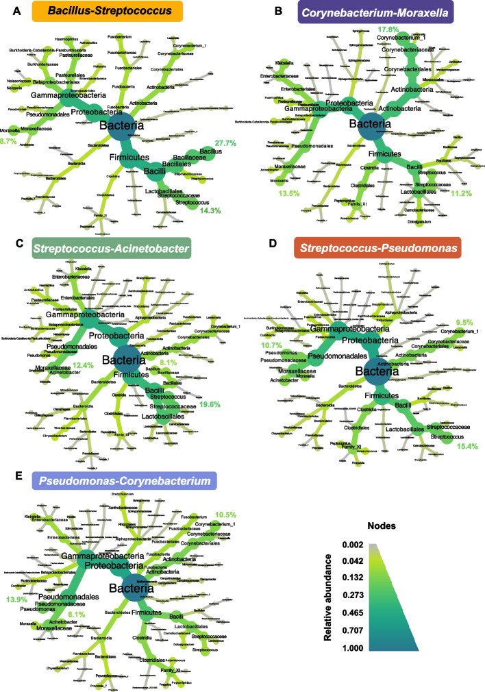
We identified five clusters in the nasopharynx and named them after the two most abundant genera found in each cluster. The most frequent cluster was named Bacillus-Streptococcus and included 48.2% (n = 80) of the total samples from the nasopharynx. The three most abundant genera in this cluster were Bacillus (27.7%), Streptococcus (14.3%), and Moraxella (8.7%) (Fig. 3A). This cluster was the least diverse and least even of all nasopharyngeal clusters (Fig. S4A). The other clusters were named Corynebacterium-Moraxella (16.3%, n = 27), Streptococcus-Acinetobacter (13.3%, n = 22), Streptococcus-Pseudomonas (12.0%, n = 20), and Pseudomonas-Corynebacterium (10.2%, n = 17) (Fig. 3B–E).
Fig. 3.
Nasopharyngeal microbiota clusters. Taxonomic heat trees for the five clusters identified. A Bacillus-Streptococcus (48.2% of the total samples), B Corynebacterium-Moraxella in dark blue (16.3%), C Streptococcus-Acinetobacter (13.3%), D Streptococcus-Pseudomonas (12.0%), and E Pseudomonas-Corynebacterium (10.2%). The center of each tree represents the kingdom and has a relative abundance of 1. In the extremities, the relative abundances at the genus level are represented. From the center to the extremities, each taxonomic level from kingdom to genus is indicated. The gradient of colors represents relative abundance. In each cluster, the relative abundance of the three most common genera is specified
Clusters in which Streptococcus were not dominant had a higher effective number of genera (supplementary information “Bacterial profiles in the oropharynx and nasopharynx” and Fig. S4 therein).
Association between nasopharyngeal microbiota profiles and variables under study
A mixed general linear model was used to investigate potential associations between the nasopharyngeal microbiota profiles and the pneumococcal carrier state and sociodemographic and environmental characteristics. The cluster Bacillus-Streptococcus was used as a reference since this cluster accounted for the highest number of samples (Table 3). In winter, the nasopharyngeal microbiota was less likely to be described by the Streptococcus-Acinetobacter cluster (OR = 0.18; 95% CI 0.02–0.85) and more likely to be described by the Corynebacterium-Moraxella cluster (OR = 28.1, 95% CI 4.4–307.2) compared to the reference. Males were more likely to have a nasopharyngeal microbiota described by clusters Corynebacterium-Moraxella (OR = 227.0, 95% CI 25.2–679.6) or Pseudomonas-Corynebacterium (OR = 12.5, 95% CI 1.5–310.3). Having contact with children increased the likelihood of having a nasopharyngeal microbiota described by the cluster Pseudomonas-Corynebacterium (OR = 15.6, 95% CI 1.8–376.1), whereas being a smoker decreased the likelihood of having that cluster (OR = 0.13, 95% CI 0.02–0.72). Of note, Pseudomonas-Corynebacterium included only one pneumococcal carrier (Table 3), which may indicate that this microbiota profile has a protective role against pneumococcal carriage.
Table 3.
Association between nasopharyngeal microbiota profiles and variables under study
| Characteristics | Microbiota profiles | ||||||||
|---|---|---|---|---|---|---|---|---|---|
|
Bacillus-Streptococcus N = 80 |
Streptococcus-Pseudomonas N = 20 |
Streptococcus-Acinetobacter N = 22 |
Corynebacterium-Moraxella N = 27 |
Pseudomonas-Corynebacterium N = 17 |
|||||
| n (%) | n (%) | ORadj (95% CI) | n (%) | ORadj (95% CI) | n (%) | ORadj (95% CI) | n (%) | ORadj (95% CI) | |
| Pneumococcal carrier, yes | 21 (26.3) | 3 (18.7) | 0.26 (0.04–1.21) | 3 (18.7) | 0.22 (0.03–1.18) | 5 (18.5) | 0.71 (0.04–10.59) | 1 (5.8) | NA |
| Age, > 37 years | 43 (53.7) | 9 (20.0) | 0.64 (0.14–2.62) | 9 (20.0) | 0.58 (0.15–2.06) | 7 (25.9) | 0.68 (0.10–3.89) | 8 (47.1) | 1.08 (0.12–10.15) |
| Gender, male | 22 (27.5) | 6 (37.5) | 1.19 (0.35–3.77) | 6 (37.5) | 0.74 (0.21–2.40) | 26 (96.3) | 226.97 (25.16–679.59) | 14 (82.6) | 12.52 (1.51–310.27) |
| Children, yes | 41 (52.2) | 7 (43.7) | 4.70 (1.04–25.82) | 7 (43.7) | 0.60 (0.15–2.39) | 7 (25.9) | 0.24 (0.02–1.99) | 10 (58.8) | 15.59 (1.83–376.07) |
| Smoker, yes | 39 (48.7) | 7 (43.7) | 0.56 (0.16–1.81) | 7 (43.7) | 0.41 (0.12–1.23) | 11 (40.7) | 0.65 (0.12–3.26) | 4 (23.5) | 0.13 (0.02–0.72) |
| Season | |||||||||
| Spring | 28 (35.0) | 12 (75.0) | Ref | 12 (75.0) | Ref | 3 (11.1) | Ref | 0 (0.0) | Ref |
| Summer | 22 (27.5) | 8 (50.0) | 3.54 (0.94–15.86) | 8 (50.0) | 0.78 (0.23–2.53) | 2 (7.4) | 0.50 (0.04–4.75) | 4 (23.5) | NA |
| Autumn | 11 (13.7) | 0 (0.0) | 2.55 (0.38–16.32) | 0 (0.0) | NA | 2 (7.4) | 0.71 (0.06–7.38) | 12 (70.1) | NA |
| Winter | 19 (23.8) | 2 (12.5) | 1.11 (0.21–5.76) | 2 (12.5) | 0.18 (0.02–0.85) | 20 (74.1) | 28.1 (4.38–307.16) | 1 (5.9) | NA |
Ref, variable used as reference. NA, nonadmissible as there are no data in the reference. Bold indicates statistically significant results. The Bacillus-Streptococcus profile was the most frequent and thus was used as a reference against which the other profiles were compared
Nasopharyngeal profiles of subpopulations of pneumococcal carriers, adults who have close contact with children, and smokers
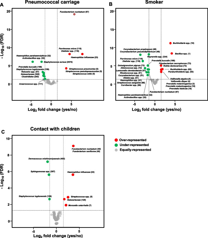
To identify which ASVs differed between the nasopharyngeal microbiota based on the pneumococcal carrier state, smoking habits, and contact with children, ZIGM models were fitted. The nasopharyngeal microbiota between individuals identified as pneumococcal carriers and noncarriers showed significant differences (PERMANOVA, P = 0.001). Among pneumococcal carriers, four ASVs were found to be overrepresented (Fig. 4A, Table S2). These were identified as presumptive H. influenzae (ASV23), Fusobacterium nucleatum (ASV87), Parvimonas micra or Dialister spp. (ASV116), and S. pneumoniae, S. pseudopneumoniae, or S. mitis (ASV5). In addition, eight ASVs were found to be underrepresented. These were identified as presumptive Haemophilus parahaemolyticus or Actinobacillus spp. (ASV32), Staphylococcus lugdunensis (ASV159), and Staphylococcus aureus (ASV2970).
Fig. 4.
Volcano plots representing ASVs that showed differential abundance in the nasopharynx. A Effect of pneumococcal carrier status. B Effect of smoking status. C Effect of having contact with children. Bacterial taxa overrepresented among pneumococcal carriers, smokers, and adults who have regular contact with children are represented by red circles on the right side of each corresponding plot. Bacterial taxa underrepresented among pneumococcal carriers, smokers, and adults who have regular contact with children are represented by green circles on the left side of each corresponding plot. Gray circles indicate bacterial taxa that were not differentially abundant
The nasopharyngeal microbiota between individuals identified as smokers and nonsmokers also showed significant differences (PERMANOVA, P = 0.001). Among smokers, seven ASVs were found to be overrepresented (Fig. 4B, Table S3). Of these, the three with the highest FCs were ASV1 (log2FC = 4.19), ASV10 (log2FC = 3.85), and ASV63 (log2FC = 1.88). These were identified as presumptive Bacillus spp. (ASV1), Burkholderia spp. (ASV10), and Burkholderia spp. or Paraburkholderia spp. (ASV63). Ten ASVs were found to be underrepresented. Of these, ASV41 (log2FC = − 2.05), ASV46 (log2FC = − 3.64), and ASV72 (log2FC = − 2.46) showed the lowest FCs (Fig. 4B, Table S3). These were identified as presumptive H. parahaemolyticus or Haemophilus sputorum (ASV41), Corynebacterium propinquum or Corynebacterium pseudodiphtericum (ASV46), and D. pigrum or uncultured Alloiococcus spp. (ASV72).
Finally, differences between the nasopharyngeal microbiota of adults who had regular contact with children compared to adults who did not have frequent contact with children were also significant (PERMANOVA, P = 0.04). Among adults who had contact with children, five ASVs were overrepresented (Fig. 4C, Table S4). Of these, ASV5 (log2FC = 2.57), ASV23 (log2FC = 3.61), and ASV40 (log2FC = 3.69) had the highest FCs. The latter was identified as presumptive F. nucleatum or Fusobacterium naviforme. ASV159 (log2FC = − 1.51), ASV307 (log2FC = − 1.62), and ASV402 (log2FC = − 1.94) were found to be underrepresented. These were identified as presumptive S. lugdunensis (ASV159), Sphingomonas spp. (ASV307), and Dermacoccus nishinomiyaensis (ASV402).
Diversity of nasopharyngeal profiles of subpopulations of pneumococcal carriers, adults who have close contact with children, and smokers
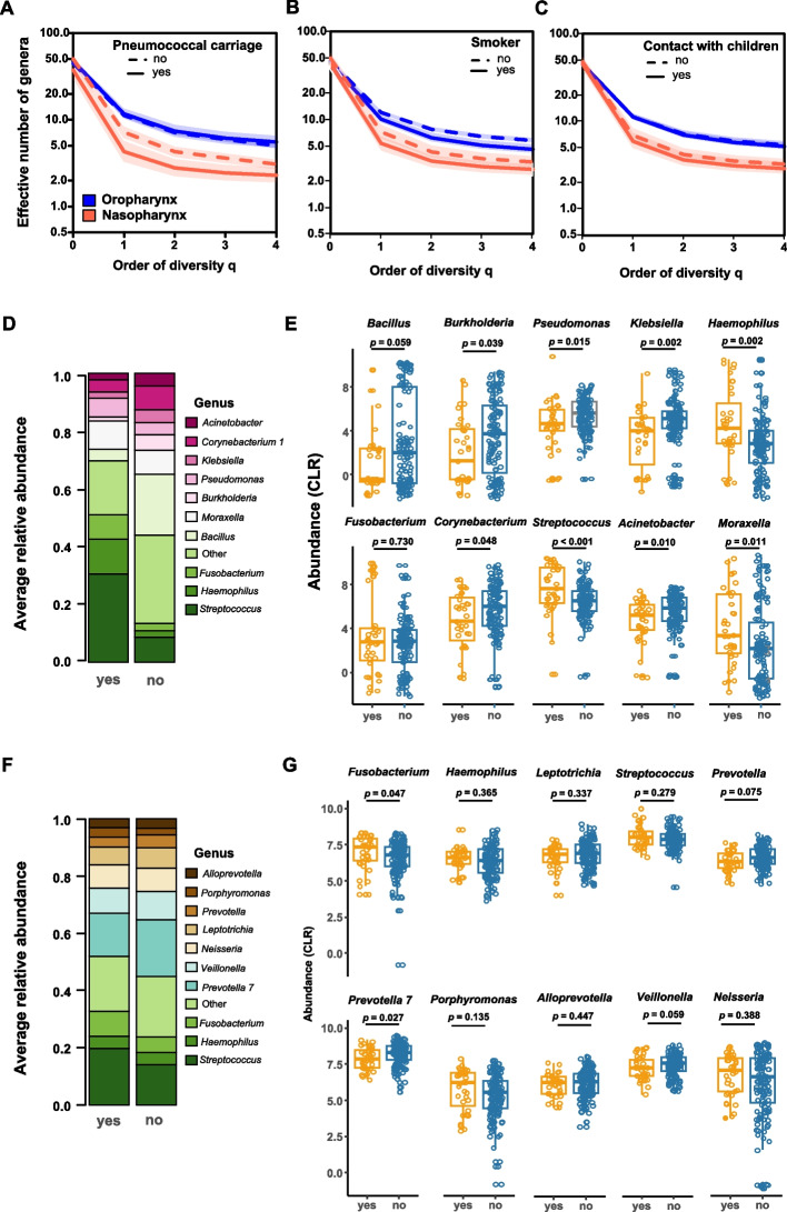
Diversity, at the taxonomic level of genus, was significantly lower in the nasopharynx of pneumococcal carriers (0D = 37.6, 1D = 4.3, 2D = 2.8) than in the nasopharynx of noncarriers (0D = 49.7, 1D = 7.1, 2D = 4.3) for each diversity number (P = 0.037, P = 0.003, and P = 0.006, respectively) (Fig. 5A and Fig. S5A). In addition, the nasopharynx microbiota of pneumococcal carriers was less even than the nasopharynx microbiota of noncarriers, supporting the higher dominance of the most abundant species found in this niche (Fig. S5A).
Fig. 5.
Diversity profiles. A Diversity of nasopharyngeal and oropharyngeal microbiota given by Hill numbers of order 0 to 4 in pneumococcal carriers and noncarriers. B Diversity of nasopharyngeal and oropharyngeal microbiota given by the Hill numbers of order 0 to 4 of smokers and nonsmokers. C Diversity of nasopharyngeal and oropharyngeal microbiota given by the Hill numbers of order 0 to 4 of individuals who have regular contact with children and those who do not have. D Average relative abundance of the ten most frequent genera found in the microbiota of the nasopharynx represented by stacked bar plots. The remaining less abundant genera were grouped as a single bar (other). E Abundance of the ten most abundant genera found in the nasopharynx of carriers and noncarriers. F Average relative abundance of the ten most frequent genera found in the microbiota of the oropharynx represented by stacked bar plots. The remaining less abundant genera were grouped as a single bar (other). G Abundance of the ten most abundant genera found in the oropharynx of carriers and noncarriers. P-values determined by the Wilcoxon rank-sum test
In parallel, the proportion of reads classified as Streptococcus in the nasopharynx of pneumococcal carriers compared to noncarriers was significantly higher (P < 0.001); among carriers, Streptococcus accounted for much as 30% on average of all genera (summing up to more than 50% of all genera in 29% of carriers); among noncarriers, Streptococcus accounted, on average, for 10% of all genera (summing up to more than 50% of all genera in only 3% of noncarriers) (Fig. 5D − E and Fig. S6).
Only marginal differences between the diversity exhibited by the nasopharynx of smokers (0D = 42.7, 1D = 5.4, 2D = 3.4) compared to nonsmokers (0D = 50.2, 1D = 7.3, 2D = 4.3) were found when comparing each diversity number (P = 0.063, P = 0.019, and P = 0.032, respectively) (Fig. 5B and Fig. S5B), and no significant differences were observed when the diversity exhibited by the microbiota of the nasopharynx of individuals who had regular contact with children (0D = 45.4, 1D = 5.9, 2D = 3.6) was compared with those without regular contact (0D = 48.4, 1D = 6.9, 2D = 4.1) (Fig. 5C and Fig. S5C).
Characterization of oropharyngeal microbiota clusters
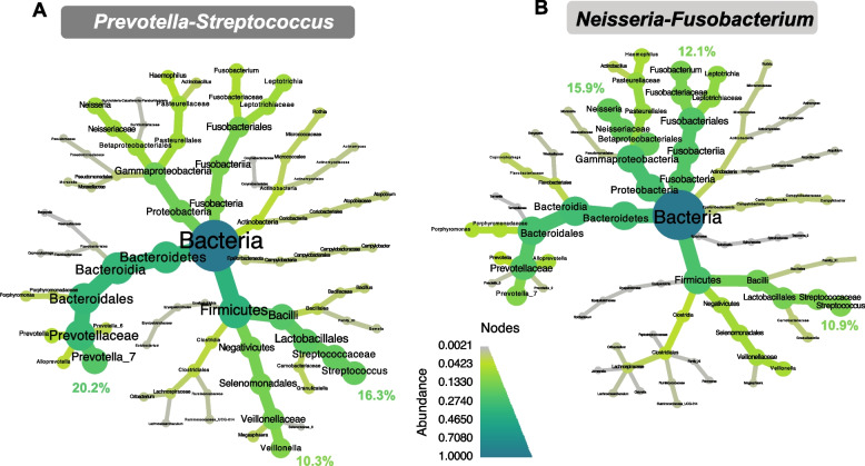
The two oropharyngeal microbiota clusters were named Prevotella-Streptococcus and Neisseria-Fusobacterium. The Prevotella-Streptococcus cluster accounted for 83.9% (n = 151) of all samples from the oropharynx. The three most abundant genera in this cluster were Prevotella (20.2%), Streptococcus (16.3%), and Veillonella (10.3%) (Fig. 6A). The Neisseria-Fusobacterium cluster accounted for 16.1% (n = 29) of all samples, and the three most abundant genera were Neisseria (15.9%), Fusobacterium (12.1%), and Streptococcus (10.9%) (Fig. 6B). Both clusters had comparable diversity (Fig. S4B) and a high abundance of ASV2, classified as presumptive Streptococcus spp., which included, among other species, presumptive S. pneumoniae (supplementary information “Bacterial profiles in the oropharynx and nasopharynx”).
Fig. 6.
Oropharyngeal microbiota clusters. Taxonomic heat trees for the two clusters identified. A Prevotella-Streptococcus (83.9% of the total samples) and B Neisseria-Fusobacterium (16.1%). The center of each tree represents the kingdom and has a relative abundance of 1. In the extremities, the relative abundances at the genus level are represented. From the center to the extremities, each taxonomic level from kingdom to genus is indicated. The gradient of colors represents relative abundance. In each cluster, the relative abundance of the three most common genera is specified
Association between oropharyngeal microbiota profiles and variables under study
Mixed general linear models were used to investigate potential associations between the oropharyngeal microbiota profiles and the pneumococcal carrier status of the oropharynx and sociodemographic and environmental characteristics. The cluster Prevotella-Streptococcus was used as a reference since this cluster accounted for the highest number of samples (Table 4). Pneumococcal carriers were 3.6-fold (95% CI 1.3–12.1) more likely to have their oropharyngeal microbiota described by cluster Neisseria-Fusobacterium compared to Prevotella-Streptococcus, whereas smokers were 86% less likely to have their oropharyngeal microbiota described by cluster Neisseria-Fusobacterium (95% CI 0.04–0.4).
Table 4.
Association between oropharyngeal microbiota profiles and variables under study
| Characteristics | Microbiota profiles | ||
|---|---|---|---|
|
Prevotella-Streptococcus N = 142 |
Neisseria-Fusobacterium N = 29 |
||
| n (%) | n (%) | ORadj (95% CI) | |
| Pneumococcal carrier, yes | 24 (16.9) | 11 (37.9) | 3.6 (1.3–12.1) |
| Age, > 37 years | 73 (51.4) | 7 (24.1) | 0.4 (0.1–1.1) |
| Gender, male | 66 (46.5) | 14 (48.4) | 2.1 (0.4–3.0) |
| Children, yes | 70 (49.3) | 12 (41.4) | 0.6 (0.2–1.9) |
| Smoker, yes | 67 (47.2) | 4 (13.7) | 0.14 (0.04–0.4) |
| Season | |||
| Spring | 40 (28.2) | 6 (20.7) | Ref |
| Summer | 40 (28.2) | 8 (27.6) | 1.56 (0.5–5.6) |
| Autumn | 23 (16.2) | 5 (17.2) | 1.93 (0.4–9.0) |
| Winter | 39 (27.4) | 10 (34.5) | 1.23 (0.4–4.4) |
Ref, variable used as reference. Bold indicates statistically significant results. The Prevotella-Streptococcus profile was the most frequent and thus was used as a reference against which the other profile was compared
Oropharyngeal profiles of subpopulations of pneumococcal carriers, adults who have close contact with children, and smokers
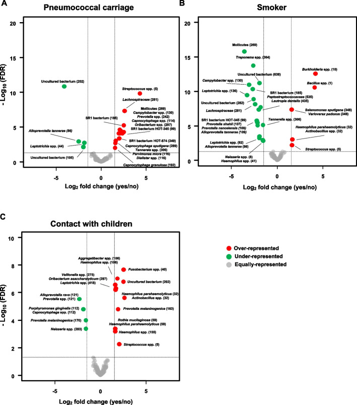
Significant differences between the oropharyngeal microbiota of pneumococcal carriers and nonpneumococcal carriers were found using PERMANOVA (P = 0.002). Eleven ASVs were overrepresented among pneumococcal carriers (Fig. 7A, Table S5), with ASV5 (log2FC = 4.31) and ASV281 (log2FC = 2.63) showing the highest FC. The latter was identified as Lachnospiraceae. On the other hand, ASV44 (log2FC = − 1.85) and ASV86 (log2FC = − 2.49) were underrepresented (Fig. 7A, Table S5). These were identified as presumptive Leptotrichia spp. and Alloprevotella tannerae, respectively.
Fig. 7.
Volcano plots representing ASVs that showed differential abundance in the oropharynx. A Effect of pneumococcal carrier status. B Effect of smoking status. C Effect of having contact with children. Bacterial taxa overrepresented among pneumococcal carriers, smokers, and adults who have regular contact with children are represented by red circles on the right side of each corresponding plot. Bacterial taxa underrepresented among pneumococcal carriers, smokers, and adults who have regular contact with children are represented by green circles on the left side of each corresponding plot. Gray circles indicate bacterial taxa that were not differentially abundant
Differences between the oropharyngeal microbiota of smokers and nonsmokers were also observed (P = 0.001). Five ASVs were overrepresented among smokers (Fig. 7B, Table S6). Among these, the ones with the highest FC were ASV1 (log2FC = 4.08) and ASV10 (log2FC = 4.17). Fifteen ASVs were underrepresented among smokers. Of these, ASV130 (log2FC = − 3.06), ASV136 (log2FC = − 3.30), and ASV269 (log2FC = − 3.59) showed the lowest FCs and were identified as presumptive Campylobacter showae or Campylobacter rectus, Leptotrichia spp., and Mollicutes, respectively.
Finally, differences between the oropharyngeal microbiota of individuals who had regular contact with children compared to those who did not (P = 0.004) were observed. Ten ASVs were overrepresented in individuals who had contact with children (Fig. 7C, Table S7), with ASV32 (log2FC = 2.62) and ASV40 (log2FC = 2.54) showing the highest FC. On the other hand, four ASVs were underrepresented (Fig. 7C, Table S7): ASV112 (log2FC = − 2.32), ASV121 (log2FC = − 1.87), ASV170 (log2FC = − 1.60), and ASV283 (log2FC = − 1.60). These were identified as presumptive Porphyromonas gingivalis or Capnocytophaga spp., Alloprevotella rava or Prevotella spp., Prevotella melaninogenica, and Neisseria spp., respectively.
Diversity of oropharyngeal profiles of subpopulations of pneumococcal carriers, adults who have close contact with children, and smokers
Diversity at the genus level was not significantly different when the oropharyngeal microbiota of pneumococcal carriers (0D = 47.3, 1D = 11.7, 2D = 7.4) and noncarriers (0D = 44.9, 1D = 11.2, 2D = 7.1) were compared (Fig. 5A and Fig. S7A). There was also no difference (P = 0.129) between the average proportion of reads belonging to the genus Streptococcus found in the oropharynx of carriers (19.8%) vs. noncarriers (14.4%) (Fig. 5F − G and Fig. S6).
In contrast, the oropharynx of smokers (0D = 43.5, 1D = 10.2, 2D = 6.3) was significantly less diverse than the oropharynx of nonsmokers (0D = 46.7, 1D = 12.2, 2D = 7.8) when comparing each diversity number (P = 0.095, P = 0.010, and P = 0.010, respectively) (Fig. 5B and Fig. S7B). Finally, no significant differences were observed between the oropharyngeal microbiota of individuals who had regular contact with children (0D = 45.1, 1D = 11.3, 2D = 7.0) and those who did not (0D = 45.6, 1D = 11.3, 2D = 7.2) (Fig. 5C and Fig. S7C).
Dynamics of microbiota carriage
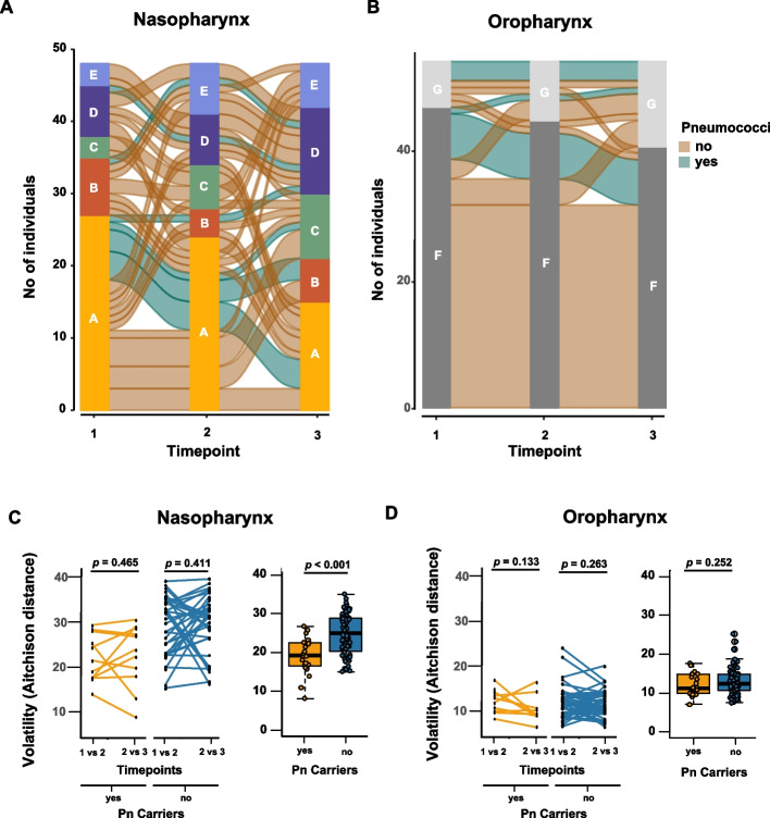
While 71.2% of the individuals maintained the same oropharyngeal cluster across the three time points, only 15.7% maintained the same nasopharyngeal cluster (chi-square test, P < 0.001) (Fig. 8A − B). On average, there was a higher volatility in the nasopharynx than in the oropharynx (Fig. 8C − D). In addition, in the nasopharynx, individuals carrying pneumococci were more likely to maintain the same nasopharyngeal cluster than those not carrying pneumococci (41.6% vs. 8.8%, chi-square test, P = 0.015), suggesting a higher stability in the former case (Fig. 8A). This result was supported by a lower volatility of the nasopharyngeal microbiota of pneumococcal carriers vs. noncarriers (Wilcoxon rank-sum test, P < 0.001) (Fig. 8C). In the oropharynx, this was not observed (Fig. 8D). Other factors, such as having contact with children, being a smoker, gender, age, and season, did not impact the dynamics of carriage.
Fig. 8.
Dynamics of the nasopharyngeal and oropharyngeal microbiota. A Alluvial plot representing the change in individuals’ nasopharyngeal clusters, stratified by carriage of pneumococcus, at the three sampling times. B Alluvial plot representing the change in individuals’ oropharyngeal clusters, stratified by carriage of pneumococcus, at the three sampling times. C Volatility of the nasopharyngeal microbiota depending on pneumococcal carrier state. D Volatility of the oropharyngeal microbiota depending on pneumococcal carrier state. In C and D, left graphics show volatility per individual calculated as the Aitchison distance between microbiota at consecutive time points: first vs. second and second vs. third. Lines connect volatility values of each individual. In C and D, the graphics on the right show boxplots of aggregated volatility. Yellow indicates pneumococcal carriers; blue indicates pneumococcal noncarriers
Discussion
Few metagenomic studies focusing on adults have been published, with the majority about microbiota dysbiosis in relation to disease [15, 35, 36]. Here, we took advantage of a longitudinal study conducted among immunocompetent healthy adults aged between 25 and 50 years old [9] to study the nasopharyngeal and oropharyngeal microbiota. In addition, we also compared the nasopharynx and oropharynx microbiota based on S. pneumoniae carrier status, smoking habits, and regular contact with children.
We found several differences between the microbiota of the nasopharynx vs. the oropharynx. We observed a higher bacterial load in oropharyngeal samples and a more homogeneous microbiota across individuals with just two clusters compared to five clusters identified in the nasopharynx. These observations are in line with a study that shows that the oropharynx has a high bacterial load, and that it varies little between individuals [37].
The oropharyngeal microbiota and the nasopharynx revealed continuity and niche-specific characteristics: the bacteria thriving in the oropharynx were obligatory anaerobes (e.g., Prevotellaceae, Veillonellaceae, or Leptotrichiaceae), whereas the bacteria thriving in the nasopharynx were mostly facultative anaerobes (e.g., Moraxellaceae and Corynebacteriaceae). Streptococcaceae, on the other hand, were common in both sites.
In the five nasopharyngeal microbiota clusters, the genus Streptococcus was one of the most abundant genera in three of these clusters, and the genus Corynebacterium was the most abundant in the remaining two clusters. Of note, although these genera showed high abundances, they were never equally abundant in the same cluster. This result agrees with the observation of antagonistic relationships between species of these genera. For example, it has been shown that C. accolens is able to produce lipases and modify triacylglycerols present in the human skin, including the human nostrils, into free fatty acids, thus inhibiting the growth of S. pneumoniae [38]. A healthy nasopharyngeal microbiota has been frequently associated with Corynebacterium, Dolosigranulum, and/or Moraxella-dominated profiles [39–41], which coincides with two of the identified clusters in this study: cluster Corynebacterium-Moraxella and cluster Pseudomonas-Corynebacterium, which, together, were observed in 26.5% of the samples identified in the nasopharyngeal microbiota clusters. Nonetheless, in this study, the majority (73.5%) of nasopharyngeal microbiota samples were represented by clusters codominated by Streptococcus. As the participants in the study were healthy individuals, this suggests a broader range of clusters associated with a healthy state. Since season (winter or summer) was associated with two clusters codominated by Corynebacterium and Streptococcus, we hypothesized that nasopharyngeal clusters may be very dynamic and may shift between clusters codominated by different genera. An alternation between different nasopharyngeal microbiota profiles in children due to changes associated with seasonality was previously described for healthy youth and infants, which further supports our own observations [42, 43].
In the oropharynx, we found only two microbiota clusters. The most abundant genera comprised Prevotella, Streptococcus, Neisseria, and Fusobacterium, which have already been described in the healthy oropharyngeal microbiota of adults [1, 44].
We found that the microbiota composition of the nasopharynx and oropharynx could depend on population demographic characteristics (i.e., age and gender) and/or environmental factors (i.e., smoking habits, contact with children, and season). Indeed, we found that by comparing to a reference microbiota profile, there were two out of five nasopharyngeal microbiota profiles that could be associated with pneumococcal carrier state, smoking status, contact with children, sampling season, and gender. Regarding the oropharyngeal microbiota profiles, we found that the two clusters were associated with pneumococcal carrier status, smoking habits, and age. These results are in line with previous studies that also found that demographic characteristics and environmental factors can affect the microbiota of the upper respiratory tract [1, 42].
We observed that the nasopharyngeal microbiota among pneumococcal carriers had a lower evenness than that among nonpneumococcal carriers. This raises the possibility of a specialization of bacteria able to thrive in this niche.
The presence of S. pneumoniae in the oropharyngeal niche seems not to disrupt the normal microbiota since the microbiota of both carriers and noncarriers are very similar. As reported, the oropharynx showed higher bacterial diversity, and as an ecosystem, a higher diversity contributes to niche stability [45].
There are several reports of a synergistic relationship between pneumococcus and H. influenzae [46]. Both bacteria are part of the nasopharyngeal niche of healthy humans. However, these are also pathobionts that can cause several infections, such as bronchitis, pneumonia, otitis media, septicemia, and meningitis. To date, it is not yet known whether this interaction is strain and/or serotype-specific or their molecular mechanisms [47]. Cope et al. [48] showed that biofilms with both species had higher cell densities, and that these bacteria can modulate each other’s virulence gene expression, leading to a persistent biofilm. Aside from this interaction, Horiuchi et al. [49] also reported a synergistic interaction between P. micra and F. nucleatum. This type of interaction may explain the increased abundance of these specific ASVs in pneumococcal carriers.
On the other hand, the nasopharyngeal microbiota of the nonpneumococcal carriers also showed several bacterial taxa that were overrepresented or even unique in the nasopharyngeal microbiota. Among these were Neisseria spp. (ASV24, ASV57, and ASV131), S. aureus (ASV2970), and S. lugdunensis (ASV159), for example. Several reports have observed a negative relationship between pneumococcus and S. aureus and identified mechanisms possibly associated with it [50, 51]. Additionally, Brozyna et al. observed that S. aureus is able to enhance the growth of S. lugdunensis [52].
Along with Streptococcus (ASV5), we found other bacterial taxa associated with the oropharyngeal microbiota of pneumococcal carriers. These were either Lachnospiraceae (ASV281) or Mollicutes (ASV269), which are frequently identified as being part of a healthy microbiota [53]. However, Capnocytophaga (ASV114 and ASV192), Oribacterium (ASV297), and Tannerella (ASV306) were also detected. These genera are frequently associated with diseases such as periodontitis [54].
The oropharyngeal microbiota of nonpneumococcal carriers comprised a higher abundance of genera such as Alloprevotella (ASV86) and Leptotrichia (ASV44). The first was found in the human oral cavity [55], whereas the latter was found to be present in the oropharynx of healthy adults [13, 56].
We have also looked for differences in the upper respiratory tract microbiota based on two population characteristics that we know from our previous study [9] to be important for pneumococcal acquisition, namely, smoking habits and having contact with children. These characteristics were associated with differences in the microbiota in both niches. Although different, both the nasopharynx and oropharynx of smokers showed high abundances of Bacillus (ASV1 and ASV48) and Burkholderia (ASV10 and ASV63). These genera comprise a high range of human pathogenic species and have been frequently associated with environmental contamination. Nonetheless, in this study, they were spread across individuals and were most likely present due to the high proportion of individuals with smoking habits in our sample. In fact, both genera have been reported as being part of the bacterial metagenome of cigarettes, providing evidence that the source of these pathogenic bacteria may be the cigarettes themselves [57]. Furthermore, we also found that Rothia dentocariosa (ASV73), Prevotella melaninogenica (ASV3 and ASV18), and Veillonella atypica (ASV4) were present in high abundance in the nasopharyngeal microbiota and Selenomonas sputigena (ASV348) in the oropharyngeal microbiota of smokers. These are all bacterial taxa usually found to be associated with oral diseases such as caries [58–61], which may be expected in smokers [62].
Finally, the upper respiratory tract microbiota of adults with contact with children was found to be different from that of adults without contact with children. For example, Streptococcus (ASV5), M. catarrhalis (ASV7), and H. influenzae (ASV23) were overrepresented in the nasopharyngeal microbiota of individuals who have contact with children. Interestingly, these are the most common pathobionts known to colonize the upper respiratory tract of children [18, 46]. These bacteria are capable of causing infections such as bronchitis, otitis media, sinusitis, and pneumonia in both children and adults, although they are more frequent in the first age group [1]. Thus, colonization and, consequently, infection in young adults, albeit low, may be due to the transmission of these bacteria through contact with children.
The oropharyngeal microbiota of adults with and without contact with children, as expected, was found to be mostly represented by genera already described as part of this niche [39]. Examples of these genera were Fusobacterium (ASV40), Leptotrichia (ASV418), Haemophilus (ASV155 and ASV106), Veillonella (ASV275), Prevotella (ASV121, ASV170 and ASV163), and Neisseria (ASV283). Although we observed an increase in several pathobionts in the nasopharyngeal microbiota of individuals who have regular contact with children, regarding the oropharyngeal microbiota, this increase was only noticeable for Streptococcus (ASV5).
In terms of dynamics, our results are in agreement with others that showed that the microbiota of the oropharynx is stable [37]. In addition, we observed that the nasopharyngeal microbiota of adults is much less stable. However, pneumococcal carriers tend to have a more stable nasopharyngeal microbiota than noncarriers. This may be the result of S. pneumoniae dominance, reflected by the lower evenness in the nasopharyngeal community of S. pneumoniae carriers.
Our study has some limitations. First, the original study aimed to investigate the dynamics of carriage of S. pneumoniae in immunocompetent healthy adults; therefore, STGG medium was used to store the samples. Although this may not be the ideal medium for such studies, it has been successfully used and validated previously by others [24]. Second, we were unable to use STGG as a negative control in our analyses, as no aliquots from the original study had been stored. Nevertheless, several unsupervised methods have been used to remove possible contaminants. Also, the current comparison of the upper respiratory microbiota based on the S. pneumoniae carrier state was performed exclusively based on the previous identification of this bacterium by culture methods and/or qPCR [1]. Finally, to meet our established criteria for the selection of individuals, we only used 12 out of 25 pneumococcal carrier individuals identified in a previous study [9] (leading to 72 paired samples out of 224 nasopharyngeal samples and 240 oropharyngeal samples). However, when taking into account the total number of individuals and samples included in this study, it is currently one of the largest studies performed in healthy adults.
Our study also has some strengths. First, to the best of our knowledge, it is a microbiota study with one of the largest number of samples from the upper respiratory tract regarding immunocompetent healthy adults. Second, it is the first study that aimed to understand the impact of S. pneumoniae colonization on the microbiota of both the nasopharyngeal and oropharyngeal niches.
Conclusions
In conclusion, our study revealed notable differences between the nasopharyngeal and oropharyngeal microbiota, with the nasopharyngeal niche exhibiting lower diversity. The presence of S. pneumoniae in the nasopharyngeal niche led to a microbiota shift not observed at the genus level in the oropharyngeal niche. Moreover, we identified various bacterial taxa that differ in prevalence between pneumococcal carriers and noncarriers, indicating potential interactions influencing the microbiota composition. Although some of these interactions are known, there may be additional unidentified factors playing a crucial role. For instance, P. micra was present in both the nasopharyngeal and oropharyngeal microbiota of pneumococcal carriers, hinting at intricate relationships yet to be fully elucidated. Additionally, our study highlighted differences in the upper respiratory tract microbiota based on smoking status and contact with children. Smokers’ microbiota exhibited an excess of pathogenic bacteria often found in cigarette metagenomes and are associated with periodontal diseases. Adults with contact with children showed higher abundances of pathobionts frequently found in children, such as Streptococcus, H. influenzae, and M. catarrhalis.
In summary, our findings contribute to increase our understanding of how different factors shape the upper respiratory tract microbiota of adults opening the possibility of using such information to design strategies aimed to promote a healthy respiratory microbiota.
Supplementary Information
Additional file 1: Fig. S1. Quality plot of the reverse and forward read sequences resulting from FastQC. Forward sequences were trimmed at position (cycle) 240 and reverse sequences were trimmed at position 230 to reach a quality score (QS) of 30 at the 25th percentile. Fig. S2. Oropharynx and nasopharynx microbiota profiles. Barplot representing the relative abundances of the taxonomic level Class in the oropharynx and nasopharynx and of samples misclassified in each site. Fig. S3. Identification of balances between groups of taxa associated with discrimination of the oropharynx and nasopharynx. The components defining the selected balance are specified on top of the boxplot that represents the distribution of the balance score for each of the groups. On the right the Receiver Operator Curve (ROC) with its AUC value and the density curve for each group is shown. TPR indicates true positive rate and FPR indicates false positive rate. Fig. S4. Diversity profiles of nasopharyngeal and oropharyngeal clusters. Fig. S5. Diversity of nasopharyngeal microbiota, for each Hill number. A. Comparison between pneumococcal carriers and non-carriers. B. Comparison between smokers and non-smokers. C. Comparison between individuals who have contact with children with adults who do not have contact. Fig. S6. Ten most abundant genera in the nasopharynx and oropharynx depending on pneumococcal carrier state. Fig. S7. Alpha diversity for the oropharyngeal microbiota calculated with the Hill numbers. A according to their pneumococcal carriage status. B. smoking and C contact with children. Table S1. Differences in abundance at the taxonomic level of family between nasopharynx and oropharynx. Table S2. Bacteria (ASV) differentially present in the nasopharyngeal microbiota of pneumococcal carriers and non-carriers. Table S3. Bacteria (ASV) differentially present in the nasopharyngeal microbiota of smokers and non-smokers. Table S4. Bacteria (ASV) differentially present in the nasopharyngeal microbiota of individuals that have and do not have regular contact with children. Table S5. Bacteria (ASV) differentially present in the oropharyngeal microbiota of pneumococcal carriers and non-carriers. Table S6. Bacteria (ASV) differentially present in the oropharyngeal microbiota of smokers and non-smokers. Table S7. Bacteria (ASV) differentially present in the oropharyngeal microbiota of individuals that have and do not have regular contact with children.
Abbreviations
- ASV
Amplicon sequence variant
- AUC
Area under the ROC curve
- CI
Confidence interval
- CLR
Centered log10 ratio
- Ct
Cycle threshold
- SD
Standard deviation
- FC
Fold change
- FPR
False-positive rate
- OR
Odds ratio
- PERMANOVA
Permutational analysis of variance
- qPCR
Real-time polymerase chain reaction
- ROC
Receiver operating characteristic
- STGG
Skim milk, tryptone, glucose, and glycerin
- TPR
True-positive rate
- URT
Upper respiratory tract
- WHO
World Health Organization
- ZIGMM
Zero-inflated Gaussian mixed model
Authors’ contributions
ACP, JL, STA, MH and RSL conceived the study. STA and JL did DNA extraction and quantification. ACP and JL did metagenomic and data analysis. ACP, JL and RSL interpreted the results. ACP, JL and RSL wrote the manuscript. All authors read and critically revised the manuscript.
Funding
This work was supported by Fundação para a Ciência e a Tecnologia (FCT), I.P., through MOSTMICRO-ITQB R&D Unit (UIDB/04612/2020, UIDP/04612/2020) and LS4FUTURE Associated Laboratory (LA/P/0087/2020). JL and STA were supported by FCT grants UI/BD/153385/2022 and SFRH/BD/108380/2015, respectively. The funders had no role in the study design, data collection and analysis, decision to publish, or preparation of the manuscript.
Availability of data and materials
Metagenomic sequencing data as well as meta information were deposited at NCBI’s Sequence Read Archive under the BioProject accession number PRJNA962709.
Declarations
Ethics approval and consent to participate
The study was approved by the ethical committee of Instituto de Higiene e Medicina Tropical, Universidade Nova de Lisboa, and was registered at the National Commission of Data Protection (ref. 3803/2014). Signed informed consent was obtained from all participants; samples and questionnaires were processed anonymously.
Consent for publication
Not applicable.
Competing interests
The authors declare no competing interests.
Footnotes
Publisher’s Note
Springer Nature remains neutral with regard to jurisdictional claims in published maps and institutional affiliations.
A. Cristina Paulo and João Lança are co-first authors.
Contributor Information
A. Cristina Paulo, Email: acpaulo@itqb.unl.pt.
Raquel Sá-Leão, Email: rsaleao@itqb.unl.pt.
References
- 1.Man WH, de Steenhuijsen Piters WA, Bogaert D. The microbiota of the respiratory tract: gatekeeper to respiratory health. Nat Rev Microbiol. 2017;15(5):259–270. doi: 10.1038/nrmicro.2017.14. [DOI] [PMC free article] [PubMed] [Google Scholar]
- 2.Henriques-Normark B, Tuomanen EI. The pneumococcus: epidemiology, microbiology, and pathogenesis. Cold Spring Harb Perspect Med. 2013;3(7):a010215. doi: 10.1101/cshperspect.a010215. [DOI] [PMC free article] [PubMed] [Google Scholar]
- 3.Brooks LRK, Mias GI. Virulence and host immunity: aging, diagnostics, and prevention. Front Immunol. 2018;9:1366. doi: 10.3389/fimmu.2018.01366. [DOI] [PMC free article] [PubMed] [Google Scholar]
- 4.Siegel SJ, Weiser JN. Mechanisms of bacterial colonization of the respiratory tract. Annu Rev Microbiol. 2015;69:425–444. doi: 10.1146/annurev-micro091014-104209. [DOI] [PMC free article] [PubMed] [Google Scholar]
- 5.Nunes S, Félix S, Valente C, Simões AS, Tavares DA, Almeida ST, et al. The impact of private use of PCV7 in 2009 and 2010 on serotypes and antimicrobial resistance of Streptococcus pneumoniae carried by young children in Portugal: comparison with data obtained since 1996 generating a 15-year study prior to PCV13 introduction. Vaccine. 2016;34(14):1648–1656. doi: 10.1016/j.vaccine.2016.02.045. [DOI] [PubMed] [Google Scholar]
- 6.Félix S, Handem S, Nunes S, Paulo AC, Candeias C, Valente C, et al. Impact of private use of the 13-valent pneumococcal conjugate vaccine (PCV13) on pneumococcal carriage among Portuguese children living in urban and rural regions. Vaccine. 2021;39(32):4524–4533. doi: 10.1016/j.vaccine.2021.06.035. [DOI] [PubMed] [Google Scholar]
- 7.Wyllie AL, Rümke LW, Arp K, Bosch AATM, Bruin JP, Rots NY, et al. Molecular surveillance on Streptococcus pneumoniae carriage in nonelderly adults; little evidence for pneumococcal circulation independent from the reservoir in children. Sci Rep. 2016;6:34888. doi: 10.1038/srep34888. [DOI] [PMC free article] [PubMed] [Google Scholar]
- 8.Trzciński K, Bogaert D, Wyllie A, Chu ML, van der Ende A, Bruin JP, et al. Superiority of trans-oral over trans-nasal sampling in detecting Streptococcus pneumoniae colonization in adults. PLoS One. 2013;8(3):e60520. doi: 10.1371/journal.pone.0060520. [DOI] [PMC free article] [PubMed] [Google Scholar]
- 9.Almeida ST, Paulo AC, Froes F, de Lencastre H, Sá-Leão R. Dynamics of pneumococcal carriage in adults: a new look at an old paradigm. J Infect Dis. 2021;223(9):1590–1600. doi: 10.1093/infdis/jiaa558. [DOI] [PubMed] [Google Scholar]
- 10.Sá-Leão R, Nunes S, Brito-Avô A, Alves CR, Carriço JA, Saldanha J, et al. High rates of transmission of and colonization by Streptococcus pneumoniae and Haemophilus influenzae within a day care center revealed in a longitudinal study. J Clin Microbiol. 2008;46(1):225–234. doi: 10.1128/JCM.01551-07. [DOI] [PMC free article] [PubMed] [Google Scholar]
- 11.Almeida ST, Nunes S, Santos Paulo AC, Valadares I, Martins S, Breia F, et al. Low prevalence of pneumococcal carriage and high serotype and genotype diversity among adults over 60 years of age living in Portugal. PLoS One. 2014;9(3):90974. doi: 10.1371/journal.pone.0090974. [DOI] [PMC free article] [PubMed] [Google Scholar]
- 12.Greenberg D, Givon-Lavi N, Broides A, Blancovich I, Peled N, Dagan R. The contribution of smoking and exposure to tobacco smoke to Streptococcus pneumoniae and Haemophilus influenzae carriage in children and their mothers. Clin Infect Dis. 2006;42(7):897–903. doi: 10.1086/500935. [DOI] [PubMed] [Google Scholar]
- 13.Stearns JC, Davidson CJ, McKeon S, Whelan FJ, Fontes ME, Schryvers AB, et al. Culture and molecular-based profiles show shifts in bacterial communities of the upper respiratory tract that occur with age. ISME J. 2015;9(5):1268. doi: 10.1038/ismej.2015.49. [DOI] [PMC free article] [PubMed] [Google Scholar]
- 14.De Boeck I, Wittouck S, Wuyts S, Oerlemans EFM, van den Broek MFL, Vandenheuvel D, et al. Comparing the healthy nose and nasopharynx microbiota reveals continuity as well as niche-specificity. Front Microbiol. 2017;8:2372. doi: 10.3389/fmicb.2017.02372. [DOI] [PMC free article] [PubMed] [Google Scholar]
- 15.Smit LAM, Boender GJ, de Steenhuijsen Piters WAA, Hagenaars TJ, Huijskens EGW, Rossen JWA, et al. Increased risk of pneumonia in residents living near poultry farms: does the upper respiratory tract microbiota play a role? Pneumonia (Nathan) 2017;9:3. doi: 10.1186/s41479-017-0027-0. [DOI] [PMC free article] [PubMed] [Google Scholar]
- 16.van den Munckhof EHA, Hafkamp HC, de Kluijver J, Kuijper EJ, de Koning MNC, Quint WGV, et al. Nasal microbiota dominated by Moraxella spp. is associated with respiratory health in the elderly population: a case control study. Respir Res. 2020;21(1):181. doi: 10.1186/s12931-020-01443-8. [DOI] [PMC free article] [PubMed] [Google Scholar]
- 17.Yadav M, Chauhan NS. Microbiome therapeutics: exploring the present scenario and challenges. Gastroenterol Rep (Oxf) 2022;10:goab046. doi: 10.1093/gastro/goab046. [DOI] [PMC free article] [PubMed] [Google Scholar]
- 18.Bogaert D, Keijser B, Huse S, Rossen J, Veenhoven R, van Gils E, et al. Variability and diversity of nasopharyngeal microbiota in children: a metagenomic analysis. PLoS One. 2011;6(2):e17035. doi: 10.1371/journal.pone.0017035. [DOI] [PMC free article] [PubMed] [Google Scholar]
- 19.Walters W, Hyde ER, Berg-Lyons D, Ackermann G, Humphrey G, Parada A, et al. Improved bacterial 16S rRNA gene (V4 and V4–5) and fungal internal transcribed spacer marker gene primers for microbial community surveys. mSystems. 2016;1(1):e00009. doi: 10.1128/mSystems.00009-15. [DOI] [PMC free article] [PubMed] [Google Scholar]
- 20.Callahan BJ, McMurdie PJ, Rosen MJ, Han AW, Johnson AJ, Holmes SP. DADA2: high-resolution sample inference from Illumina amplicon data. Nat Methods. 2016;13(7):581–583. doi: 10.1038/nmeth.3869. [DOI] [PMC free article] [PubMed] [Google Scholar]
- 21.Team RC. R: A language and environment for statistical computing.. Vienna, Austria. URL https://www.R-project.org/: R Foundation for Statistical Computing; 2019.
- 22.Yilmaz P, Parfrey LW, Yarza P, Gerken J, Pruesse E, Quast C, et al. The SILVA and “All-species Living Tree Project (LTP)” taxonomic frameworks. Nucleic Acids Res. 2014;42(Database issue):D643–8. doi: 10.1093/nar/gkt1209. [DOI] [PMC free article] [PubMed] [Google Scholar]
- 23.Davis NM, Proctor DM, Holmes SP, Relman DA, Callahan BJ. Simple statistical identification and removal of contaminant sequences in marker-gene and metagenomics data. Microbiome. 2018;6(1):226. doi: 10.1186/s40168-018-0605-2. [DOI] [PMC free article] [PubMed] [Google Scholar]
- 24.Verhagen LM, Rivera-Olivero IA, Clerc M, Chu MLJN, van Engelsdorp GJ, Kristensen MI, et al. Nasopharyngeal microbiota profiles in rural Venezuelan children are associated with respiratory and gastrointestinal infections. Clin Infect Dis. 2020 doi: 10.1093/cid/ciaa015. [DOI] [PMC free article] [PubMed] [Google Scholar]
- 25.Ding T, Song T, Zhou B, Geber A, Ma Y, Zhang L, et al. Microbial composition of the human nasopharynx varies according to influenza virus type and vaccination status. mBio. 2019;10(4):e01296. doi: 10.1128/mBio.01296-19. [DOI] [PMC free article] [PubMed] [Google Scholar]
- 26.Gloor GB, Macklaim JM, Pawlowsky-Glahn V, Egozcue JJ. Microbiome datasets are compositional: and this is not optional. Front Microbiol. 2017;8:2224. doi: 10.3389/fmicb.2017.02224. [DOI] [PMC free article] [PubMed] [Google Scholar]
- 27.Lahti L, Shetty S. Tools for microbiome analysis in R. Version 2.1.26. http://microbiome.github.com/microbiome (2017). Accessed May 2020.
- 28.Aitchison J, Barceló-Vidal C, Martín-Fernández JA, Pawlowsky-Glahn V. Logratio analysis and compositional distance. Math Geol. 2000;32(3):271–275. doi: 10.1023/A:1007529726302. [DOI] [Google Scholar]
- 29.Tibshirani R, Walter G, Hastie T. Estimating the number of clusters in a dataset via the gap statistic. J R Statist Soc B. 2002;63:411–423. doi: 10.1111/1467-9868.00293. [DOI] [Google Scholar]
- 30.Alberdi A, Gilbert MTP. A guide to the application of Hill numbers to DNA-based diversity analyses. Mol Ecol Resour. 2019;19(4):804–817. doi: 10.1111/1755-0998.13014. [DOI] [PubMed] [Google Scholar]
- 31.Xia Y, Sun J, Chen D. Statistical analysis of microbiome data with R. Singapore: Springer; 2018. [Google Scholar]
- 32.Zhang X, Guo B, Yi N. Zero-inflated Gaussian mixed models for analyzing longitudinal microbiome data. PLoS One. 2020;15(11):e0242073. doi: 10.1371/journal.pone.0242073. [DOI] [PMC free article] [PubMed] [Google Scholar]
- 33.Rivera-Pinto J, Egozcue JJ, Pawlowsky-Glahn V, Paredes R, Noguera-Julian M, Calle ML. Balances: a new perspective for microbiome analysis. mSystems. 2018;3(4):e00053. doi: 10.1128/mSystems.00053-18. [DOI] [PMC free article] [PubMed] [Google Scholar]
- 34.Bastiaanssen TFS, Gururajan A, van de Wouw M, Moloney GM, Ritz NL, Long-Smith CM, et al. Volatility as a concept to understand the impact of stress on the microbiome. Psychoneuroendocrinology. 2021;124:105047. doi: 10.1016/j.psyneuen.2020.105047. [DOI] [PubMed] [Google Scholar]
- 35.Kaul D, Rathnasinghe R, Ferres M, Tan GS, Barrera A, Pickett BE, et al. Microbiome disturbance and resilience dynamics of the upper respiratory tract during influenza a virus infection. Nat Commun. 2020;11(1):2537. doi: 10.1038/s41467-020-16429-9. [DOI] [PMC free article] [PubMed] [Google Scholar]
- 36.de SteenhuijsenPiters WAA, Jochems SP, Mitsi E, Rylance J, Pojar S, Nikolaou E, et al. Interaction between the nasal microbiota and S. pneumoniae in the context of live-attenuated influenza vaccine. Nat Commun. 2019;10(1):2981. doi: 10.1038/s41467-019-10814-9. [DOI] [PMC free article] [PubMed] [Google Scholar]
- 37.Lemon KP, Klepac-Ceraj V, Schiffer HK, Brodie EL, Lynch SV, Kolter R. Comparative analyses of the bacterial microbiota of the human nostril and oropharynx. mbio. 2010;1(3):e00129. doi: 10.1128/mBio.00129-10. [DOI] [PMC free article] [PubMed] [Google Scholar]
- 38.Bomar L, Brugger SD, Yost BH, Davies SS, Lemon KP. Corynebacterium accolens releases antipneumococcal free fatty acids from human nostril and skin surface triacylglycerols. mBio. 2016;7(1):e01725–15. doi: 10.1128/mBio.01725-15. [DOI] [PMC free article] [PubMed] [Google Scholar]
- 39.de SteenhuijsenPiters WA, Sanders EA, Bogaert D. The role of the local microbial ecosystem in respiratory health and disease. Philos Trans R Soc Lond B Biol Sci. 2015;370(1675):20140294. doi: 10.1098/rstb.2014.0294. [DOI] [PMC free article] [PubMed] [Google Scholar]
- 40.Brugger SD, Eslami SM, Pettigrew MM, Escapa IF, Henke MT, Kong Y, et al. Dolosigranulum pigrum cooperation and competition in human nasal microbiota. mSphere. 2020;5(5):e00852. doi: 10.1128/mSphere.00852-20. [DOI] [PMC free article] [PubMed] [Google Scholar]
- 41.Brugger SD, Bomar L, Lemon KP. Commensal-pathogen interactions along the human nasal passages. PLoS Pathog. 2016;12(7):e1005633. doi: 10.1371/journal.ppat.1005633. [DOI] [PMC free article] [PubMed] [Google Scholar]
- 42.Zhao H, Chen S, Yang F, Wu H, Ba Y, Cui L, et al. Alternation of nasopharyngeal microbiota in healthy youth is associated with environmental factors: implication for respiratory diseases. Int J Environ Health Res. 2022;32(5):952–962. doi: 10.1080/09603123.2020.1810209. [DOI] [PubMed] [Google Scholar]
- 43.Mika M, Mack I, Korten I, Qi W, Aebi S, Frey U, et al. Dynamics of the nasal microbiota in infancy: a prospective cohort study. J Allergy Clin Immunol. 2015;135(4):905–12.e11. doi: 10.1016/j.jaci.2014.12.1909. [DOI] [PubMed] [Google Scholar]
- 44.Charlson ES, Chen J, Custers-Allen R, Bittinger K, Li H, Sinha R, et al. Disordered microbial communities in the upper respiratory tract of cigarette smokers. PLoS One. 2010;5(12):e15216. doi: 10.1371/journal.pone.0015216. [DOI] [PMC free article] [PubMed] [Google Scholar]
- 45.Arese Lucini F, Morone F, Tomassone MS, Makse HA. Diversity increases the stability of ecosystems. PLoS One. 2020;15(4):e0228692. doi: 10.1371/journal.pone.0228692. [DOI] [PMC free article] [PubMed] [Google Scholar]
- 46.Chien YW, Vidal JE, Grijalva CG, Bozio C, Edwards KM, Williams JV, et al. Density interactions among Streptococcus pneumoniae, Haemophilus influenzae and Staphylococcus aureus in the nasopharynx of young Peruvian children. Pediatr Infect Dis J. 2013;32(1):72–77. doi: 10.1097/INF.0b013e318270d850. [DOI] [PMC free article] [PubMed] [Google Scholar]
- 47.Tikhomirova A, Kidd SP. Haemophilus influenzae and Streptococcus pneumoniae: living together in a biofilm. Pathog Dis. 2013;69(2):114–126. doi: 10.1111/2049-632X.12073. [DOI] [PubMed] [Google Scholar]
- 48.Cope EK, Goldstein-Daruech N, Kofonow JM, Christensen L, McDermott B, Monroy F, et al. Regulation of virulence gene expression resulting from Streptococcus pneumoniae and nontypeable Haemophilus influenzae interactions in chronic disease. PLoS One. 2011;6(12):e28523. doi: 10.1371/journal.pone.0028523. [DOI] [PMC free article] [PubMed] [Google Scholar]
- 49.Horiuchi A, Kokubu E, Warita T, Ishihara K. Synergistic biofilm formation by Parvimonas micra and Fusobacterium nucleatum. Anaerobe. 2020;62:102100. doi: 10.1016/j.anaerobe.2019.102100. [DOI] [PubMed] [Google Scholar]
- 50.Regev-Yochay G, Lipsitch M, Basset A, Rubinstein E, Dagan R, Raz M, et al. The pneumococcal pilus predicts the absence of Staphylococcus aureus cocolonization in pneumococcal carriers. Clin Infect Dis. 2009;48(6):760–763. doi: 10.1086/597040. [DOI] [PMC free article] [PubMed] [Google Scholar]
- 51.McNally LM, Jeena PM, Gajee K, Sturm AW, Tomkins AM, Coovadia HM, et al. Lack of association between the nasopharyngeal carriage of Streptococcus pneumoniae and Staphylococcus aureus in HIV-1-infected South African children. J Infect Dis. 2006;194(3):385–390. doi: 10.1086/505076. [DOI] [PubMed] [Google Scholar]
- 52.Brozyna JR, Sheldon JR, Heinrichs DE. Growth promotion of the opportunistic human pathogen, Staphylococcus lugdunensis, by heme, hemoglobin, and coculture with Staphylococcus aureus. Microbiologyopen. 2014;3(2):182–195. doi: 10.1002/mbo3.162. [DOI] [PMC free article] [PubMed] [Google Scholar]
- 53.Chen WP, Chang SH, Tang CY, Liou ML, Tsai SJ, Lin YL. Composition analysis and feature selection of the oral microbiota associated with periodontal ddsease. Biomed Res Int. 2018;2018:3130607. doi: 10.1155/2018/3130607. [DOI] [PMC free article] [PubMed] [Google Scholar]
- 54.Wu X, Chen J, Xu M, Zhu D, Wang X, Chen Y, et al. 16S rDNA analysis of periodontal plaque in chronic obstructive pulmonary disease and periodontitis patients. J Oral Microbiol. 2017;9(1):1324725. doi: 10.1080/20002297.2017.1324725. [DOI] [PMC free article] [PubMed] [Google Scholar]
- 55.Downes J, Dewhirst FE, Tanner ACR, Wade WG. Description of Alloprevotella rava gen. nov., sp. nov., isolated from the human oral cavity, and reclassification of Prevotella tannerae Moore et al. 1994 as Alloprevotella tannerae gen. nov., comb. nov. Int J Syst Evol Microbiol. 2013;63(4):1214–8. doi: 10.1099/ijs.0.041376-0. [DOI] [PMC free article] [PubMed] [Google Scholar]
- 56.Eribe ERK, Olsen I. Leptotrichia species in human infections II. J Oral Microbiol. 2017;9(1):1368848. doi: 10.1080/20002297.2017.1368848. [DOI] [PMC free article] [PubMed] [Google Scholar]
- 57.Sapkota AR, Berger S, Vogel TM. Human pathogens abundant in the bacterial metagenome of cigarettes. Environ Health Perspect. 2010;118(3):351–356. doi: 10.1289/ehp.0901201. [DOI] [PMC free article] [PubMed] [Google Scholar]
- 58.Karabudak S, Ari O, Durmaz B, Dal T, Basyigit T, Kalcioglu MT, et al. Analysis of the effect of smoking on the buccal microbiome using next-generation sequencing technology. J Med Microbiol. 2019;68(8):1148–1158. doi: 10.1099/jmm.0.001003. [DOI] [PubMed] [Google Scholar]
- 59.Tanaka S, Yoshida M, Murakami Y, Ogiwara T, Shoji M, Kobayashi S, et al. The relationship of Prevotella intermedia, Prevotella nigrescens and Prevotella melaninogenica in the supragingival plaque of children, caries and oral malodor. J Clin Pediatr Dent. 2008;32(3):195–200. doi: 10.17796/jcpd.32.3.vp657177815618l1. [DOI] [PubMed] [Google Scholar]
- 60.Nagpal D, Prakash S, Bhat KG, Singh G. Detection and comparison of Selenomonas sputigena in subgingival biofilms in chronic and aggressive periodontitis patients. J Indian Soc Periodontol. 2016;20(3):286–291. doi: 10.4103/0972-124X.181247. [DOI] [PMC free article] [PubMed] [Google Scholar]
- 61.Khan ST, Ahamed M, Musarrat J, Al-Khedhairy AA. Anti-biofilm and antibacterial activities of zinc oxide nanoparticles against the oral opportunistic pathogens Rothia dentocariosa and Rothia mucilaginosa. Eur J Oral Sci. 2014;122(6):397–403. doi: 10.1111/eos.12152. [DOI] [PubMed] [Google Scholar]
- 62.Jiang X, Wang Y, Huang R. Correlation between tobacco smoking and dental caries: a systematic review and meta-analysis. Tob Induc Dis. 2019;17:34. doi: 10.18332/tid/106117. [DOI] [PMC free article] [PubMed] [Google Scholar]
Associated Data
This section collects any data citations, data availability statements, or supplementary materials included in this article.
Supplementary Materials
Additional file 1: Fig. S1. Quality plot of the reverse and forward read sequences resulting from FastQC. Forward sequences were trimmed at position (cycle) 240 and reverse sequences were trimmed at position 230 to reach a quality score (QS) of 30 at the 25th percentile. Fig. S2. Oropharynx and nasopharynx microbiota profiles. Barplot representing the relative abundances of the taxonomic level Class in the oropharynx and nasopharynx and of samples misclassified in each site. Fig. S3. Identification of balances between groups of taxa associated with discrimination of the oropharynx and nasopharynx. The components defining the selected balance are specified on top of the boxplot that represents the distribution of the balance score for each of the groups. On the right the Receiver Operator Curve (ROC) with its AUC value and the density curve for each group is shown. TPR indicates true positive rate and FPR indicates false positive rate. Fig. S4. Diversity profiles of nasopharyngeal and oropharyngeal clusters. Fig. S5. Diversity of nasopharyngeal microbiota, for each Hill number. A. Comparison between pneumococcal carriers and non-carriers. B. Comparison between smokers and non-smokers. C. Comparison between individuals who have contact with children with adults who do not have contact. Fig. S6. Ten most abundant genera in the nasopharynx and oropharynx depending on pneumococcal carrier state. Fig. S7. Alpha diversity for the oropharyngeal microbiota calculated with the Hill numbers. A according to their pneumococcal carriage status. B. smoking and C contact with children. Table S1. Differences in abundance at the taxonomic level of family between nasopharynx and oropharynx. Table S2. Bacteria (ASV) differentially present in the nasopharyngeal microbiota of pneumococcal carriers and non-carriers. Table S3. Bacteria (ASV) differentially present in the nasopharyngeal microbiota of smokers and non-smokers. Table S4. Bacteria (ASV) differentially present in the nasopharyngeal microbiota of individuals that have and do not have regular contact with children. Table S5. Bacteria (ASV) differentially present in the oropharyngeal microbiota of pneumococcal carriers and non-carriers. Table S6. Bacteria (ASV) differentially present in the oropharyngeal microbiota of smokers and non-smokers. Table S7. Bacteria (ASV) differentially present in the oropharyngeal microbiota of individuals that have and do not have regular contact with children.
Data Availability Statement
Metagenomic sequencing data as well as meta information were deposited at NCBI’s Sequence Read Archive under the BioProject accession number PRJNA962709.


