Abstract
Background
Inflammatory bowel disease (IBD) is a global disease with a growing public health concern and is associated with a complex interplay of factors, including the microbiota and immune system. Resveratrol, a natural anti-inflammatory and antioxidant agent, is known to relieve IBD but the mechanism involved is largely unexplored.
Methods
This study examines the modulatory effect of resveratrol on intestinal immunity, microbiota, metabolites, and related functions and pathways in the BALB/c mice model of IBD. Mouse RAW264.7 macrophage cell line was used to further explore the involvement of the macrophage-arginine metabolism axis. The treatment outcome was assessed through qRT-PCR, western blot, immunofluorescence, immunohistochemistry, and fecal 16S rDNA sequencing and UHPLC/Q-TOF–MS.
Results
Results showed that resveratrol treatment significantly reduced disease activity index (DAI), retained mice weight, repaired colon and spleen tissues, upregulated IL-10 and the tight junction proteins Occludin and Claudin 1, and decreased pro-inflammatory cytokines IL-1β, IL-6, and TNF-α. Resveratrol reduced the number of dysregulated metabolites and improved the gut microbial community structure and diversity, including reversing changes in the phyla Bacteroidetes, Proteobacteria, and Firmicutes, increasing ‘beneficial’ genera, and decreasing potential pathogens such as Lachnoclostridium, Acinobacter, and Serratia. Arginine–proline metabolism was significantly different between the colitis-treated and untreated groups. In the colon mucosa and RAW264.7 macrophage, resveratrol regulated arginine metabolism towards colon protection by increasing Arg1 and Slc6a8 and decreasing iNOS.
Conclusion
This uncovers a previously unknown mechanism of resveratrol treatment in IBD and provides the microbiota-macrophage-arginine metabolism axis as a potential therapeutic target for intestinal inflammation.
Supplementary Information
The online version contains supplementary material available at 10.1186/s40001-023-01257-6.
Keywords: Inflammatory bowel disease, Resveratrol, Microbiota, Arginine metabolism, Macrophage
Background
Inflammatory bowel disease (IBD), which includes ulcerative colitis (UC) and Crohn’s disease (CD), is a global disease with growing public health concerns due to increasing incidence. In 2020, it was asserted that developing countries are in the emergence stage of IBD evolution, newly industrialized countries are in the Acceleration in Incidence stage, while Western countries are in the Compounding Prevalence stage [1]. Although the exact cause of IBD is unknown, it is believed to be associated with a complex interplay of microbiota, immune system, environmental, and genetic factors [2, 3]. IBD is linked with compositional and metabolic alterations in the intestinal microbiota (dysbiosis) coupled with immune dysregulation, where chronic inflammation shapes the gut microbiota and vice versa, contributing to the development and progression of IBD [4]. Therefore, the restoration of microbial composition and diversity, and their derived metabolites in the gut is a promising therapeutic approach in IBD.
Macrophages are the gatekeepers of intestinal immune homeostasis as they discriminate between innocuous antigens and potential pathogens to maintain tolerogenic immunity, thus, their impairment leads to chronic relapsing immune dysregulation and pathologies of the gastrointestinal tract, including IBD [5]. Large numbers of macrophages are found in the colon mucosa of IBD patients and animal models, participating in the initiation and resolution of inflammation. Macrophages are polarized into either pro-inflammatory M1 phenotypes via induction by interferon γ (IFN-γ), tumor necrosis factor-alpha (TNF-α), and bacterial lipopolysaccharide (LPS) to produce a wide range of proinflammatory cytokines such as interleukin (IL)-1β and IL-6, or anti-inflammatory M2 phonotypes via induction by IL-4 and IL-13 to express arginase 1 (Arg1) and anti-inflammatory cytokines, including IL-10 [6, 7]. In effect, M2 macrophages promote intestinal tissue repair and inflammation resolution and are found to also interact with the microbiota [8, 9]. The anti-inflammatory effect of macrophages is linked with arginine metabolism, as the two arginine catalytic enzymes, inducible nitric oxide synthase (iNOS) and Arg1, are well-characterized hallmark molecules of the classically and alternatively activated macrophages, respectively [10, 11]. In a related process, the depletion of intracellular creatine by downregulating the creatine transporter Slc6a8 (solute carrier family 6 member 8) alters macrophage-mediated immune responses since creatine inhibits iNOS by suppressing IFN-γ-JAK-STAT1 signaling and promotes IL-4-STAT6-activated Arg1 expression [11]. Because macrophages also express Arg1 and Slc6a8, a therapeutic substance that modulates arginine metabolism towards increases in Arg1 and Slc6a8, would be promising in the treatment of IBD.
As a potential therapy for IBD, resveratrol, a type of natural phenol and phytoalexin that acts against pathogens and possesses anti-inflammatory and antioxidant activity, has been widely studied [12, 13]. Resveratrol has been reported to relieve IBD in animal models by regulating immune responses and signaling pathways [14, 15] as well as the gut microbiome [16]. However, the mechanisms involved in these effects remain largely unknown. This study examines the treatment effect of resveratrol in the modulation of intestinal immune response, mucosal tissue repair, gut microbiota community structure and related functions, and metabolites and their associated pathways in the mice model of IBD. With the progress in next-generation sequencing technology, 16S rDNA of microbiota and UHPLC/Q-TOF–MS (ultra-high-performance liquid chromatography-quadrupole time-of-flight mass spectrometry) detection of metabolomic was used to identify various changes of the gut microbiota composition. The involvement of the macrophage-arginine metabolism axis was also examined.
Material and methods
Resveratrol
Resveratrol, a non-flavonoid polyphenol organic compound, is an antitoxin produced by many plants when stimulated. Its chemical name is 3,4',5-trihydroxy-1,2-diphenyl ethylene, and the chemical formula is C14H12O3. The resveratrol used in this study was purchased from Sigma, USA (CAS 501-36-0, PubChem Chemical No.24278055).
Animal model
Male BALB/c mice (6–8 weeks old, 20 ± 2 g) were purchased from the Animal Research Center of Jiangsu University (Zhenjiang, China). All mice were treated accordingly in the SPF laboratory. Mice were randomly divided into 3 groups (n = 5/group); and the negative control group (NC group), the Dextran sulfate sodium (DSS)-induced colitis group (DSS group), and the Resveratrol-treated colitis group (RSV group). The NC group was given autoclaved purified water, while the DSS and RSV groups were given autoclaved purified water containing 3% DSS. In addition, mice in the RSV group were given resveratrol solvent (100 mg/kg) by oral gavage every day. The mice were weighed daily and their fecal characteristics were recorded. When the mice in the DSS group showed obvious bloody stool and weight loss, the feces of the mice in each group were collected aseptically for subsequent 16S rDNA sequencing and UHPLC/Q-TOF–MS. On the 10th day, all mice were sacrificed and colon and spleen tissues were collected for subsequent experiments.
Cell culture
Mouse leukemic monocyte/macrophage cell line (RAW264.7) was purchased from Beiner Biotechnology Company (Beijing, China) and cultured in DMEM medium (Hyclone) containing 10% fetal calf serum (FBS; Excell,Uruguay) at 37 °C in humid air with 5% CO2. The RAW264.7 was cultured in an LPS-induced inflammatory environment and treated with or without resveratrol (80 nmol/ml) for 12 h.
Western-blot
RIPA (Radio-Immunoprecipitation Assay) lysis buffer was added to colon tissue and RAW264.7 cells and the protein concentration was measured by the BCA (Bicinchoninic acid) method. A total of 200 μg of protein sample was separated by 10% SDS-PAGE. The isolated proteins were transferred onto PVDF membranes, blocked in 5% skim milk powder (dissolved in TBST buffer) for 1 h. PVDF membranes were incubated with primary antibodies (PCNA-1:1000, Abcam;Occludin-1:5000, Proteintech; Claudin1-1:1000, Proteintech; iNOS-1:500, Proteintech; Arg1-1:1000, Proteintech; Slc6a8-1:400, Proteintech) at 4 °C overnight, and then incubated with secondary antibodies at room temperature for 1 h to visualize protein bands and generate images.
Quantitative Real-time (qRT)-PCR
Total RNA was extracted from mouse colons and RAW264.7 cells by chloroform extraction using Trizol reagent (Vazyme, Nanjing, China). cDNA was synthesized using the HiScript 1st Strand cDNA Synthesis Kit (Vazyme, Nanjing, China). β-actin was used as an internal control to detect the expression of target genes. Primer sequences used are shown in Table 1.
Table 1.
Sequence of primers used for qRT-PCR
| Primer name | Sequence (5' to 3') |
|---|---|
| mouse-IL-1β-F | CACTACAGGCTCCGAGATGAACAAC |
| mouse-IL-1β-R | TGTCGTTGCTTGGTTCTCCTTGTAC |
| mouse-IL6-F | CTCCCAACAGACCTGTCTATAC |
| mouse-IL6-R | CCATTGCACAACTCTTTTCTCA |
| mouse-TNF-α-F | ATGTCTCAGCCTCTTCTCATTC |
| mouse-TNF-α-R | GCTTGTCACTCGAATTTTGAGA |
| mouse-IL10-F | TTCTTTCAAACAAAGGACCAGC |
| mouse-IL10-R | GCAACCCAAGTAACCCTTAAAG |
| mouse iNOS-F | ATCTTGGAGCGAGTTGTGGATTGTC |
| mouse iNOS-R | TAGGTGAGGGCTTGGCTGAGTG |
| mouse Arg1-F | AATCTGGTTGTGTATCCTCGTT |
| mouse Arg1-R | AGAGGTGTATTAATGTCCGCAT |
| mouse Slc6a8-F | GCCCTACCTCCTCTCCTTCTTTCC |
| mouse Slc6a8-R | TTTCCCTCCTCTCCTGTTACCCAAG |
Immunohistochemistry (IHC)
Paraffin-embedded colon tissue Sections (4 μm thick) were processed for hematoxylin and eosin (H&E) staining or deparaffinized for immunohistochemistry (IHC). In IHC, sections were dewaxed and placed in 3% hydrogen peroxide solution, followed by incubation at room temperature for 30 min before thermal repair of antigens through boiling for 30 min in citrate buffer. After blocking with 5% BSA for 30 min, the primary antibody (Slc6a8- 1:200, Proteintech) was incubated at 4 °C overnight, followed by the secondary antibody (Wuhan Boster Biological Technology, Wuhan, China) at room temperature for 1 h. StreptAvidin Biotin Complex (SABC) was added and incubated at 37 °C for 30 min. Finally, diaminobenzidine substrate (DAB) was applied to sections and counter-stained with hematoxylin for microscopic examination.
Immunofluorescence
The sections were dewaxed to block nonspecific antigens as described in IHC. Sections were incubated with iNOS antibody (1:200) and Arg1 antibody (1:300) at 4° overnight, and then incubated with the corresponding fluorescent secondary antibody for 2 h at room temperature. The specimens were stained with hoechest33342 (1:300; Sigma–Aldrich) for 10 min at room temperature and observed under a microscope. During the experiment, exposure to light was avoided to prevent fluorescence quenching.
16S rDNA gene sequencing
The experimental process for the fecal analysis of the microbiota community and function prediction involved extraction of genome DNA, amplicon generation, PCR products quantification and qualification, product mixing and purification, and library preparation and sequencing. The sample quality was strictly controlled in each link from DNA extraction to computer sequencing to ensure the authenticity of sequencing data. For the combined analysis of 16S metabolomics, the process involved 16S rDNA amplicon sequencing of significantly different microbiota and significantly different metabolites, followed by various correlation analyses. To confirm differences in the abundance of individual taxonomy between the groups, STAMP software was utilized. LEfSe was used for the quantitative analysis of biomarkers within different groups. To identify differences in microbial communities between the groups, ANOSIM and ADONIS were performed based on the Bray–Curtis dissimilarity distance matrices.
LC–MS/MS Analysis
Analyses were performed using a UHPLC (1290 Infinity LC, Agilent Technologies) coupled to a quadrupole time-of-flight (AB SciexTripleTOF 6600) in Shanghai Applied Protein Technology Co., Ltd.
Statistics Analysis
All data were expressed as mean ± SD. Comparisons between multiple groups were assessed by student t-test. P < 0.05 was considered statistically significant.
Results
Resveratrol relieves the features of colitis in mice
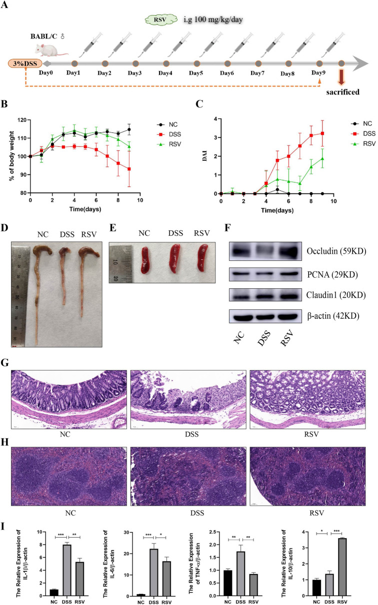
The male BALB/c mice were randomly divided into 3 groups (n = 5/group); the negative control (NC) group, the DSS-induced colitis (DSS) group, and the Resveratrol-treated colitis (RSV) group. After the induction of colitis in the DSS and RSV groups, the RSV group was treated with 100 mg/kg resveratrol solvent by gavage every day (Fig. 1A). Weight assessment indicated that while the DSS group decreased in average weight, the RSV group significantly retained weight (Fig. 1B). The disease activity index (DAI) of the RSV group was reduced compared to the DSS group (Fig. 1C). Moreover, resveratrol treatment significantly restored the gross morphological appearance of mice colon and spleen tissues (Fig. 1D, E), as further confirmed with H&E staining (Fig. 1G, H) indicating reduced damage to intestinal villus structure in the RSV group relative to the DSS group. To examine whether these tissue and cellular changes translated into molecular level changes, we measured the expression level of the tight junction proteins Occludin and Claudin 1, as well as proliferating cell nuclear antigen (PCNA), an essential molecule in nucleic acid metabolism as a component of the replication and repair machinery. Results showed that resveratrol treatment increased the protein expression of Occludin, Claudin 1, and PCNA in mice colon compared to the untreated group (Fig. 1F). Cytokine analysis via qRT-PCR also showed increased anti-inflammatory cytokine IL-10 but decreased pro-inflammatory cytokines IL-1β, IL-6, and TNFα in the RSV group compared to the DSS group (Fig. 1I). These observations indicate the ability of resveratrol to reduce intestinal mucosal inflammation, repair tissue damage, and restore tight junction molecules, thus relieving the microscopic and macroscopic features of colitis in mice.
Fig. 1.
Resveratrol relieves the features of colitis in mice. A Schematic diagram of animal model construction. B Percentage of weight loss in each group during modeling. C Disease Activity Index score. D: General view of the colorectum. E General view of spleen. F Western blot detection of PCNA、Occludin、Claudin1 expression in colon tissues. G HE staining of colon tissue (200 ×). H HE staining of spleen tissue (100 ×). I qRT-PCR detection of cytokines expression in colon tissues. * represents p < 0.05, ** represents p < 0.01 and *** represents p < 0.001
Resveratrol improves the general community structure and diversity of gut microbiota
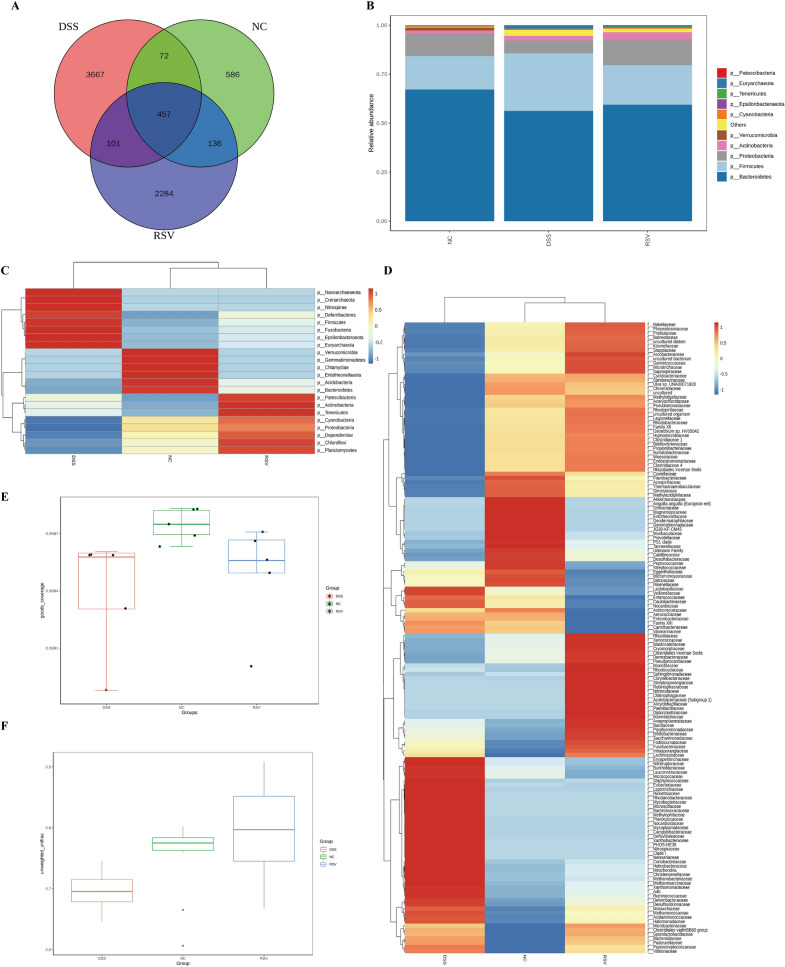
The effect of the treatment on the microbiota structure and diversity was examined through 16S rDNA gene sequencing of mice fecal samples. Quality control checks revealed adequacy of sample size and species richness, adequate sequencing data to sufficiently reflect the microbial information in the samples, and uniform distribution of species (Additional file 1: Fig. S1F–H). Venn analysis of OTUs showed that compared to the NC group (586 OTUs), the DSS group had increased OTU abundance (3667 OTUs), which was reduced in the RSV group (2284 OTUs). Moreover, the RSV group shared more common OTUs with the control group compared to the DSS group (136 vs 72) (Fig. 2A). Previous studies indicate that the phyla Firmicutes, Bacteroidetes, Proteobacteria, and Actinobacteria form approximately 99% of the microbiota, with Bacteroidetes and Firmicutes contributing about 90% [17, 18], thus, the Bacteroidetes/Firmicutes ratio is crucial in maintaining normal intestinal homeostasis. Our results showed that while the DSS group had reduced the abundance of Bacteroidetes and Proteobacteria and increased Firmicutes, resveratrol reversed these changes (Fig. 2B). The abundance of other phyla such as Nanoarchaeaeota, Crenarchaeota, Nitrospirae, Deferribacteres, Fusobacteria, Epsilonbacteraeota, and Euryarchaeota was also significantly restored by resveratrol (Fig. 2C). The cluster heatmap of abundant families further reveals the restoration effect of resveratrol treatment on the microbiota community structure as many clusters in the RSV group closely resemble the NC group compared to the DSS group (Fig. 2D). There was reduced alpha-diversity (Fig. 2E) in the DSS group (DSS vs NC p-value = 0.008) but a non-significant difference between the RSV and NC groups (p-value = 0.056). In addition, while there was a significant beta-diversity difference (Fig. 2F) between the DSS and NC groups (p-value = 0.026), there was a non-significant difference between the RSV and NC groups (p-value = 0.202). These findings show the ability of resveratrol to improve the colitis-induced microbiota community changes and significantly restore the alpha and beta diversity.
Fig. 2.
Resveratrol improves gut microbial general community structure and diversity. A Venn diagram of OTUs between the DSS group, the RSV group, and the control. B Community abundance of the top 10 phyla within the groups. C Cluster heatmap of species abundance at the family level within the groups. D Cluster heatmap of species abundance at the phyla level within the groups. E Goods coverage box chart of the difference between α diversity index of the groups. F Group difference analysis of β diversity based on Unweighted Unifrac distance
Resveratrol modulates specific bacteria species and restores functional dysregulation
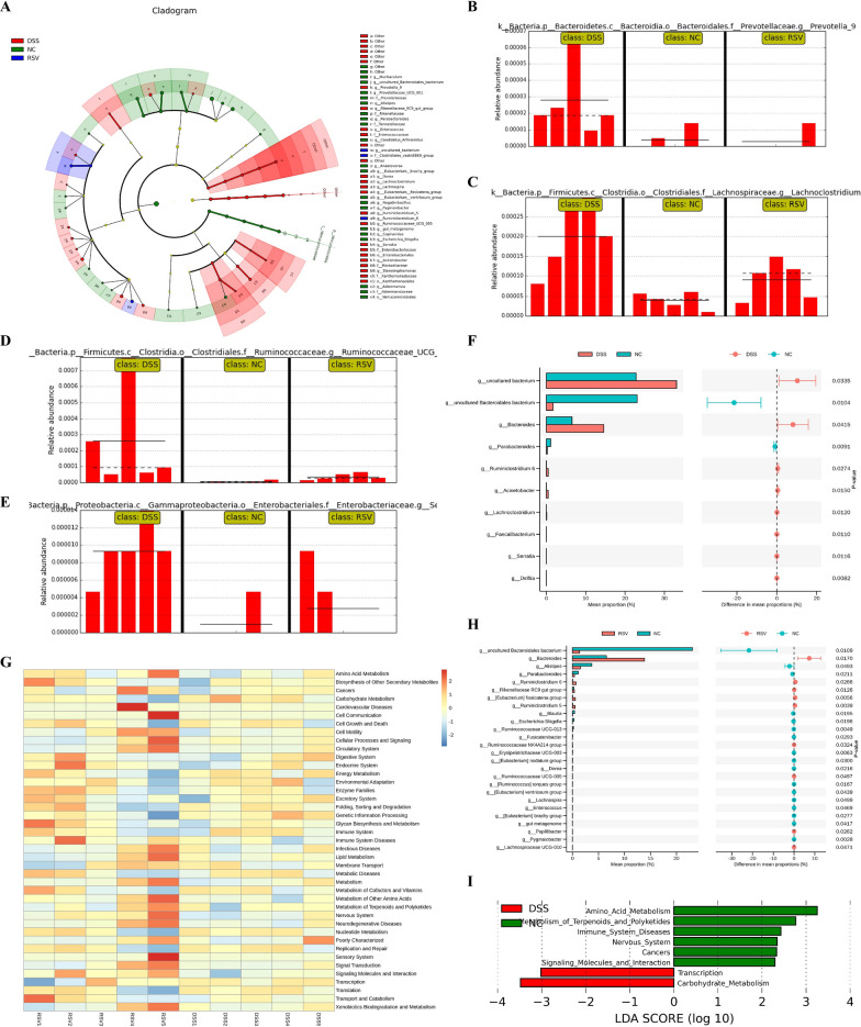
Differential analysis of significantly abundant bacteria among the groups revealed specific bacteria markers that characterize each group. Compared to the DSS group, resveratrol treatment was associated with the restoration of overpopulated genera such as Provotella 9, Lachnoclostridium, Ruminococcaceae UCG 005, and Serratia (Fig. 3A–E). Other overpopulated genera reduced by resveratrol included Ruminoclostridium 5, Stenotrophomonas, Eubacterium fissicatena group, and Acinetobacter (Fig. 3A). Further analysis revealed increased potential pathogenic genera in the DSS group, including Lachnoclostridium, Acinetobacter, and Serratia, that significantly differentiated the DSS group from the NC group (Fig. 3F) but failed to differentiate the RSV group from the NC groups (Fig. 3H). Earlier studies have identified Lachnoclostridium to be significantly enriched in the fecal samples of colitis [19], colitis-associated colorectal cancer [20] in mice, and as a potential marker in adenoma patients [21]. Moreover, Acinetobacter is enriched in actively inflamed colitis tissue [22] and Serratia has been long reported as an opportunistic pathogen [23]. Our results also showed that the RSV group had an increased abundance of ‘beneficial’ genera such as Rikenellaceae RC9 gut group, Ruminococcaceae NK4A214 group, Ruminococcaceae UCG-005, Ruminiclostridium 5, and [Eubacterium] fissicatena group compared with the NC group (Fig. 3H). Thus, resveratrol treatment not only decreases the overpopulation of bacteria groups but also increases certain beneficial species. The implication of these modulations on differential gut microbiota functional changes was examined via the KEGG heatmap functional prediction analysis and KEGG LFfSe LDA (Fig. 3G, I). The comparison between the DSS and NC groups revealed eight significantly dysregulated functions in the DSS group, including downregulated amino acid metabolism, metabolism of terpenoids and polyketides, and signaling molecules and interaction, but upregulated transcription and carbohydrate metabolism (Fig. 3I). Interestingly, KEGG LFfSe LDA revealed no significantly differential dysregulated function between the RSV group and the NC group, implicating the ability of resveratrol to restore colitis-induced functional alterations of the microbiota.
Fig. 3.
Resveratrol modulates specific bacteria species and restores functional dysregulation. A Cladogram (evolutionary branch diagram) of statistically different microbiota within each group. B–E Comparison of abundances of bacterial markers with significant differences between NC, DSS, and RSV groups (Provotella_9,Lachnoclostridium, Ruminococcaceae_UCG_005, and Serratia). F STAMP t-test of species with significant differences at the genus level between the DSS and NC groups. G KEGG heatmap prediction of the function of differential bacteria between the RSV and DSS group. H STAMP t-test of species with significant differences at the genus level between the RSV and NC groups. I KEGG LDA score of predicted functions of the DSS vs NC group
Resveratrol reduces dysregulation of gut metabolites in the mitigation of colitis
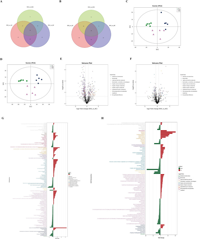
Gut microbiota-derived metabolites play a crucial role in the onset and development of many diseases, including IBD [24–26]. Our earlier studies revealed that gut metabolites are significantly altered between healthy controls and IBD in both humans and mice [27, 28]. Using the UHPLC/Q-TOF–MS technique, samples were quality controlled (Additional file 1: Fig. S1B–E) and a total of 1163 metabolites belonging to 12 superclass chemical classifications (Additional file 1: Fig. S1A) were identified in the current study. While 233 of these metabolites were dysregulated between the DSS vs NC groups, this was reduced to 147 between the RSV vs NC groups (Fig. 4A, B). Relative to the control group, analysis of differential metabolites in the positive ion mode revealed a reduced number of dysregulated metabolites in the resveratrol-treated group (87 metabolites) than the untreated group (163 metabolites) (Fig. 4B). PCA was used to help observe the cluster location of metabolites in each group. Results showed a reduced PCA score in the DSS group compared to the NC group, with the RSV group’s score located between the NC and DSS groups in both positive and negative ion modes (Fig. 4C, D). Volcanic plot analysis revealed a decrease in the concentration of significantly differential metabolites between the RSV vs NC groups as compared to the DSS vs NC groups (Fig. 4E, F). An intuitive expression of all differential metabolites classified into their respective superclass in the positive ion mode between the DSS vs NC and RSV vs NC groups are presented in Fig. 4G, H.
Fig. 4.
Resveratrol reduces gut metabolites dysregulation. A Venn diagram of differential metabolites within different groups in negative ion mode. B Venn diagram of differential metabolites within different groups in positive ion mode. C PCA score map in negative ion mode. D PCA score map in positive ion mode. E Volcano plot of differential metabolites between the DSS and NC groups in positive ion mode. F Volcano plot of differential metabolites between the RSV and NC groups in positive ion mode. G Multiple analysis of significant differences in metabolite expression in positive ion mode between the DSS and NC groups. H Multiple analysis of significant differences in metabolite expression in positive ion mode between the RSV and NC groups
Resveratrol modulates the arginine and proline metabolic pathway in the mitigation of colitis
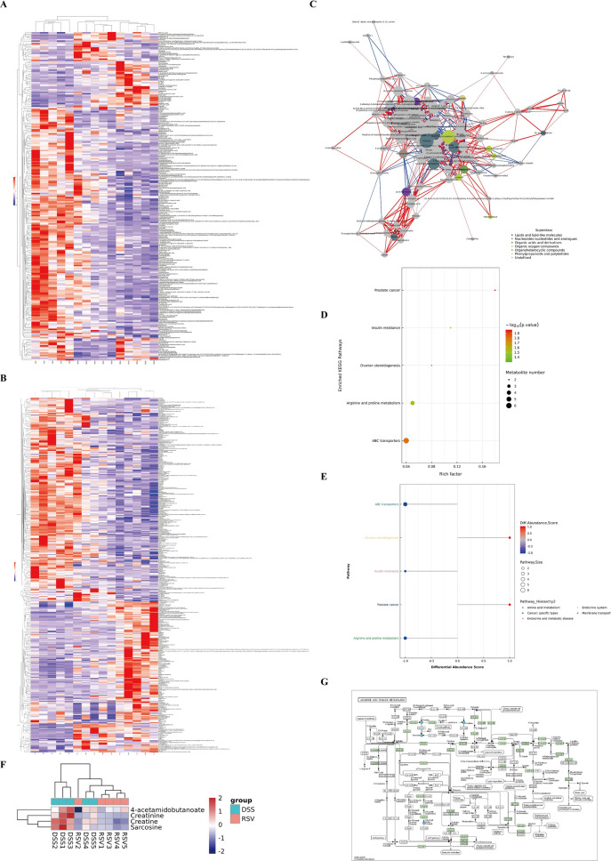
We further performed functional analysis of the differentially expressed metabolites through hierarchical clustering heatmap, network, and KEGG pathway analyses to determine key metabolites and their associated pathways crucial in the resveratrol-mediated mitigation of colitis. The clustering heatmap showed unique clusters of differential metabolites between the three groups (Fig. 5A, B, Additional file 1: Fig. S1I, J) while the correlation network showed the interaction between six defined superclasses of molecules between the resveratrol treated and untreated groups, including lipids and lipid-like molecules, organic acids and derivatives, organoheterocyclic compounds, and organic oxygen compounds (Fig. 5C). Among the key molecules at the center of the network was creatine and its derivative, creatinine, which belongs to organic acids and derivatives. KEGG enrichment pathway analysis between the resveratrol-treated (RSV) and untreated (DSS) groups revealed the two most enriched metabolic pathways as arginine and proline metabolism and ABC transporters (Fig. 5D), which was further confirmed with differential abundance score plot (Fig. 5E). Concerning the arginine and proline metabolic pathway, creatine, creatinine, and two other molecules (sarcosine and 4-acetamidobutanoate) were the differentially significant molecules expressed between the RSV and DSS groups (Fig. 5F), where resveratrol treatment reduced their overexpression. The detailed connection between these molecules in arginine and proline metabolism and other related metabolic pathways is shown in Fig. 5G. These findings implicate the microbiota-arginine/proline metabolism axis in the treatment effect of resveratrol in colitis.
Fig. 5.
Resveratrol modulates the arginine/proline metabolic pathway. A Hierarchical clustering heatmap of significantly differential metabolites in negative ion mode. B Hierarchical clustering heatmap of significantly differential metabolites in positive ion mode. C Differential metabolite network diagram between the RSV group and the DSS group in positive ion mode. D KEGG enrichment pathway diagram (bubble diagram). E Differential abundance score plot of all enriched metabolic pathways between the RSV group and the DSS group. F Clustering heatmap of differential metabolites in the KEGG pathway for arginine and proline metabolism between the DSS and the RSV groups. G KEGG pathway diagram of arginine and proline metabolism containing differential metabolites
Resveratrol modulates the microbiota-arginine/proline metabolic axis during the mitigation of colitis
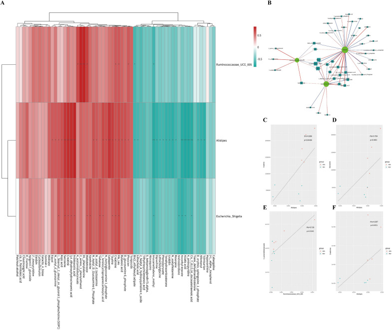
Having established the ability of resveratrol to modulate the microbiota and its metabolites during the mitigation of colitis, correlation analysis of the significantly differential microbiota and metabolites was performed to assess their interaction. Between the RSV and DSS groups, three bacteria genera (Ruminococcaceae UCG 005, Alistipes, and Escherichia/Shigella) and 63 metabolites were correlated (Fig. 6A), and a network diagram between the three bacteria genera and 42 metabolites was constructed (Fig. 6B). It was revealed that creatinine significantly correlated with all three bacteria genera, creatine and sarcosine correlated with two genera, and 4-acetamidobutanoate correlated with one genus. This indicates potential interaction points between the arginine and proline metabolic pathway and these gut bacteria genera. A scatter plot further showed the significant regulatory effect of resveratrol on the arginine and proline metabolism-associated molecules (Fig. 6C–F), which could also serve as potential markers for successful resveratrol-treated colitis.
Fig. 6.
Resveratrol modulates the microbiota-arginine/proline metabolic axis. A Spearman correlation analysis of hierarchical clustering heatmap of significant difference microbiota and significant difference metabolites betweenthe RSV and DSS groups. P-value reflects the significant level of correlation and was defined by P < 0.05 as *, P < 0.01 as **, P < 0.001 as ***. B Spearman correlation analysis network of significant difference flora and metabolites between the RSV and DSS groups. In the correlation network diagram, the color of the line represents the positive and negative value of the correlation coefficient between the two (blue represents negative correlation and red represents positive correlation), and the thickness of the line is directly proportional to the absolute value of the correlation coefficient. The node size is positively correlated with its degree, that is, the greater the degree, the larger the node size; C–F Representative scatter diagram of correlation (Creatine, Sarcosine,4-acetamidobutanoate, Creatinine)
Resveratrol regulates arginine metabolism in mice colon mucosal tissue in the mitigation of colitis
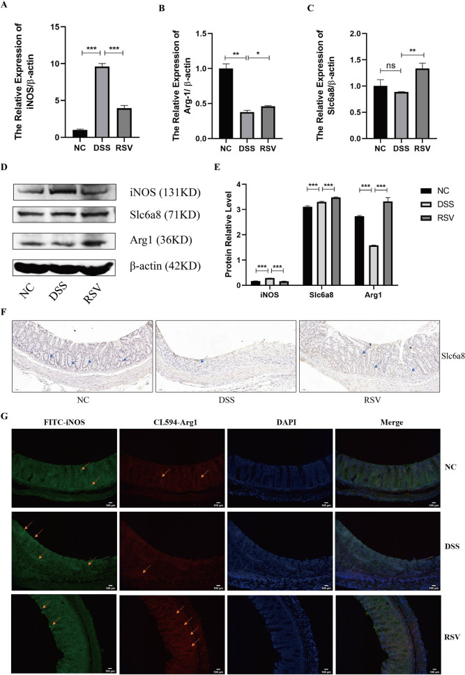
After establishing the potential role of the arginine and proline metabolic axis in the regulatory function of resveratrol during the mitigation of colitis through the colonic cytokine expression, tissue examination, as well as metagenomics and metabolomics analysis of the mice fecal samples, the specific mechanism involved in this effect was explored using mice colonic mucosal tissues. Previous studies indicate that macrophages can metabolize arginine in two major ways, i.e., breakdown arginine into iNOS to promote inflammation or produce polyamines via Arg1 to promote anti-inflammation [11, 29]. In a related process, Slc6a8, a creatine transporter highly expressed by macrophages, participates in anti-inflammatory effects by transporting creatine [11]. Therefore, we examine whether resveratrol could upregulate Slc6a8 to transport creatine into macrophages and enhance the metabolism of the Arg1 pathway, thereby reducing the iNOS-induced inflammatory response in the gut. qRT-PCR analysis of mice colon tissues showed reduced expression of iNOS but increased Slc6a8 and Arg1 in the resveratrol-treated mice (RSV group) compared to the untreated (DSS) group (Fig. 7A–C). The same expression pattern was confirmed by western blot analysis (Fig. 7D, E) while immunohistochemistry revealed significantly restored levels of Slc6a8 in RSV tissues compared to DSS tissues (Fig. 7F). Moreover, the immunofluorescence technique was used to confirmed the reduced expression of iNOS and increased levels of Arg1 in the resveratrol treated group compared to the DSS group (Fig. 7G). These observations implicate that resveratrol relieves colitis by not only inhibiting iNOS but also promoting the crucial enzyme Arg1 and the transporter Slc6a8.
Fig. 7.
Resveratrol regulates arginine metabolism in mice colon. A–C qRT-PCR analysis of the mRNA expression level of the arginine metabolism-related molecules (iNOS, Arg1, Slc6a8) in mouse colon tissues. D–E Western blot analysis of the protein expression level of arginine metabolism-related molecules in mouse colon tissues and its grayscale scanning analysis. F IHC analysis of Slc6a8 expression in mouse colon tissues (200 ×). G Representative images of IF staining for iNOS and Arg1 on sections of mouse colon tissues (200 ×). * represents p < 0.05, ** represents p < 0.01 and *** represents p < 0.001
Resveratrol regulates arginine metabolism in macrophages in vitro
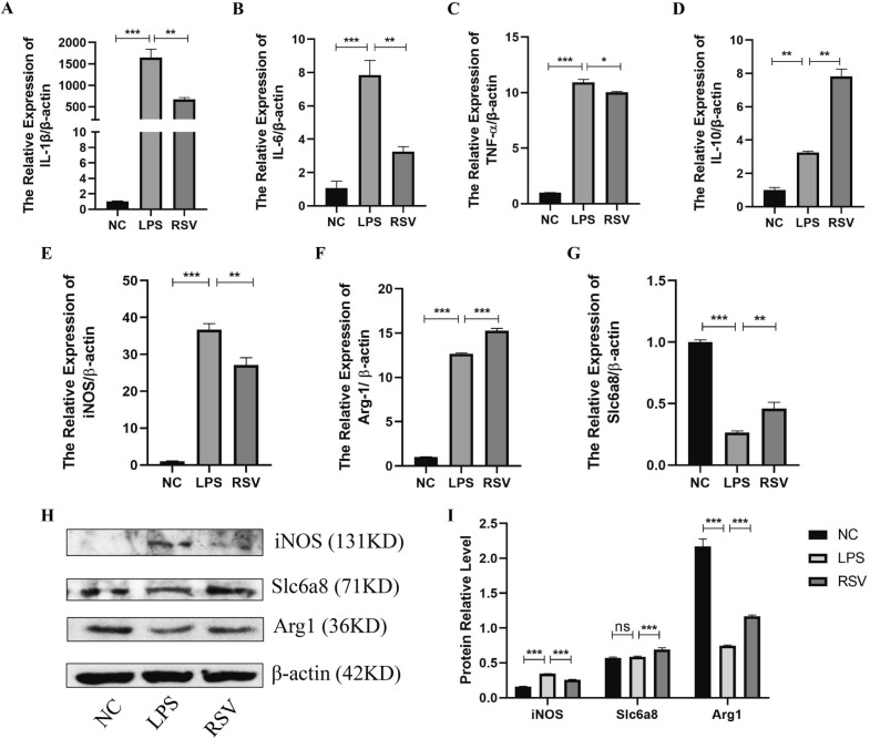
Restoring colonic immune balance by targeting macrophage polarization is crucial to mitigating colitis. The anti-inflammatory effect of macrophages is linked with arginine metabolism through increased macrophage Arg1 activity and reduced iNOS expression. After establishing the ability of resveratrol to increase Arg1 and Slc6a8 and decrease iNOS in the colonic mucosa of mice, we examined whether this effect was associated with macrophages. Therefore, we cultured RAW264.7 macrophage cell line in an LPS-induced inflammatory environment and treated it with 80 nmol/ml resveratrol for 12 h. Results of qRT-PCR showed the anti-inflammatory effect of resveratrol on macrophages as it downregulated the pro-inflammatory cytokines IL-1β, IL-6, and TNF-α but upregulated the anti-inflammatory cytokine IL-10 (Fig. 8A–D). As observed in the colonic mucosa of the mice model, resveratrol significantly downregulated iNOS and upregulated Arg1 and Slc6a8 in macrophages compared to the untreated setup (LPS) (Fig. 8E–G). Western blot confirmed the same trend of increased Arg1 and Slc6a8 and decreased iNOS in resveratrol-treated macrophages (Fig. 8H, I). These findings indicate the ability of resveratrol to relieve inflammation through the modulation of the macrophage-arginine metabolism axis.
Fig. 8.
Resveratrol regulates arginine metabolism in macrophages in vitro. A–D qRT-PCR detection of cytokines expression in RAW264.7 cells. E–G qRT-PCR analysis of the mRNA expression level of the mRNA level of arginine metabolism-related molecules (iNOS, Arg1, Slc6a8) in RAW264.7 cells. H, I Western blot analysis of the protein expression level of arginine metabolism-related molecules in RAW264.7 cells and its grayscale scanning analysis. * represents p < 0.05, ** represents p < 0.01 and *** represents p < 0.001
Discussion
IBD is characterized by chronic inflammation associated with immune dysregulation and dysbiosis. Resveratrol, a natural plant compound with anti-inflammatory and antioxidant properties, has been shown to possess therapeutic efficacy in several conditions, including IBD [30], metabolic syndromes [31], aging and age-related diseases [32], wound healing [33], vascular system conditions [34], and potentially, as a cancer therapy [35]. In this study, our initial assessment revealed the ability of resveratrol to abrogate colitis by reducing colonic pro-inflammatory markers (IL-1β, IL-6, TNFα) and DAI of mice, increasing anti-inflammatory marker IL-10, increasing colon tight junction molecules (Occludin, Claudin 1), and enhancing colon and spleen tissue repair and gross appearance. Other studies report that resveratrol reduces inflammation in patients with UC to improve their quality of life and disease clinical colitis activity [36], and in animal models of IBD [14, 37], where there are reduced pro-inflammatory markers (TNF-α, IFN-γ, IL-1β, IL-6, and IL-4) and DAI. In their randomized, double-blind, placebo-controlled pilot study, Maryam and colleagues found that the supplementation of 500 mg resveratrol daily for 6 weeks in active mild to moderate UC patients significantly reduced inflammatory markers such as plasma levels of TNF-α, high-sensitivity C-reactive protein (hs-CRP), and activity of NF-κB in PBMCs (peripheral blood mononuclear cells) [36]. The DSS-induced enteritis model in mice is very similar to human UC in terms of clinical symptoms and pathological features. However, due to species and environmental factors, we cannot fully simulate the human pathogenesis, which is also one of the shortcomings of this study, but it can still provide new ideas for the exploration of pathogenesis.
The gut microbiota is a central regulator of not only the host immune system, metabolism, and development but also influences the onset and development of several diseases, including IBD. Recent developments in genome sequencing technologies, bioinformatics, and culturomics have enabled researchers to explore the microbiota at a more detailed level than before, and in particular, their functions and association with disease and health [38]. We used 16S rDNA gene sequencing of the microbiome in fecal samples to assess the microbial changes triggered by resveratrol in the mitigation of colitis. Compared to the colitis untreated group, resveratrol treatment improved colitis-induced microbiota changes in the OTU structure and diversity. Specifically, resveratrol not only restored the alpha and beta diversity but reversed inflammation-induced changes in the abundance of the three most dominant phyla (Bacteroidetes, Proteobacteria, Firmicutes), thus significantly restoring the Bacteroidetes/Firmicutes ratio, a crucial factor in maintaining normal intestinal homeostasis [39, 40]. Resveratrol reduced the overpopulated of several bacteria genera, including potential pathogenic genera such as Lachnoclostridium, Acinetobacter, and Serratia, and increased ‘beneficial’ genera such as the Rikenellaceae RC9 gut group, Ruminococcaceae NK4A214 group, Ruminococcaceae UCG-005, Ruminiclostridium 5, and [Eubacterium] fissicatena group. Moreover, while eight microbiota-associated functions were significantly dysregulated in the untreated colitis mice, including amino acid metabolism, carbohydrate metabolism, transcription, and signaling molecules and interaction, KEGG LFfSe LDA revealed no significantly dysregulated function in the resveratrol-treated mice compared to the normal controls. This indicates that resveratrol potently mitigates diseases by improving microbiota and functional alterations in the gut as documented in experimental diseases like colitis [41, 42], obesity [43], diabetic nephropathy [44], and atherosclerosis [45], among others. Concerning colitis, studies have shown that more RSV tends to be metabolized in the intestinal flora after oral administration, and that mice with different gut microbiota composition metabolize RSV differently. Our study indicated that the intake of RSV alters the intestinal microbiota of DSS-induced mice. Moreover, the amount of metabolites such as dihydro-resveratrol increased accordingly, suggesting a specific role of RSV in the regulation of intestinal flora and its metabolites. In another study, the probiotic strain Li01 promoted larger amounts of RSV metabolizing into DHR, RES-sulfate, and RES-glucuronide, where the level of DHR increased most significantly among the metabolites [46]. Due to the extent of the exploration in this study, we only focused on RSV as a potential treatment for colitis, however, other compounds with similar properties could elicit similar responses. Thus, presenting a gap to be researched in future studies.
One of the primary means through which the gut microbiota interacts with the host is employing metabolites, which are small molecular particles produced as intermediate or end products of microbial metabolism [24]. Signals from microbial metabolites influence immune homeostasis, immune maturation, host energy metabolism, and maintenance of mucosal integrity, thus, alterations in the composition and function of the microbiota and its derived metabolites are associated with several diseases, including IBD [24–26]. In this study, fecal metabolite detection via UHPLC/Q-TOF–MS indicated that resveratrol treatment reduced the number of significantly dysregulated metabolites from 233 (DSS vs NC) to 147 (RSV vs NC). The KEGG enrichment pathway analysis revealed that the arginine–proline metabolism axis was crucial in the resveratrol-mediated mitigation of colitis, as resveratrol treatment reduced the overexpression of molecules in this axis (creatine, creatinine, sarcosine, and 4-acetamidobutanoate). Arginine metabolism is crucial in the pathophysiology of IBD, as its components, the arginine–creatine and arginine–polyamine axis are mainly protective against inflammation, relative to the pro-inflammatory arginine–nitric oxide (iNOS) axis [47]. In colon mucosa samples, we found that resveratrol not only increase Arg1 and the creatine transporter Slc6a8, which together promote anti-inflammation but also decreased iNOS. Slc6a8, which is confirmed to significantly decrease in both CD and UC, is known to regulate energy balance in intestinal epithelial cells and thereby epithelial integrity and barrier function [48]. In IBD patients, increased sarcosine is reported as a metabolomic marker linked with relapse of quiescent CD and UC [49], while 4-acetamidobutanoate is associated with subjects with high liver and kidney disease severity [50]. Therefore, the potent modulation of arginine metabolism is promising in IBD treatment and requires further exploration.
Macrophages are key in the resolution of gut inflammation and are linked with arginine metabolism through the expression of the arginine catalytic enzymes Arg1 and iNOS, and the generation of creatine as a key source of cellular energy reserve [11]. Our results showed that resveratrol downregulated macrophage expression of iNOS alongside the pro-inflammatory cytokines IL-1β, IL-6, and TNF-α but upregulated Arg1, Slc6a8, and the anti-inflammatory cytokine IL-10. Creatine reprograms macrophage polarization by inhibiting immune effector molecules such as iNOS and IFN-γ-JAK-STAT1 transcription-factor signaling while enhancing IL-4-STAT6-activated Arg1 expression. Depletion of intracellular creatine by reduced expression of Slc6a8 alters macrophage-mediated immune responses [11]. The ability of resveratrol to regulate all three key metabolic fates of arginine towards anti-inflammation might have significantly contributed to relieving inflammation in vivo and in vitro. However, the interaction between RSV and gut microbiota involves a very complex mechanism. Our study found that arginine metabolism plays an important role in this process, which only provides a new perspective and idea, and the detailed mechanism still needs to be further explored.
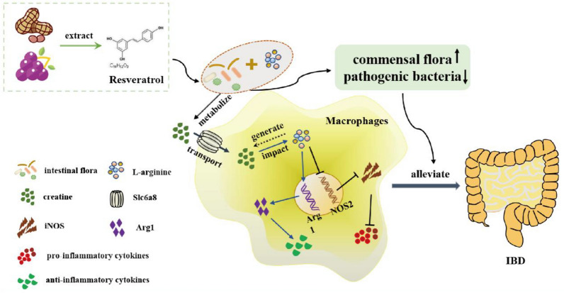
Mechanistically, resveratrol increases the expression of Slc6a8 on macrophages to transport sufficient amounts of creatine. The intracellular creatine influences arginine metabolism towards upregulated production of the Arg1 gene and decreased production of the NOS2 gene. Thus, there is decreased secretion of pro-inflammatory factors and increased anti-inflammatory factors, resulting in improved microbial community composition, diversity, and metabolites, and ultimately relieving colitis (Fig. 9).
Fig. 9.
Summary of the mechanism of resveratrol-mediated colitis
Conclusions
Resveratrol mitigates colitis by relieving clinical features of colitis, increasing tight junction molecules, improving microbiota composition and diversity, and reducing metabolite dysregulation in mice. This effect is associated with arginine metabolism-mediated macrophage polarization through the upregulation of Arg1 and Slc6a8, and the downregulation of iNOS. This uncovers a previously unknown mechanism of resveratrol in the treatment of IBD and provides the microbiota-macrophage-arginine metabolism axis as a potential therapeutic target for gut inflammation.
Supplementary Information
Additional file 1: Figure S1. A The number of identified metabolites in each chemical classification. B PCA analysis of negative ion mode population samples and quality control samples. C PCA analysis of positive ion mode population samples and quality control samples. D Correlation map of QC samples in negative ion mode. E Correlation map of QC samples in positive ion mode. F Rank abundance curve reflecting species abundance and uniform distribution of species. G Shannon curve indicating that the amount of sequencing data is large enough to reflect the vast majority of microbial information in the samples. H Species accumulation curve on the adequacy of sample size and estimation of species richness. I KEGG Hierarchical Clustering Analysis of expression changes within the groups in negative ion mode. J KEGG Hierarchical Clustering Analysis of expression changes within the groups in positive ion mode.
Acknowledgements
Not applicable.
Abbreviations
- IBD
Inflammatory bowel disease
- UC
Ulcerative colitis
- CD
Crohn’s disease
- IFN-γ
Interferon γ
- TNF-α
Tumor necrosis factor-alpha
- LPS
Lipopolysaccharide
- IL
Interleukin
- Arg1
Arginase 1
- iNOS
Inducible nitric oxide synthase
- Slc6a8
Solute carrier family 6 member 8
- UHPLC/Q-TOF–MS
Ultra-high-performance liquid chromatography-quadrupole time-of-flight mass spectrometry
- IHC
Immunohistochemistry
- DSS
Dextran sulfate sodium
- DAI
Disease activity index
- PCNA
Proliferating cell nuclear antigen
- hs-CRP
High-sensitivity C-reactive protein
- PBMCs
Peripheral blood mononuclear cells
Author contributions
XX: conception and design, collection and/or assembly of data, data analysis and interpretation, and manuscript writing. DKWO: conception and design, collection and assembly of data, data analysis and interpretation, and manuscript writing. BP: collection and/or assembly of data, data analysis and interpretation. YZ: data analysis and interpretation. NW: collection and/ or assembly of data and interpretation. ZW and FM: study design, data analysis and interpretation, manuscript writing, and final approval of manuscript. All authors read and approved the final manuscript.
Funding
This work was supported by the National Natural Science Fund of China (Grant no.82250410378), the 2022 Jiangsu Excellent postdoctoral program (Grant no.2022ZB634) the Suqian Key Laboratory of Science and Technology Project (Suqian Clinical Medical Laboratory) (Grant No. M201902), the project of the Zhenjiang Key Research and Development Plan (social development) (grant no. SH2022091) and the open project of clinical medical research center of Gynecology and Traditional Chinese Medicine of Zhenjiang (Grant no. SS202204-KFC01).
Availability of data and materials
The datasets used and/or analyzed during the current study are available from the corresponding author upon reasonable request.
Declarations
Ethics approval and consent to participate
The study was approved by the Ethical Committee of Jiangsu University (2012258). Animal experiments were carried out in accordance with the U.K. Animals (Scientific Procedures) Act, 1986 and associated guidelines.
Consent for publication
Not applicable.
Competing interests
The authors declare that they have no competing interests.
Footnotes
Publisher's Note
Springer Nature remains neutral with regard to jurisdictional claims in published maps and institutional affiliations.
Xinwei Xu, Dickson Kofi Wiredu Ocansey and Bing Pei contributed equally to this work.
Contributor Information
Zengxu Wang, Email: 13815152119@163.com.
Fei Mao, Email: maofei2003@ujs.edu.cn.
References
- 1.Kaplan GG, Windsor JW. The four epidemiological stages in the global evolution of inflammatory bowel disease. Nat Rev Gastroenterol Hepatol. 2021;18(1):56–66. doi: 10.1038/s41575-020-00360-x. [DOI] [PMC free article] [PubMed] [Google Scholar]
- 2.Ananthakrishnan AN, Bernstein CN, Iliopoulos D, et al. Environmental triggers in IBD: a review of progress and evidence. Nat Rev Gastroenterol Hepatol. 2018;15(1):39–49. doi: 10.1038/nrgastro.2017.136. [DOI] [PubMed] [Google Scholar]
- 3.Ocansey DKW, Zhang L, Wang Y, et al. Exosome-mediated effects and applications in inflammatory bowel disease. Biol Rev Camb Philos Soc. 2020;95(5):1287–1307. doi: 10.1111/brv.12608. [DOI] [PMC free article] [PubMed] [Google Scholar]
- 4.Ni J, Wu GD, Albenberg L, Tomov VT. Gut microbiota and IBD: causation or correlation? Nat Rev Gastroenterol Hepatol. 2017;14(10):573–584. doi: 10.1038/nrgastro.2017.88. [DOI] [PMC free article] [PubMed] [Google Scholar]
- 5.Na YR, Stakenborg M, Seok SH, Matteoli G. Macrophages in intestinal inflammation and resolution: a potential therapeutic target in IBD. Nat Rev Gastroenterol Hepatol. 2019;16(9):531–543. doi: 10.1038/s41575-019-0172-4. [DOI] [PubMed] [Google Scholar]
- 6.Du Y, Rong L, Cong Y, et al. Macrophage polarization: an effective approach to targeted therapy of inflammatory bowel disease. Expert Opin Ther Targets. 2021;25(3):191–209. doi: 10.1080/14728222.2021.1901079. [DOI] [PubMed] [Google Scholar]
- 7.Zhou X, Li W, Wang S, et al. YAP aggravates inflammatory bowel disease by regulating M1/M2 macrophage polarization and gut microbial homeostasis. Cell Rep. 2019;27(4):1176–1189.e5. doi: 10.1016/j.celrep.2019.03.028. [DOI] [PubMed] [Google Scholar]
- 8.Ai L, Ren Y, Zhu M, et al. Synbindin restrains proinflammatory macrophage activation against microbiota and mucosal inflammation during colitis. Gut. 2021;70(12):2261–2272. doi: 10.1136/gutjnl-2020-321094. [DOI] [PubMed] [Google Scholar]
- 9.Leonardi I, Li X, Iliev ID. Macrophage interactions with fungi and bacteria in inflammatory bowel disease. Curr Opin Gastroenterol. 2018;34(6):392–397. doi: 10.1097/mog.0000000000000479. [DOI] [PMC free article] [PubMed] [Google Scholar]
- 10.Kieler M, Hofmann M, Schabbauer G. More than just protein building blocks: how amino acids and related metabolic pathways fuel macrophage polarization. FEBS J. 2021;288(12):3694–3714. doi: 10.1111/febs.15715. [DOI] [PMC free article] [PubMed] [Google Scholar]
- 11.Ji L, Zhao X, Zhang B, et al. Slc6a8-mediated creatine uptake and accumulation reprogram macrophage polarization via regulating cytokine responses. Immunity. 2019;51(2):272–284.e7. doi: 10.1016/j.immuni.2019.06.007. [DOI] [PubMed] [Google Scholar]
- 12.Meng T, Xiao D, Muhammed A, et al. Anti-inflammatory action and mechanisms of resveratrol. Molecules. 2021 doi: 10.3390/molecules26010229. [DOI] [PMC free article] [PubMed] [Google Scholar]
- 13.Malaguarnera L. Influence of resveratrol on the immune response. Nutrients. 2019 doi: 10.3390/nu11050946. [DOI] [PMC free article] [PubMed] [Google Scholar]
- 14.Zhu F, Zheng J, Xu F, et al. Resveratrol alleviates dextran sulfate sodium-induced acute ulcerative colitis in mice by mediating PI3K/Akt/VEGFA pathway. Front Pharmacol. 2021;12:693982. doi: 10.3389/fphar.2021.693982. [DOI] [PMC free article] [PubMed] [Google Scholar]
- 15.Wang J, Zhang Z, Fang A, et al. Resveratrol attenuates inflammatory bowel disease in mice by regulating SUMO1. Biol Pharm Bull. 2020;43(3):450–457. doi: 10.1248/bpb.b19-00786. [DOI] [PubMed] [Google Scholar]
- 16.Wellington VNA, Sundaram VL, Singh S, Sundaram U. Dietary supplementation with vitamin d, fish oil or resveratrol modulates the gut microbiome in inflammatory bowel disease. Int J Mol Sci. 2021 doi: 10.3390/ijms23010206. [DOI] [PMC free article] [PubMed] [Google Scholar]
- 17.Arumugam M, Raes J, Pelletier E, et al. Enterotypes of the human gut microbiome. Nature. 2011;473(7346):174–180. doi: 10.1038/nature09944. [DOI] [PMC free article] [PubMed] [Google Scholar]
- 18.Richard ML, Sokol H. The gut mycobiota: insights into analysis, environmental interactions and role in gastrointestinal diseases. Nat Rev Gastroenterol Hepatol. 2019;16(6):331–345. doi: 10.1038/s41575-019-0121-2. [DOI] [PubMed] [Google Scholar]
- 19.Zhang W, Zou G, Li B, et al. Fecal microbiota transplantation (FMT) alleviates experimental colitis in mice by gut microbiota regulation. J Microbiol Biotechnol. 2020;30(8):1132–1141. doi: 10.4014/jmb.2002.02044. [DOI] [PMC free article] [PubMed] [Google Scholar]
- 20.Wang CS, Li WB, Wang HY, et al. VSL#3 can prevent ulcerative colitis-associated carcinogenesis in mice. World J Gastroenterol. 2018;24(37):4254–4262. doi: 10.3748/wjg.v24.i37.4254. [DOI] [PMC free article] [PubMed] [Google Scholar]
- 21.Liang JQ, Li T, Nakatsu G, et al. A novel faecal Lachnoclostridium marker for the non-invasive diagnosis of colorectal adenoma and cancer. Gut. 2020;69(7):1248–1257. doi: 10.1136/gutjnl-2019-318532. [DOI] [PMC free article] [PubMed] [Google Scholar]
- 22.Tang MS, Poles J, Leung JM, et al. Inferred metagenomic comparison of mucosal and fecal microbiota from individuals undergoing routine screening colonoscopy reveals similar differences observed during active inflammation. Gut Microbes. 2015;6(1):48–56. doi: 10.1080/19490976.2014.1000080. [DOI] [PMC free article] [PubMed] [Google Scholar]
- 23.Gupta V, Sharma S, Pal K, et al. Serratia, no longer an uncommon opportunistic pathogen—case series & review of literature. Infect Disord Drug Targets. 2021;21(7):e300821191666. doi: 10.2174/1871526521666210222125215. [DOI] [PubMed] [Google Scholar]
- 24.Lavelle A, Sokol H. Gut microbiota-derived metabolites as key actors in inflammatory bowel disease. Nat Rev Gastroenterol Hepatol. 2020;17(4):223–237. doi: 10.1038/s41575-019-0258-z. [DOI] [PubMed] [Google Scholar]
- 25.Gasaly N, de Vos P, Hermoso MA. Impact of bacterial metabolites on gut barrier function and host immunity: a focus on bacterial metabolism and its relevance for intestinal inflammation. Front Immunol. 2021;12:658354. doi: 10.3389/fimmu.2021.658354. [DOI] [PMC free article] [PubMed] [Google Scholar]
- 26.Agus A, Clément K, Sokol H. Gut microbiota-derived metabolites as central regulators in metabolic disorders. Gut. 2021;70(6):1174–1182. doi: 10.1136/gutjnl-2020-323071. [DOI] [PMC free article] [PubMed] [Google Scholar]
- 27.Xu X, Ocansey DKW, Hang S, et al. The gut metagenomics and metabolomics signature in patients with inflammatory bowel disease. Gut Pathog. 2022;14(1):26. doi: 10.1186/s13099-022-00499-9. [DOI] [PMC free article] [PubMed] [Google Scholar]
- 28.Ocansey DKW, Zhang Z, Xu X, et al. Mesenchymal stem cell-derived exosome mitigates colitis via the modulation of the gut metagenomics-metabolomics-farnesoid X receptor axis. Biomater Sci. 2022;10(17):4822–4836. doi: 10.1039/d2bm00559j. [DOI] [PubMed] [Google Scholar]
- 29.Orecchioni M, Ghosheh Y, Pramod AB, Ley K. Macrophage polarization: different gene signatures in M1(LPS+) vs. classically and M2(LPS-) vs. alternatively activated macrophages. Front Immunol. 2019;10:1084. doi: 10.3389/fimmu.2019.01084. [DOI] [PMC free article] [PubMed] [Google Scholar]
- 30.Nunes S, Danesi F, Del Rio D, Silva P. Resveratrol and inflammatory bowel disease: the evidence so far. Nutr Res Rev. 2018;31(1):85–97. doi: 10.1017/s095442241700021x. [DOI] [PubMed] [Google Scholar]
- 31.Hou CY, Tain YL, Yu HR, Huang LT. The effects of resveratrol in the treatment of metabolic syndrome. Int J Mol Sci. 2019 doi: 10.3390/ijms20030535. [DOI] [PMC free article] [PubMed] [Google Scholar]
- 32.Zhou DD, Luo M, Huang SY, et al. Effects and mechanisms of resveratrol on aging and age-related diseases. Oxid Med Cell Longev. 2021;2021:9932218. doi: 10.1155/2021/9932218. [DOI] [PMC free article] [PubMed] [Google Scholar]
- 33.Pignet AL, Schellnegger M, Hecker A, et al. Resveratrol-induced signal transduction in wound healing. Int J Mol Sci. 2021 doi: 10.3390/ijms222312614. [DOI] [PMC free article] [PubMed] [Google Scholar]
- 34.Breuss JM, Atanasov AG, Uhrin P. Resveratrol and its effects on the vascular system. Int J Mol Sci. 2019 doi: 10.3390/ijms20071523. [DOI] [PMC free article] [PubMed] [Google Scholar]
- 35.Vervandier-Fasseur D, Latruffe N. The potential use of resveratrol for cancer prevention. Molecules. 2019 doi: 10.3390/molecules24244506. [DOI] [PMC free article] [PubMed] [Google Scholar]
- 36.Samsami-Kor M, Daryani NE, Asl PR, Hekmatdoost A. Anti-inflammatory effects of resveratrol in patients with ulcerative colitis: a randomized, double-blind, Placebo-controlled. Pilot Study Arch Med Res. 2015;46(4):280–285. doi: 10.1016/j.arcmed.2015.05.005. [DOI] [PubMed] [Google Scholar]
- 37.Abdallah DM, Ismael NR. Resveratrol abrogates adhesion molecules and protects against TNBS-induced ulcerative colitis in rats. Can J Physiol Pharmacol. 2011;89(11):811–818. doi: 10.1139/y11-080. [DOI] [PubMed] [Google Scholar]
- 38.Adak A, Khan MR. An insight into gut microbiota and its functionalities. Cell Mol Life Sci. 2019;76(3):473–493. doi: 10.1007/s00018-018-2943-4. [DOI] [PMC free article] [PubMed] [Google Scholar]
- 39.Vandeputte D, Falony G, Vieira-Silva S, et al. Stool consistency is strongly associated with gut microbiota richness and composition, enterotypes and bacterial growth rates. Gut. 2016;65(1):57–62. doi: 10.1136/gutjnl-2015-309618. [DOI] [PMC free article] [PubMed] [Google Scholar]
- 40.Koliada A, Syzenko G, Moseiko V, et al. Association between body mass index and Firmicutes/Bacteroidetes ratio in an adult Ukrainian population. BMC Microbiol. 2017;17(1):120. doi: 10.1186/s12866-017-1027-1. [DOI] [PMC free article] [PubMed] [Google Scholar]
- 41.Li F, Han Y, Cai X, et al. Dietary resveratrol attenuated colitis and modulated gut microbiota in dextran sulfate sodium-treated mice. Food Funct. 2020;11(1):1063–1073. doi: 10.1039/c9fo01519a. [DOI] [PMC free article] [PubMed] [Google Scholar]
- 42.Alrafas HR, Busbee PB, Nagarkatti M, Nagarkatti PS. Resveratrol modulates the gut microbiota to prevent murine colitis development through induction of Tregs and suppression of Th17 cells. J Leukoc Biol. 2019;106(2):467–480. doi: 10.1002/jlb.3a1218-476rr. [DOI] [PMC free article] [PubMed] [Google Scholar]
- 43.Wang P, Li D, Ke W, et al. Resveratrol-induced gut microbiota reduces obesity in high-fat diet-fed mice. Int J Obes (Lond) 2020;44(1):213–225. doi: 10.1038/s41366-019-0332-1. [DOI] [PubMed] [Google Scholar]
- 44.Cai TT, Ye XL, Li RR, et al. Resveratrol modulates the gut microbiota and inflammation to protect against diabetic nephropathy in mice. Front Pharmacol. 2020;11:1249. doi: 10.3389/fphar.2020.01249. [DOI] [PMC free article] [PubMed] [Google Scholar]
- 45.Chen ML, Yi L, Zhang Y, et al. Resveratrol attenuates trimethylamine-N-oxide (TMAO)-induced atherosclerosis by regulating TMAO synthesis and bile acid metabolism via remodeling of the gut microbiota. MBio. 2016;7(2):e02210–e2215. doi: 10.1128/mBio.02210-15. [DOI] [PMC free article] [PubMed] [Google Scholar]
- 46.Yao M, Fei Y, Zhang S, et al. Gut microbiota composition in relation to the metabolism of oral administrated resveratrol. Nutrients. 2022 doi: 10.3390/nu14051013. [DOI] [PMC free article] [PubMed] [Google Scholar]
- 47.Li JY, Guo YC, Zhou HF, et al. Arginine metabolism regulates the pathogenesis of inflammatory bowel disease. Nutr Rev. 2022 doi: 10.1093/nutrit/nuac070. [DOI] [PMC free article] [PubMed] [Google Scholar]
- 48.Hall CHT, Lee JS, Murphy EM, et al. Creatine transporter, reduced in colon tissues from patients with inflammatory bowel diseases, regulates energy balance in intestinal epithelial cells, epithelial integrity, and barrier function. Gastroenterology. 2020;159(3):984–998.e1. doi: 10.1053/j.gastro.2020.05.033. [DOI] [PMC free article] [PubMed] [Google Scholar]
- 49.Borren NZ, Plichta D, Joshi AD, et al. Multi-"-Omics" profiling in patients with quiescent inflammatory bowel disease identifies biomarkers predicting relapse. Inflamm Bowel Dis. 2020;26(10):1524–1532. doi: 10.1093/ibd/izaa183. [DOI] [PMC free article] [PubMed] [Google Scholar]
- 50.Mindikoglu AL, Opekun AR, Putluri N, et al. Unique metabolomic signature associated with hepatorenal dysfunction and mortality in cirrhosis. Transl Res. 2018;195:25–47. doi: 10.1016/j.trsl.2017.12.002. [DOI] [PMC free article] [PubMed] [Google Scholar]
Associated Data
This section collects any data citations, data availability statements, or supplementary materials included in this article.
Supplementary Materials
Additional file 1: Figure S1. A The number of identified metabolites in each chemical classification. B PCA analysis of negative ion mode population samples and quality control samples. C PCA analysis of positive ion mode population samples and quality control samples. D Correlation map of QC samples in negative ion mode. E Correlation map of QC samples in positive ion mode. F Rank abundance curve reflecting species abundance and uniform distribution of species. G Shannon curve indicating that the amount of sequencing data is large enough to reflect the vast majority of microbial information in the samples. H Species accumulation curve on the adequacy of sample size and estimation of species richness. I KEGG Hierarchical Clustering Analysis of expression changes within the groups in negative ion mode. J KEGG Hierarchical Clustering Analysis of expression changes within the groups in positive ion mode.
Data Availability Statement
The datasets used and/or analyzed during the current study are available from the corresponding author upon reasonable request.


