Abstract
Biological membrane channels mediate information exchange between cells and facilitate molecular recognition. While tuning the shape and function of membrane channels for precision molecular sensing via de-novo routes is complex, an even more significant challenge is interfacing membrane channels with electronic devices for signal readout, which results in low efficiency of information transfer - one of the major barriers to the continued development of high-performance bioelectronic devices. To this end, we integrate membrane spanning DNA nanopores with bioprotonic contacts to create programmable, modular, and efficient artificial ion-channel interfaces. Here we show that cholesterol modified DNA nanopores spontaneously and with remarkable affinity span the lipid bilayer formed over the planar bio-protonic electrode surface and mediate proton transport across the bilayer. Using the ability to easily modify DNA nanostructures, we illustrate that this bioprotonic device can be programmed for electronic recognition of biomolecular signals such as presence of Streptavidin and the cardiac biomarker B-type natriuretic peptide, without modifying the biomolecules. We anticipate this robust interface will allow facile electronic measurement and quantification of biomolecules in a multiplexed manner.
Subject terms: DNA nanostructures, Sensors and biosensors, Ion channels, Membranes, Electronic devices
Synthetic membrane channels have many potential applications, but interfacing membrane channels with electronic devices for efficient information transfer is challenging. Here the authors integrate membrane spanning DNA nanopores with bioprotonic contacts to create programmable, modular, and efficient artificial ion-channel interfaces.
Introduction
In biological systems, communication between cells occurs via membrane proteins and ion channels that act as size-selective filters or stimulus-responsive molecular valves to either passively allow or actively control the flow of ions across the cell membrane1. Cellular communication often surpasses information processing in electronic devices in efficiency, regulation, and specificity2,3. Augmenting electronic devices with biological components can enable one to access, analyze, and respond to intercellular information via data transduction and signal transmission1,4. Examples include metal oxide semiconductors integrated with ATPase5, carbon nanotubes6,7 and silicon nanowires to sense pH8, 2D transistors functionalized with gramicidin9, organic electrochemical devices with membrane channels10,11, and H+ selective bioprotonic devices integrated with gramicidin12, alamethicin12 and light sensitive rhodopsins13,14. Synthetic membrane channels can further increase the functionality of these devices with well-defined geometries, durability, robustness, and ease of modification15,16. Self-assembled synthetic membrane channels are particularly attractive due to their ease of fabrication16. To this end, Watson-Crick pairing based hybridization of single-stranded DNA (ssDNA) can rationally and in a bottom-up manner design self-assembled DNA nano-structures17 that mimic membrane proteins with sophisticated architectures18–21 and varied functionalities22–24.
Here, we merge synthetic self-assembled DNA nanopores based ion-channels with H+ selective Palladium (Pd)-based electrodes to create a bioprotonic device that records and modulates H+ currents traversing across the bilayer membrane (Fig. 1). Unlike previous studies that used single channel ionic current measurements in membrane spanning nanopores25,26, our device architecture enables biomolecular recognition as a function of ensemble measurement of the overall conductance change of the membrane that is an average over many ion-channels spread over several nanopore states. Utilizing programmable DNA nanopores, we showcase the versatility of the device in detecting specific biomolecules through distinct electronic signals, eliminating the need for additional pre-processing of the biomolecules.
Fig. 1. Schematics of bioprotonic devices.
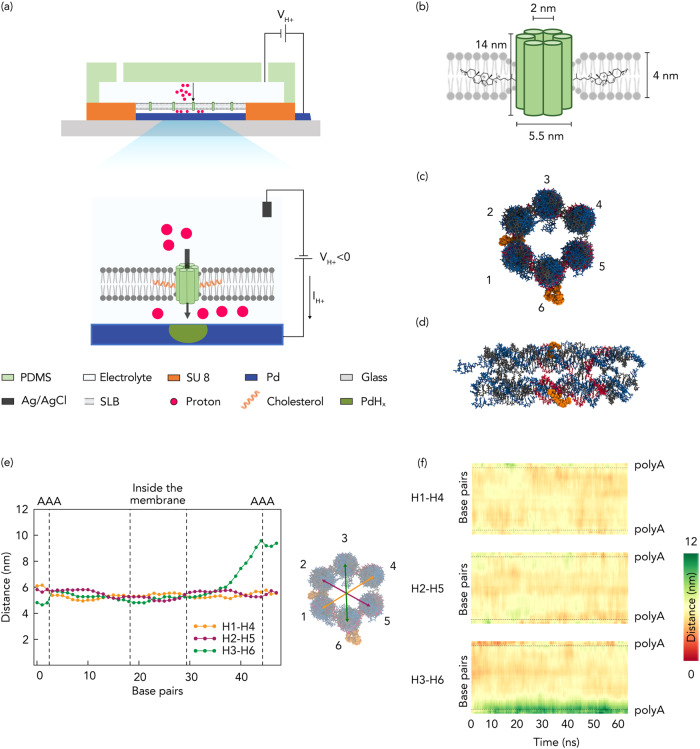
a Schematic depiction of the bioprotonic device. b Schematic representation of a DNA nanopore comprising six-helix bundles and 2-cholesterol anchors. c Top view of DNA nanopores with positioned cholesterol anchors. d Lateral view of DNA nanopores with positioned base pairs. e Simulation of the average distance between the diametrically opposite strands across the length of the nanopore. Yellow for distance between strands 1 and 4, green for distance between strands 2 and 5 and magenta for distance between strands 3 and 6. f Average distance heatmap for the pairs indicated in (c) and (e).
Results
DNA Nanopore Bioprotonics
The DNA nanopore bioprotonic device comprises DNA nanopore ion channels spanning a supported lipid bilayer membrane (SLB) that is atop a Pd contact integrated with a microfluidic architecture (Fig. 1a and Supplementary Fig.1). A voltage (VH+) between the Pd contact and the Ag/AgCl reference electrode positioned in the solution causes a current of H+ between the Pd contact and the solution depending on polarity12,27. As previously reported, this flow of H+ induces the electrochemical formation or dissolution of PdHx that results in a measurable current (IH+) in the electronic circuit12–14. Although this strategy does not afford enough temporal nor spatial resolution to investigate individual ion channel states, we used it to measure the average change in membrane conductance due to ion channel insertion and activity. To create biomimicking ion channels that enable H+ transfer across the SLBs, we formed 14 nm long barrel shaped DNA nanopores via bottom-up rational design and directed self-assembly (Fig. 1b). While simple ion-channels made out of DNA duplexes that lack a central hollow pore have been previously demonstrated as effective membrane spanning ion-conduction pathways28, we chose a DNA nanostructure geometry consisting of a central physical pore to closely mimic the membrane29–31 and enable a larger range of signal differentiation upon varied degrees of blockage of the pore. To design the nanostructure, we adapted the single stranded tile assembly method proposed by Seeman et al.32 to self-assemble a nanobarrel-like structure with a hollow lumen from equimolar amounts of 13 short ssDNA strands. To design the strands, we first defined the desired geometry in a hexagonal lattice-based DNA design software caDNAno33 and filled the shape from top to bottom with an even number of parallel double helices, held together by periodic crossovers of the strands. The sequences were randomly generated and then rationally down selected to maximize primary interactions as designed and minimize secondary and tertiary complex formations. The resulting 13 ssDNA strands (Supplementary Data 1) were mixed in equimolar amounts to enable one-pot self-assembly into 6 inter-linked Helix Bundles (6HB) that form the walls of the nanopore (Fig. 1b, c, d). We functionalized the DNA nanopore with Tetri-ethylene Glycol–Cholesterol (TEG-Chol) to provide an anchor for insertion of the hydrophilic DNA nanopores into the hydrophobic environment of the SLB (Fig. 1b, c, d and Supplementary Fig. 2 and 3). Next, we conducted transmission electron microscopy (Supplementary Fig. 3), dynamic light scattering (DLS) (Supplementary Fig. 3c and d) analysis as well as molecular dynamics (MD) simulations to investigate the dimensions and the stability of the 6HB inside the SLB and its pore size under dynamic environments (Fig. 1e and f). The average distance between the diametrically opposite DNA helices across the length of the nanopore is analyzed, as depicted in Fig. 1e, providing insights into the pore size. Additionally, Fig. 1f illustrates the dynamic behavior of these distances, as they change with time on a base pair level within the DNA nanopore. Our analysis revealed that inside the membrane, the center-to-center distances of the opposite helices ranged between 5 and 6 nm. Given that the radius of a DNA double helix is 1 nm, this indicated that the average pore size fluctuated between 3 and 4 nm. Outside the membrane, the DNA helices exhibited increased mobility, resulting in some helices moving apart from one another (Fig. 1e green plot). However, as seen in the Fig. 1f, this phenomenon did not impact the stability of the pore as the TEG-Chol anchors stabilized the DNA nanopore inside the SLB. Therefore, we infer that the length of the nanopore provides sufficient area for decoration with hydrophobic anchors to enable spontaneous insertion while projecting further beyond the SLB to enable desired interactions at the lip of the nanopore without disrupting its stability within the bilayer. The small inner lumen size facilitates proton transport across the channel while obstructing proteins and other larger biomolecules to remain on the cis (negatively charged) side of the nanopore29.
Control of H+ flow with DNA nanopore bioprotonics
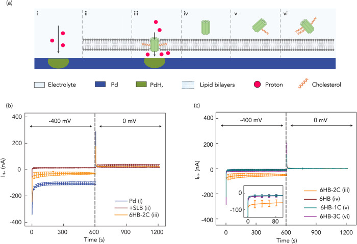
To validate the DNA nanopore ion-channel is indeed a H+ conductor, we measured the dependence of IH+ to VH+ in the DNA bioelectronic device (Fig. 2a). First, we verified that the bare Pd contact transfers H+ at the solution interface (Fig. 2a-i). To do so, we recorded IH+ as a function of VH+ with the following sequence as previously reported12. In the first step, VH+ = −400 mV for 600 s induces H+ to flow from the solution into the Pd contact to form PdHx (Fig. 2a–i) as indicated by IH+ = −125 ± 11 nA (Fig. 2b). In the second step, VH+ = 0 mV transferred H+ from the PdHx contact into the solution12. Here, IH+ indicates the prior formation of PdHx that allows H+ to transfer from the surface back into the solution even at VH+ = 0 mV because at a neutral pH, the protochemical potential of H+ in the PdHx contact is higher than the protochemical potential of H+ in the solution34,35. We confirmed the characteristics of the Pd contact with Electrochemical Impedance Spectroscopy (EIS) (Supplementary Fig. 4 and Supplementary Table 1). Second, we confirmed that the SLBs create barriers and block H+ transport from the solution into the Pd surface to make sure that when we inserted the DNA nanopore we measured H+ transport across the nanopore (Fig. 2a-ii), as indicated by IH+ = −7 ± 1 nA (Fig. 2b). To verify the formation of SLBs, we repeated the current measurements and reported the current which are shown as the IH+ of 0 nM DNA in (see below) and obtained fluorescence imaging of fluorescence recovery after photobleaching measurements (FRAP) (Supplementary Fig. 5). The measured current, referred to as the leakage current, indicates that few H+ diffuse and leak across the bilayer membrane, possibly through the surface defects and are reduced at the Pd surface. After addition of 15 nM DNA nanopores modified with two cholesterol handles (6HB-2C) to the solution, we expected that the DNA nanopores spontaneously insert into the lipid bilayer (Fig. 2a-iii) to form membrane spanning ion channels. This insertion resulted in IH+ = −52 ± 4 nA for VH+ = −400 mV (Fig. 2b), which is much larger than IH+ for the SLBs coated Pd indicating that the DNA nanopores provide a pathway for H+ to move across the SLBs. For all measurements, to avoid the accumulation of protons on Pd contact, in the second sequence, we set the VH+ to 0 mV. The higher photochemical potential than the electrolyte leads to the release of protons into the electrolyte and with positive IH+. As predicted, DNA nanopores without any cholesterol handles (Fig. 2a-iv) did not insert into the SLBs as corroborated by the same observed IH+ as recorded for the naked SLBs (Fig. 2c). Nanopores with one or three cholesterol handles (6HB-1C, 6HB-3C) (Fig. 2a-v, vi) also did not insert into the SLBs (Fig. 2c). It is likely that 6HB-1C did not insert into the SLBs because one cholesterol handle is not enough to drive the hydrophilic DNA nanopore into the hydrophobic SLBs in a membrane-spanning configuration36. However, with the same reasoning one would we expected to see even better insertion for 6HB-3C compared to 6HB-2C. It is likely that the increased hydrophobicity of 6HB-3C drove its aggregation in solution to minimize its interaction with water and made the hydrophobic handles unavailable for insertion into the SLB. This aggregation is confirmed by multiple bands observed for 6HB-3C in gel electrophoresis (Supplementary Fig. 3a) and a hydrodynamic radius eight times larger for 6HB-3C compared to 6HB as measured by DLS (Supplementary Fig. 3c).
Fig. 2. Schematics of control of H+ by membrane spanning DNA nanopores.
a i) Pd contact with electrolyte solution; ii) Pd contact coated with SLBs; iii) DNA nanopores with 2-cholesterol anchors (6HB-2C); iv) DNA nanopores without cholesterol anchor (6HB); v) DNA nanopores with 1-cholesterol anchor (6HB-1C); vi) DNA nanopores with 3-cholesterol anchors (6HB-3C). b IH+ versus time plot for V = −400 mV and V = 0 mV. Blue trace Pd, red trace SLB, Orange trace 6HB-2C. At V = −400 mV, the IH+ = −125 ± 11 nA with bare Pd decreased to −7 ± 1 nA with SLBs that indicates formed bilayers inhibit H+ transfer from the bulk solution to the Pd/solution interface. The IH+ = −52 ± 4 nA with 6HB-2C confirmed that the nanopore channels support the H+ transport. Error bars are 1 s.d. (n = 3). c IH+ versus time plot in different situations of Fig.2a under V = −400 mV and V = 0 mV. Orange trace 6HB-2C (2a-iii), red trace 6HB (2a-iv), cyan trace 6HB-1C (2a-v), and purple trace 6HB-3C (2a-vi). Under −400 mV, we also measured IH+ = −11 ± 5 nA, −13 ± 7 nA, and −12 ± 4 nA with 6HB, 6HB-1C and 6HB-3C, respectively. Error bars are 1 s.d. (n = 3). Only 6HB-2C provides created pathway to facilitate the flow of H+ to the Pd/solution interface. For all measurements, we switched the voltage to 0 mV, roughly after 600 s from the first instance of measurement, the H absorbed in Pd is oxidized to H+ and released back into the solution, allowing the current measured to return to 0 nA.
Programming the DNA nanopores for biomolecular sensing
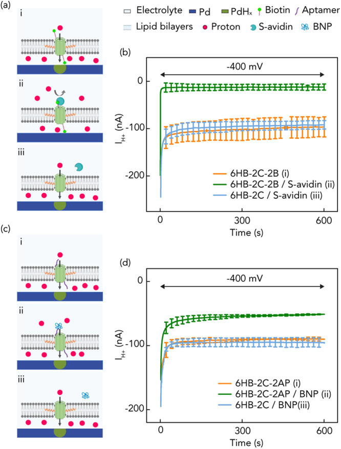
DNA self-assembly allows for programming a desired functionality in the DNA nanopores by designing ad-hoc DNA sequences. As proof-of-concept biomolecular sensing, we programmed DNA nanopores for the detection of two proteins Streptavidin (S-avidin) and a cardiac biomarker B-type natriuretic peptide (BNP) by including a biotin handle37 or a DNA aptamer (AP) that has been down selected using the in-vitro SELEX technology respectively on the nanopores. We did so by functionalizing 6HB-2C nanopores using two ssDNAs modified with either a Biotin handle or the AP handle at their 5' ends followed by DNA hybridization to obtain the formation of 6HB-2C-2B (Fig. 3a) and 6HB-2C-2AP (see below) nanopores respectively containing the specific tags at either end of the nano barrel. To get a clear current comparison before and after the addition of S-avidin or BNP, we increased the concentration of 6HB-2C-2B and 6HB-2C-2AP to 30 nM for the current measurements. As expected, 6HB-2C-2B nanopores inserted themselves into the SLB and resulted in a large ensemble current IH+ = −96 ± 21 nA at VH+ = −400 mV, indicating that the nanobarrel inside the DNA nanopore aids H+ transport across the SLB (Fig. 3a-i and b). However, when S-avidin was introduced into the environment in five times excess concentration with respect to the nanopore concentration, the binding event of the 5 nm sized S-avidin with the biotin handle38 on the DNA nanopore effectively occluded the nanobarrel impeding H+ transport across the SLB as indicated by a reduction of IH+ = −12 ± 6 nA (Fig. 3a-ii and b) to the level of SLB leakage IH+. This is an interesting result because prior research has shown that DNA structures without interior pores create H+ conduction pathways across the SLB28 To confirm that S-avidin is indeed blocking the pore rather than plugging conduction pathways around the DNA structure, we exposed non-biotinylated 6HB-2C to the same S-avidin concentration (6HB-2C/S-avidin) in solution and did not observe appreciable change in IH+ = −92 ± 9 nA (Fig. 3a-iii and b) because the S-avidin has no binding site available on the DNA nanopore that would result in the occlusion of the nanobarrel. Our observation and the results from ref. 37 are not necessarily contradictory because the two DNA structures are different from each other, ours contain cholesterol handles, and may interact in a different manner with the SLB. To further confirm that the current drop was indeed caused by the binding between S-avidin and biotin handle of the nanopore leading to the pathway occlusion, we also performed fluorescence imaging experiments prior to and after the addition of the proteins. To fabricate fluorescent nanopores, we modified some of the ssDNA strands with Atto 488 tags on their 5' ends (Supplementary Data 1) prior to the single pot hybridization of the nanostructures. The fluorescent images of DNA nanopores before and after the addition of S-avidin (Supplementary Fig. 6) show the same number of DNA nanopores spanning the SLBs, exhibiting that binding between S-avidin and biotin indeed blocked the channels and resulted in the decreased ensemble current rather than other possible phenomenon such as nanopore aggregation or bulk dissociation of DNA nanopores from the SLB membrane. Additionally, we measured the dependence of the IH+ on the relative concentration of S-avidin with respect to the concentration of the biotin tagged nanopores. With the increase of S-avidin’s concentration, more nanopores interacted with S-avidin leading to more blocked channels and thus a higher current decrease (Supplementary Fig. 7).
Fig. 3. Schematics of bioprotonic devices with biotin-Streptavidin and aptamer-peptide.

a i) 2-cholesterol handled DNA nanopores with biotin in the absence of streptavidin (6HB-2C-2B). Created pathway facilitates H+ transfer without inhibition of binding due to absence of streptavidin; ii) 2-cholesterol-handled DNA nanopores with binding of biotin-streptavidin (6HB-2C-2B/S-avidin). H+ transfer is inhibited by blocked pore channels; iii) 2-cholesterol-handled DNA nanopores without biotin in the presence of streptavidin (6HB-2C/S-avidin). The pores are not blocked by binding due to lacking biotin. b IH+ versus time plot for V = −400 mV. Orange trace 6HB-2C-2B (3a-i), green trace 6HB-2C-2B/S-avidin (3a-ii) and blue trace 6HB-2C/S-avidin (3a-iii). We measured IH+ = −96 ± 21 nA, −12 ± 6 nA and −92 ± 9 nA with 6HB-2C-2B, 6HB-2C-2B/S-avidin and 6HB-2C/S-avidin, respectively. Error bars are 1 s.d. (n = 3). c i) 2-cholesterol handled DNA nanopores with SELEX based DNA aptamer in the absence of B-type natriuretic peptide (6HB-2C-2AP). Created pathway facilitates H+ transfer without inhibition of binding due to absence of peptide; ii) 2-cholesterol-handled DNA nanopores with binding of aptamer-peptide (6HB-2C-2AP/BNP). H+ transfer is slightly inhibited by blocked pore channels; iii) 2-cholesterol-handled DNA nanopores without aptamer in the presence of peptide (6HB-2C/BNP). d IH+ versus time plot for V = −400 mV. Orange trace 6HB-2C-2AP (3c-i), green trace 6HB-2C-2AP/BNP (3c-ii) and blue trace 6HB-2C/BNP (3c-iii). We measured IH+ = −90 ± 3 nA, −51 ± 1 nA and −96 ± 9 nA with 6HB-2C-2AP, 6HB-2C-2AP/BNP and 6HB-2C/BNP, respectively. Error bars are 1 s.d. (n = 3).
Similar experiments and controls were conducted with 6HB-2C-2AP nanopores. For the same concentration of 6HB-2C-2AP (Fig. 3c) nanopores as that of 6HB-2C-2B nanopores, the IH+ observed at VH+ = −400 mV was −90 ± 3 nA (Fig. 3c-i and d). Alike the biotin tagged nanopores, the DNA aptamer tagged nanopores inserted themselves into the SLB to form membrane-spanning ion channels and resulted in H+ transport across the SLB. When BNP protein was introduced into the environment in five times excess concentration with respect to the nanopore concentration, a reduced IH+ of −51 ± 1 nA was observed (Fig. 3c-ii and d) at VH+ = −400 mV. This showed that the affinity interactions between AP-BNP at the lip of the ion-channels blocked the transport of H+. The smaller reduction in current in the case of AP-BNP compared to the dramatic reduction observed in Biotin-S-avidin is attributed to the weaker interaction affinity (kD of 12 ± 1.5 nM)39 compared against strong affinity of biotin-Streptavidin (kD of 10−5 nM)40. Similarly, as control, exposing non-aptamer modified 6HB-2C nanopores to the same BNP concentration in solution (6HB-2C/BNP) did not cause any appreciable change in IH+ of −96 ± 9 nA (Fig. 3c-iii and d) because the BNP has no binding site available on the DNA nanopore that would have resulted in the occlusion of the nanobarrel. By leveraging the programmability offered by the DNA nanostructures, we engineered the nanopores to demonstrate an electronic sensing response to a specific analyte in in-vitro environments without the need for modifying the analyte.
A model for the rate constant of association and dissociation of DNA nanopores self-insertion
To better understand the dynamics of DNA nanopore insertion with the SLB, we created a model based on Langmuir’s equation and absorption/desorption kinetics41,42 to analyze the insertion process of the DNA nanopore in the lipid bilayer. In this model, we describe DNA nanopores in solution (n) and lipid bilayer sites where the nanopores can be absorbed (l) as being initially separate (Eq. 1 left side). Upon insertion of the DNA nanopore into the lipid bilayer, the DNA nanopore and the lipid bilayer sites are conjoined together and we describe this entity as nl (Eq. 1 right side).
| 1 |
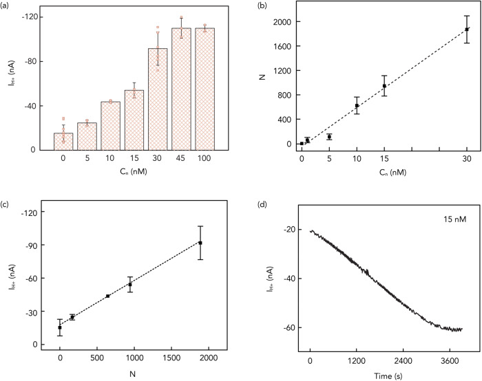
The rate constant ka (M-1s-1) describes the absorption reaction of the DNA nanopore into the lipid bilayer and the rate constant kd (s-1) describes the desorption reaction. From this model, we expect that more DNA nanopores in solution (n) correspond to a higher number of DNA nanopores inserted into the lipid bilayer (nl) resulting in IH+ to increase as a function of DNA nanopore concentration (Cn) (Fig. 4a). Given the large number of DNA nanopores compared to the absorption area of the lipid bilayer, we assume Cn to be constant throughout the absorption process. To fully understand the absorption and desorption kinetics, we need to now derive ka and kd. To do so, we introduce the differential form of the Langmuir equation:
| 2 |
where Cn and Cnl are the DNA nanopore concentrations in solution and lipid bilayers, respectively, and Cu represents the unoccupied site concentration in the SLBs. Since Cu is an unknown that we are not able to derive experimentally, we write (2) as:
| 3 |
Where Cmax = Cu + Cnl and Cmax is the maximum value of Cnl. We derive Cnl by counting the number of inserted DNA nanopores (N = CnlVlA, Vl = the volume of lipids, A = Avogadro’s number) as a function of Cn at equilibrium using fluorescent microscopy on fluorescently tagged nanopores (Fig. 4b and Supplementary Fig. 8). Unfortunately, we are not able to measure Cmax using fluorescent microscopy for Cn > 30 nM because the inserted DNA nanopores are too close to each other and difficult to count. In Fig. 4a, we have shown that IH+ increases with increasing Cn for Cn < 45 nM and then IH+ plateaus even if we increase Cn up to100 nM. We assume that for Cn > 45 nM, Cnl = Cmax. To calculate Cmax from IH+, we then model the DNA nanopores as resistors in parallel:
| 4 |
and from Eq. 4 and the slope of Fig. 4c, we calculate Rm = 1 × 10¹⁰ Ω and Gm = 1/Rm = 100 pS, the resistance and conductivity of each individual nanopore. These values are consistent with the conductivity of artificial and natural membrane channels43,44. Using the calculated value of Rm, Nmax = 2350 nanopores per device and Cmax = 2 nM. To conclude the derivation of ka, we then observe experimentally Cnl/dt by recording IH+ as a function of time introducing the DNA nanopores in the solution for t = 0 (Fig. 4d). We can thus assume Cnl = 0 and Eq. 3 simplifies to:
| 5 |
Fig. 4. Illustration of DNA nanopores characteristics.
a IH+ versus the introduced 6HB-2C concentration (Cn) plot (VH+ = −400 mV) for 6HB-2C nanopores. We measured IH+ = −15 ± 8 nA, −25 ± 3 nA, −44 ± 1 nA, −52 ± 4 nA, −92 ± 15 nA, −111 ± 9 nA and−110 ± 3 nA with 0 nM, 5 nM, 10 nM, 15 nM, 30 nM, 45 nM and 100 nM of 6HB-2C, respectively. Error bars are 1 s.d. (n = 10, 3, 3, 3, 6, 3 and 3 for 0 nM, 5 nM, 10 nM, 15 nM, 30 nM, 45 nM and 100 nM respectively). b The number of inserted 6HB-2C nanopores (N) versus the introduced 6HB-2C concentration (Cn) plot. The value of the slope in the plot is 64. Error bars are 1 s.d. (n = 3). c IH+ versus Number of inserted 6HB-2C nanopores (N) under −400 mV. The value of the slope in the plot is 4 × 10-11. Error bars are 1 s.d. (n = 3). d IH+ versus time plot during 15 nM 6HB-2C insertion process under −400 mV.
Using N = CnlVl A and Eq. 3 we can express dCnl/dt as:
| 6 |
Combing Eqs. 5 and 6 we can express ka as:
| 7 |
From the slope of Fig. 4d at t = 0, we calculate ka = 8.5 × 10³ M-1 s-1.
We then look at time t when the system reaches dynamic equilibrium and dCnl/dt = 0 and write:
| 8 |
where Cnl,e is the adsorbate concentration in bilayers at equilibrium. We derive Cnl,e from IH+ and calculate kd = 1.9 × 10⁻⁴ s-1. We then calculate the apparent dissociation constant to be kD = kd/ka = 22 nM. The apparent dissociation constant indicates a high affinity of the 6HB-2C to the SLBs and is higher than the affinity of most protein-ligand interactions (100 μM–100 nM)43,45,46.
Discussion
We have successfully demonstrated a programmable bio-protonic device with membrane-spanning DNA nanopore ion channels as molecularly precise interconnects, which measure and control the H+ transfer across the lipid bilayer interface. Leveraging the programmability of DNA constructs to custom design the nanopores and modify their surfaces, we introduced a class of self-assembling membrane-spanning molecular signal transducers. These are able to interface with bio-protonic contacts to electronically sense specific biomolecules in-vitro, bypassing the need for additional preprocessing of the biomolecules. With our device architecture, we demonstrated that the ensemble electronic current signals instead of single channel recordings can be effectively used for the electronic recognition of specific biomolecules. This approach enables the simultaneous collection of responses from multiple channels, which could potentially yield more reliable and accurate information about the target. Our ensemble method compensates for any variability or outliers in individual channel recordings, resulting in data that is more consistent and reliable, thereby enhancing the robustness and reliability in sensing the targets. In addition, this strategy greatly simplifies the device fabrication process and the recording of signals by eliminating the necessity for high precision equipment and individual tailoring associated with single-molecule devices. Furthermore, we provided valuable insights into the kinetics of DNA nanopores by conducting ensemble experiments and developing a dynamic model. These findings lay the foundations to explore potential future applications of this DNA nanopore architecture in the field of biosensing.
Methods
Materials
1,2-dioleoyl-sn-glycerol-3-phosphocholine (DOPC, Avanti Polar Lipids), 1,2-dipalmitoyl-sn-glycerol-3-phosphoethanolamine-N-(lissamine rhodamine B sulfonyl) (Fluorescent liposomes, Avanti Polar Lipids) were used as received for formation of supported lipid bilayers. Unmodified ssDNA oligos in 25 nmole scale with standard purification, 3' TEG-Chol modified ssDNA oligos in 100 nmole scale with HPLC purification, 5'Bn modified ssDNA oligos in 25 nmole scale with standard purification, AP-modified ssDNA oligos in 100 nmole scale with PAGE purification and 5'Atto modified ssDNA oligos in 100 nmole scale with HPLC purification were obtained from Integrated DNA Technologies. For sequences, refer to Supplementary Data 1. The recombinant human BNP protein (ab87200) was purchased from Abcam and Streptavidin was purchased from Thermo Fisher Scientific. TE buffer 10× (pH = 8.0), MgCl2 ∙ 6H2O, 3-aminopropyl-triethoxy-silane (APTEs) and PBS (pH = 7.5) were purchased from Sigma-Aldrich. The Ag/AgCl reference electrode (RE) and counter electrode (CE) were from Warner Instruments. Glass wafers, 4-in diameter, were obtained from University Wafer Inc.
Device architecture, fabrication and characterization
Bioprotonic devices were fabricated with conventional soft- and photo-lithography on a 500 μm thick layer of glass. The SU-8 insulating channel is 10 μm thick and the PDMS microfluidic channel is 100 μm thick on each chip. The Pd contacts, which served as electrodes for our tests, have a contact area of 0.25 mm2 (500 × 500 μm) with a thickness of 100 nm for significant interfacing with lipid solution. Pd was deposited on top of 5 nm Cr adhesion layer via electron beam evaporation. The microfluidic channel confines the flow of liquid to the top of the Pd contact and provides space to insert RE and CE (Supplementary Fig. 1).
Electrical measurements
We used a potential cut-off of −400 mV for proton current measurement via Autolab. In the first step, we applied −400 mV for 600 seconds to induce H+ to flow from the solution into the Pd contact to form PdHx and measured the proton current IH+. In the second step, VH+ = 0 mV transferred H+ from the PdHx contact into the solution to show that at a neutral pH, the protochemical potential of H+ in the PdHx contact is higher than the protochemical potential of H+ in the solution.
EIS measurements were performed with Autolab, recording impedance spectra in the frequency range between 0.1 Hz–100 kHz. An AC voltage of 0.01 V and a DC voltage of 0 V versus OCP (open circuit potential) were applied (Supplementary Fig. 4 and Supplementary Table 1).
SLB formation
DOPC liposomes were extracted and dried from a vial containing DOPC and chloroform via using nitrogen to blow it. And thus, the vial was put into a vacuum chamber for at least 6 h to dry DOPC extremely. Followingly, PBS buffer solution (pH = 7.5) was added into the vial for rehydration with the exact density (1 mg ml-1). Sonication and vortex promote dissolution of DOPC in buffer solution and then 220 nm sterilizing filters purchased from Millex confine the size of vesicles. Before the deposition of SLBs on Pd contacts, the surface was hydrophilized by oxygen plasma47. The vesicle solution was introduced and dispensed in the microfluidic channel and the device was gently agitated for at least 8 h in high relative humidity (~95% RH) to ensure vesicle fusion48,49 and the SLB formation, followed by rinsing with buffer solution to wash away vesicle residue that was unfused. Essentially, the SLBs mimic cell membranes, electrically insulate the Pd contact and divide the solution into two volumes: SLBs are not in direct contact with the surface of the solid substrate because of a very thin hydration layer of 1–2 nm thickness between Pd contact and SLBs on this cis-side50. The separation offered by this thin layer facilitates the insertion of ion channels, such as the DNA nanopores by supplying lubrication and mobility to the SLBs51.
Fluorescence imaging and fluorescence recovery after photobleaching measurements (FRAP)
The formation and quality of SLBs are validated by Fluorescence Recovery After Photobleaching (FRAP) (Supplementary Fig. 5), where fluorescent liposomes were combined with the DOPC lipids to form the bilayer. The samples were flushed with PBS buffer several times to remove excess fluorophores. Fluorescence imaging and FRAP experiments were performed on confocal microscopy (Leica, SP5 Confocal Microscope) with a 63× water immersion objective. DNA nanopores tagged with 488 Atto fluorophores on either end of the nano barrel were used for the fluorescence imaging experiments. 488 nm Ar laser was used for fluorescence imaging, and 543 nm and 594 nm HeNe laser was used for photobleaching. A 20 μm diameter spot in the supported lipid bilayer was photobleached, and its fluorescence intensity recovery was monitored for 30 min. The fluorescence intensity of diameter and changes over time were analyzed with Image J and fitted using a Gaussian function52. The diffusion coefficient was calculated with the below equation:
where Rn is the nominal radius from the user defined spot, Re is the effective radius from the bleached radius right after the bleaching process, T1/2 is half time to recovery and the diffusion coefficient was 8.52 μm2/sec.
DNA nanopores folding and characterization
The 6HB-2C DNA nanopore was assembled by heating and cooling an equimolar mixture of 11 unmodified and 2 TEG-Chol modified DNA strands (for sequences, see Supplementary Data 1). 10 μL of each of 1 μM ssDNA were mixed along with 6 μL of 200 mM MgCl2, 10 μL of 10× TE (pH = 8.0) and MQ water to prepare a 100 μL folding mixture. The mixture was divided into 50 μL aliquots so that the solution maintains an even contact with the heating elements of the thermocycler. They were first heated to a temperature of 95 °C and then sequentially cooled to 16 °C by reducing the temperature at a rate of 0.13 °C per minute. For 6HB control nanopores without cholesterol anchors (6HB) and other variations such as 6HB-1C, and 6HB-3C, 6HB-2B, 6HB-2C, 6HB-2C-2B, 6HB-2C-2AP and fluorescent tagged nanopores, the sequences were appropriately modified (for sequences, see Supplementary Data 1).
The self-assembled structures were then characterized to confirm the correct and successful formation of the DNA nanopore. Since the structures were formed from equimolar ratios of ssDNA strands, purification was not necessary. The concentration of the resulting double stranded DNA nanostructures was analyzed with a spectrophotometer using UV absorbance spectra. Native gel electrophoresis was performed to verify the completeness of the folded structure and to verify the migration of the control nanopores without any cholesterol vs. migration of 6HB-1C, 6HB-2C, and 6HB-3C nanopores (Supplementary Fig. 3a). The 6HB-2C nanopores yielded a band, which migrated to the similar height as a control nanopore without any cholesterol anchors (Supplementary Fig. 3a, lanes 3 and 5 respectively, main band migrating at 300 bp marker). Furthermore, DLS established the monomeric nature of the nanobarrels, as only a single peak (volume-based size distribution) with an average hydrodynamic radius of 9.73 nm was observed (blue curve in Supplementary Fig. 3c). Intensity based size-correlograms (Supplementary Fig. 3d) and the polydispersity index values (Supplementary Table 2) for the 6HB and 6HB-3C nanopores show presence of multiple-size distributions containing possible higher-order aggregates since unpurified samples were used directly for the experiments. For biotin modified nanopores, 6HB-2B and 6HB-2B-2C, the gel electrophoresis showed the accessibility of the biotin tags as slower migration patterns and dimer/quadrate aggregation patterns were observed in presence of excess Streptavidin protein (1x:20x concentration ratios) (Supplementary Fig. 3b lanes 6 and 8). No such migration pattern changes were observed in non-biotinylated nanopores, showing they were appropriate as controls (Supplementary Fig. 3b lanes 7).
Simulation
We perform molecular dynamics (MD) simulations with NAMD software53,54 using periodic boundary conditions. 6HB DNA nanopore design is generated with caDNAno and converted into all atom structures using automated conversation program which is available at the nanoHub web site. We covalently bound cholesterol-TEG (chol-TEG) extensions to 3'ends of designed staple strands by using the “patches” provided in the NAMD tutorial55. We, then, inserted the chol-TEG conjugated 6HB DNA nanopore into the pre-equilibrated DOPC lipid bilayer membrane using the CHARMM-GUI website56. CHARMM 3657 and CGenFF58 force fields were used to define chol-TEG conjugated DNA nanostructure. We placed the whole system inside 0.15 KCl electrolyte after removing overlapping lipid and water molecules. For water molecules and ions TIP3P59 force field was used. After generating the initial system, we minimized the energy of lipid molecules for 50,000 steps by keeping chol-TEG conjugated DNA nanostructure fixed. Next, we minimized the energy of the whole system while keeping the chol-TEG conjugated DNA nanopore harmonically restrained using the exponent of two for the harmonic constraint energy function for another 50,000 steps. We released all the harmonic constraints and equilibrated the whole system for 3 ns prior MD production runs. Finally, the whole system was simulated for 64 ns at 295 K with a 2 fs timestep by saving the coordinates at every 4 fs. During the simulations, the VDW cutoff value is taken to be 12 Å. Electrostatic interactions are computed using the PME method60, and the SHAKE algorithm is applied to keep H bonds rigid.
Statistical & Reproducibility
In this study, current measurements were conducted at least three times independently, and the results presented here are representative of these repeated experiments. The statistical analysis was done by origin software and Microsoft Excel. The sample size for all experiments was not predetermined but was kept consistent across all trials. No data were excluded from the analysis. The experiments were not randomized.
Imaging was conducted more than three times independently, and the results presented here are representative of these repeated experiments. Each image was analyzed using Image J, and statistical distribution was performed using the Gaussian fitting of the Origin software. The sample size for all experiments was not predetermined but was kept consistent across all trials. No data were excluded from the analysis. The experiments were not randomized.
For the dynamic light scattering experiments, each independent sample was measured 5 times in the Malvern zetasizer instrument and the software presented the average results of all the trials for each sample. No statistical method was used to predetermine sample size or the number of repeats of the experiment, but was kept consistent across different samples. Randomization was not used and no data was excluded from the analysis.
Reporting summary
Further information on research design is available in the Nature Portfolio Reporting Summary linked to this article.
Supplementary information
Description of Additional Supplementary Files
Source data
Acknowledgements
L.L., S.M., Y.P., B.D., J.S., M.P.A., E.E.O., A.G. and M.R. acknowledge the main financial support of the National Science Foundation (NSF 20-518). L.L. thanks to Dr. Tom Yuzvinsky in BSOE at University of California, Santa Cruz for his help about the fabrication of bioprotonic devices. B.D. acknowledges the support of the Hyak supercomputer system at the University of Washington and financial support from the TUBITAK International Doctoral Research Fellowship program (TUBITAK 2214-A). J.S. acknowledges the support of the University of California, Santa Cruz Research Experiences for Undergraduates (REU) in sustainable materials, which is supported by the National Science Foundation (NSF 22-601).
Author contributions
L.L., S.M. and Y.P. contributed equally to this work. A.G., M.R. and M.P.A conceived the interface concept. S.M. and L.L. designed the experiments. S.M. designed and characterized the DNA nanopores. L.L. designed the bioprotonic devices and built the supported lipid bilayers. L.L. and J.S. built the integrated electronic devices. L.L. and Y.P. conducted and analyzed the electronic and optical measurements. L.L., Y.P. and S.M. analyzed the data and formulated the analytical model with M.R. B.D. developed and analyzed the nanopore-bilayer simulation models. A.G. and M.R. supervised the experiments. E.E.O and M.P.A supervised the nanopore simulations. L.L., S.M., Y.P., B.D., and M.R. wrote the manuscript. M.R. and A.G. edited the manuscript and all authors read the manuscript.
Peer review
Peer review information
Nature Communications thanks the anonymous reviewer(s) for their contribution to the peer review of this work. A peer review file is available.
Data availability
Source data for figures are provided with this paper. Source data are provided with this paper.
Competing interests
The authors declare no competing interests.
Footnotes
Publisher’s note Springer Nature remains neutral with regard to jurisdictional claims in published maps and institutional affiliations.
These authors contributed equally: Le Luo, Swathi Manda, Yunjeong Park.
Contributor Information
Ashwin Gopinath, Email: agopi@mit.edu.
Marco Rolandi, Email: mrolandi@ucsc.edu.
Supplementary information
The online version contains supplementary material available at 10.1038/s41467-023-40870-1.
References
- 1.Alberts B., et al. Principles of Membrane Transport. (Garland Science, 2002).
- 2.Szaciłowski K. Digital information processing in molecular systems. Chem. Rev. 2008;108:3481–3548. doi: 10.1021/cr068403q. [DOI] [PubMed] [Google Scholar]
- 3.Waser, R. Nanoelectronics and information technology: advanced electronic materials and novel devices. (John Wiley & Sons, 2012).
- 4.Noy A. Bionanoelectronics. Adv. Mater. 2011;23:807–820. doi: 10.1002/adma.201003751. [DOI] [PubMed] [Google Scholar]
- 5.Roseman JM, Lin J, Ramakrishnan S, Rosenstein JK, Shepard KL. Hybrid integrated biological–solid-state system powered with adenosine triphosphate. Nat. Commun. 2015;6:10070. doi: 10.1038/ncomms10070. [DOI] [PMC free article] [PubMed] [Google Scholar]
- 6.Chopra N, Gavalas VG, Bachas LG, Hinds BJ, Bachas LG. Functional one‐dimensional nanomaterials: applications in nanoscale biosensors. Anal. Lett. 2007;40:2067–2096. [Google Scholar]
- 7.Feigel IM, Vedala H, Star A. Biosensors based on one-dimensional nanostructures. J. Mater. Chem. 2011;21:8940–8954. [Google Scholar]
- 8.Misra N, et al. Bioelectronic silicon nanowire devices using functional membrane proteins. Proc. Natl Acad. Sci. 2009;106:13780–13784. doi: 10.1073/pnas.0904850106. [DOI] [PMC free article] [PubMed] [Google Scholar]
- 9.Park Y, et al. Bionanoelectronic platform with a lipid bilayer/CVD-grown MoS2 hybrid. Biosens. Bioelectron. 2019;142:111512. doi: 10.1016/j.bios.2019.111512. [DOI] [PubMed] [Google Scholar]
- 10.Kawan M, et al. Monitoring supported lipid bilayers with n-type organic electrochemical transistors. Mater. Horiz. 2020;7:2348–2358. [Google Scholar]
- 11.Lu Z, et al. Understanding electrochemical properties of supported lipid bilayers interfaced with organic electronic devices. J. Mater. Chem. C. 2022;10:8050–8060. [Google Scholar]
- 12.Hemmatian Z, et al. Electronic control of H(+) current in a bioprotonic device with gramicidin A and alamethicin. Nat. Commun. 2016;7:12981. doi: 10.1038/ncomms12981. [DOI] [PMC free article] [PubMed] [Google Scholar]
- 13.Soto-Rodríguez J, Hemmatian Z, Josberger EE, Rolandi M, Baneyx F. A palladium-binding deltarhodopsin for light-activated conversion of protonic to electronic currents. Adv. Mater. 2016;28:6581–6585. doi: 10.1002/adma.201600222. [DOI] [PubMed] [Google Scholar]
- 14.Soto-Rodríguez J, Hemmatian Z, Black J, Rolandi M, Baneyx F. Two-Channel Bioprotonic Photodetector. ACS Appl. Bio. Mater. 2019;2:930–935. doi: 10.1021/acsabm.8b00789. [DOI] [PubMed] [Google Scholar]
- 15.Tunuguntla RH, et al. Enhanced water permeability and tunable ion selectivity in subnanometer carbon nanotube porins. Science. 2017;357:792–796. doi: 10.1126/science.aan2438. [DOI] [PubMed] [Google Scholar]
- 16.Jiang T, et al. Single-chain heteropolymers transport protons selectively and rapidly. Nature. 2020;577:216–220. doi: 10.1038/s41586-019-1881-0. [DOI] [PubMed] [Google Scholar]
- 17.Rothemund PWK. Folding DNA to create nanoscale shapes and patterns. Nature. 2006;440:297–302. doi: 10.1038/nature04586. [DOI] [PubMed] [Google Scholar]
- 18.Fu D, Reif J. 3D DNA nanostructures: the nanoscale architect. Appl. Sci. 2021;11:2624. [Google Scholar]
- 19.Langecker M, Arnaut V, List J, Simmel FC. DNA nanostructures interacting with lipid bilayer membranes. Acc. Chem. Res. 2014;47:1807–1815. doi: 10.1021/ar500051r. [DOI] [PubMed] [Google Scholar]
- 20.List J, Weber M, Simmel FC. Hydrophobic actuation of a DNA origami bilayer structure. Angew. Chem. Int. Ed. 2014;53:4236–4239. doi: 10.1002/anie.201310259. [DOI] [PubMed] [Google Scholar]
- 21.Shetty RM, Brady SR, Rothemund PWK, Hariadi RF, Gopinath A. Bench-top fabrication of single-molecule nanoarrays by DNA origami placement. ACS Nano. 2021;15:11441–11450. doi: 10.1021/acsnano.1c01150. [DOI] [PMC free article] [PubMed] [Google Scholar]
- 22.Frtús A, et al. The interactions between DNA nanostructures and cells: a critical overview from a cell biology perspective. Acta Biomater. 2022;146:10–22. doi: 10.1016/j.actbio.2022.04.046. [DOI] [PMC free article] [PubMed] [Google Scholar]
- 23.Lacroix A, Sleiman HF. DNA nanostructures: current challenges and opportunities for cellular delivery. ACS Nano. 2021;15:3631–3645. doi: 10.1021/acsnano.0c06136. [DOI] [PubMed] [Google Scholar]
- 24.Douglas SM, et al. Self-assembly of DNA into nanoscale three-dimensional shapes. Nature. 2009;459:414–418. doi: 10.1038/nature08016. [DOI] [PMC free article] [PubMed] [Google Scholar]
- 25.Göpfrich K, et al. DNA-tile structures induce ionic currents through lipid membranes. Nano Lett. 2015;15:3134–3138. doi: 10.1021/acs.nanolett.5b00189. [DOI] [PubMed] [Google Scholar]
- 26.Diederichs T, et al. Synthetic protein-conductive membrane nanopores built with DNA. Nat. Commun. 2019;10:5018. doi: 10.1038/s41467-019-12639-y. [DOI] [PMC free article] [PubMed] [Google Scholar]
- 27.Hemmatian Z, et al. Taking electrons out of bioelectronics: bioprotonic memories, transistors, and enzyme logic. J. Mater. Chem. C. 2015;3:6407–6412. [Google Scholar]
- 28.Göpfrich K, et al. Ion channels made from a single membrane-spanning DNA duplex. Nano Lett. 2016;16:4665–4669. doi: 10.1021/acs.nanolett.6b02039. [DOI] [PMC free article] [PubMed] [Google Scholar]
- 29.Jap BK, Walian PJ. Structure and functional mechanism of porins. Physiol. Rev. 1996;76:1073–1088. doi: 10.1152/physrev.1996.76.4.1073. [DOI] [PubMed] [Google Scholar]
- 30.Bayley H, Braha O, Gu L-Q. Stochastic sensing with protein pores. Adv. Mater. 2000;12:139–142. [Google Scholar]
- 31.Maingi V, et al. Stability and dynamics of membrane-spanning DNA nanopores. Nat. Commun. 2017;8:14784. doi: 10.1038/ncomms14784. [DOI] [PMC free article] [PubMed] [Google Scholar]
- 32.Seeman NC, Sleiman HF. DNA nanotechnology. Nat. Rev. Mater. 2017;3:17068. [Google Scholar]
- 33.Douglas SM, et al. Rapid prototyping of 3D DNA-origami shapes with caDNAno. Nucleic Acids Res. 2009;37:5001–5006. doi: 10.1093/nar/gkp436. [DOI] [PMC free article] [PubMed] [Google Scholar]
- 34.Miyake T, Rolandi M. Grotthuss mechanisms: from proton transport in proton wires to bioprotonic devices. J. Phys. Condens. Matter. 2016;28:023001. doi: 10.1088/0953-8984/28/2/023001. [DOI] [PubMed] [Google Scholar]
- 35.Deng Y, et al. H+-type and OH− -type biological protonic semiconductors and complementary devices. Sci. Rep. 2013;3:2481. doi: 10.1038/srep02481. [DOI] [PMC free article] [PubMed] [Google Scholar]
- 36.Ohmann A, et al. Controlling aggregation of cholesterol-modified DNA nanostructures. Nucleic Acids Res. 2019;47:11441–11451. doi: 10.1093/nar/gkz914. [DOI] [PMC free article] [PubMed] [Google Scholar]
- 37.Dundas CM, Demonte D, Park S. Streptavidin–biotin technology: improvements and innovations in chemical and biological applications. Appl. Microbiol. Biotechnol. 2013;97:9343–9353. doi: 10.1007/s00253-013-5232-z. [DOI] [PubMed] [Google Scholar]
- 38.Kuzuya A, Numajiri K, Kimura M, Komiyama M. Single-molecule accommodation of streptavidin in nanometer-scale wells formed in DNA nanostructures. Nucleic Acids Symp. Ser. 2008;52:681–682. doi: 10.1093/nass/nrn344. [DOI] [PubMed] [Google Scholar]
- 39.Wang Y, et al. Magnetic microparticle-based SELEX process for the identification of highly specific aptamers of heart marker–brain natriuretic peptide. Microchimica Acta. 2015;182:331–339. [Google Scholar]
- 40.Holmberg A, et al. The biotin-streptavidin interaction can be reversibly broken using water at elevated temperatures. Electrophoresis. 2005;26:501–510. doi: 10.1002/elps.200410070. [DOI] [PubMed] [Google Scholar]
- 41.Jönsson U, et al. Real-time biospecific interaction analysis using surface plasmon resonance and a sensor chip technology. Biotechniques. 1991;11:620–627. [PubMed] [Google Scholar]
- 42.Islam MA, Chowdhury MA, Mozumder MSI, Uddin MT. Langmuir adsorption kinetics in liquid media: interface reaction model. ACS Omega. 2021;6:14481–14492. doi: 10.1021/acsomega.1c01449. [DOI] [PMC free article] [PubMed] [Google Scholar]
- 43.Khan A, et al. Development and application of ligand-based NMR screening assays for γ-butyrobetaine hydroxylase. Med. Chem. Commun. 2016;7:873–880. [Google Scholar]
- 44.Zhang B, et al. Role of contacts in long-range protein conductance. Proc. Natl Acad. Sci. 2019;116:5886–5891. doi: 10.1073/pnas.1819674116. [DOI] [PMC free article] [PubMed] [Google Scholar]
- 45.Braner M, Kollmannsperger A, Wieneke R, Tampé R. ‘Traceless’ tracing of proteins – high-affinity trans-splicing directed by a minimal interaction pair. Chem. Sci. 2016;7:2646–2652. doi: 10.1039/c5sc02936h. [DOI] [PMC free article] [PubMed] [Google Scholar]
- 46.Huang R, Bonnichon A, Claridge TDW, Leung IKH. Protein-ligand binding affinity determination by the waterLOGSY method: an optimised approach considering ligand rebinding. Sci. Rep. 2017;7:43727. doi: 10.1038/srep43727. [DOI] [PMC free article] [PubMed] [Google Scholar]
- 47.Weber MJ, et al. Atomic layer deposition of high-purity palladium films from Pd(hfac)2 and H2 and O2 plasmas. J. Phys. Chem. C. 2014;118:8702–8711. [Google Scholar]
- 48.Shim JS, Geng J, Ahn CH, Guo P. Formation of lipid bilayers inside microfluidic channel array for monitoring membrane-embedded nanopores of phi29 DNA packaging nanomotor. Biomed. Microdevices. 2012;14:921–928. doi: 10.1007/s10544-012-9671-6. [DOI] [PMC free article] [PubMed] [Google Scholar]
- 49.Lind TK, Cárdenas M. Understanding the formation of supported lipid bilayers via vesicle fusion-A case that exemplifies the need for the complementary method approach (review) Biointerphases. 2016;11:020801. doi: 10.1116/1.4944830. [DOI] [PubMed] [Google Scholar]
- 50.Nickel B. Nanostructure of supported lipid bilayers in water. Biointerphases. 2008;3:Fc40–46. doi: 10.1116/1.3007998. [DOI] [PubMed] [Google Scholar]
- 51.Richter RP, Brisson AR. Following the formation of supported lipid bilayers on mica: a study combining AFM, QCM-D, and ellipsometry. Biophys. J. 2005;88:3422–3433. doi: 10.1529/biophysj.104.053728. [DOI] [PMC free article] [PubMed] [Google Scholar]
- 52.Kang M, Day CA, Kenworthy AK, DiBenedetto E. Simplified equation to extract diffusion coefficients from confocal FRAP data. Traffic. 2012;13:1589–1600. doi: 10.1111/tra.12008. [DOI] [PMC free article] [PubMed] [Google Scholar]
- 53.Phillips JC, et al. Scalable molecular dynamics with NAMD. J. Comput. Chem. 2005;26:1781–1802. doi: 10.1002/jcc.20289. [DOI] [PMC free article] [PubMed] [Google Scholar]
- 54.Phillips JC, et al. Scalable molecular dynamics on CPU and GPU architectures with NAMD. J. Chem. Phys. 2020;153:044130. doi: 10.1063/5.0014475. [DOI] [PMC free article] [PubMed] [Google Scholar]
- 55.Joshi H, Li CY, Aksimentiev A. All-atom molecular dynamics simulations of membrane-spanning DNA origami nanopores. Methods Mol. Biol. 2023;2639:113–128. doi: 10.1007/978-1-0716-3028-0_7. [DOI] [PubMed] [Google Scholar]
- 56.Jo S, Kim T, Iyer VG, Im W. CHARMM-GUI: a web-based graphical user interface for CHARMM. J. Comput. Chem. 2008;29:1859–1865. doi: 10.1002/jcc.20945. [DOI] [PubMed] [Google Scholar]
- 57.Huang J, MacKerell AD., Jr. CHARMM36 all-atom additive protein force field: validation based on comparison to NMR data. J. Comput. Chem. 2013;34:2135–2145. doi: 10.1002/jcc.23354. [DOI] [PMC free article] [PubMed] [Google Scholar]
- 58.Vanommeslaeghe K, et al. CHARMM general force field: A force field for drug-like molecules compatible with the CHARMM all-atom additive biological force fields. J. Comput. Chem. 2010;31:671–690. doi: 10.1002/jcc.21367. [DOI] [PMC free article] [PubMed] [Google Scholar]
- 59.Jorgensen W, Chandrasekhar J, Madura J, Impey R, Klein M. Comparison of simple potential functions for simulating liquid water. J. Chem. Phys. 1983;79:926–935. [Google Scholar]
- 60.Ryckaert J-P, Ciccotti G, Berendsen HJC. Numerical integration of the cartesian equations of motion of a system with constraints: molecular dynamics of n-alkanes. J. Comput. Phys. 1977;23:327–341. [Google Scholar]
Associated Data
This section collects any data citations, data availability statements, or supplementary materials included in this article.
Supplementary Materials
Description of Additional Supplementary Files
Data Availability Statement
Source data for figures are provided with this paper. Source data are provided with this paper.


