Abstract
Background
To determine the operative time and the internal restoration gap in the restoration–tooth interface in the cavity floor using an incremental technique for conventional resins and the single-increment technique for the bulk fill resin.
Material and Methods
In this in vitro experimental study, the internal gaps of the restoration–tooth interfaces in the cavity floors of two conventional resins and two bulk fill resins were microscopically analyzed, and the restoration times of the single-increment technique and the incremental technique were determined.
Results
Bulk fill resins had smaller internal gap (63.31 µm) than conventional resins (333.14 µm). Regarding the restoration time, the single-increment technique obtained the best results in operative time (3.52 minutes), with significant differences relative to the incremental technique.
Conclusions
The Tetric N-Ceram bulk fill resin presented better performance than conventional resins relative to the internal gap of the restoration–tooth interface in the cavity floor. In addition, the single-increment technique presented a short clinical restoration time.
Key words:Composite, internal gap, restoration technique.
Introduction
Restorative dentistry is constantly evolving, as it requires materials that provide quality to this type of procedure due to the various causes that lead to failed restorations. For this reason, clinical practice is focused on restoring the integrity of the dental organ with the use of adhesive materials. Certain directives, such as composite resin, have been on the market since the 1950s and do not require the removal of healthy tissue for their retention, obtaining excellent long-term results.
The conventional resin has the great disadvantage of contracting during the polymerization process because the tension of the material exceeds the bonding force due to the low percentage of inorganic load in its composition (1-3). This phenomenon creates an internal gap that causes microfiltration between the restoration–tooth interface, generating microorganisms that are responsible for secondary carious lesions, pulp damage and failure in the restoration (4-11).
Bulk fill resins are materials that have been implemented in the market to meet the needs and improve the properties of conventional resins and restoration techniques. The resins are characterized by their low polymerization contraction due to their high percentage of inorganic load in their composition (quartz, zirconite or silicate) and photoinitiators with translucent characteristics that allow the passage of light into the restorations; these characteristics improve the contraction of polymerization and allow the passage of light to a great depth, improving restoration techniques (5,10,12).
Due to the uncomfortable clinical process of dental restoration, it is recently a matter of minimizing the operative time of restoration through the practice of different techniques (13). The incremental technique used in restorations with conventional composite resin promises reduced shrinkage and increased working time because it consists of applying small increments of resin within the cavity preparation accompanied by light curing at intervals, which can generate gaps between increments (6,13,14). However, bulk fill resin allows the use of the single-increment technique, improving the operative time and facilitating the restoration (6,15).
The objective of this in vitro study is to determine the operative time and the internal restoration gap at the restoration–tooth interface in the cavity floor using the incremental technique for conventional resins and the single-increment technique for the resin.
Material and Methods
-Sample calculation and selection
In the present in vitro study, 40 third molars obtained from the Dental Teaching Clinic (surgery area) of the Universidad Católica de Cuenca, Azogues were analyzed. The sample size was 10 teeth for each composite resin group; the sample size was determined prior to a pilot study.
As inclusion criteria, upper or lower third molars were used with the same Nolla stage without carious lesions, fractures, or restorations and with a complete crown. Only teeth with the possibility of fracturing or cracking were excluded during the experimental phase.
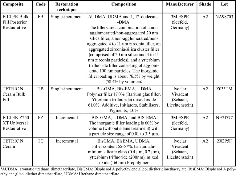
The composite resins Filtek Bulk Fill Posterior Restorative (FB) and Tetric N-Ceram Bulk Fill (TB) were analyzed for the restorations with the single-increment technique, and the Filtek Z250 XT Universal Restaurative (FZ) and Tetric N-Ceram (TC) were analyzed for the restorations with the technique. The incremental and compositional characteristics are indicated in Table 1.
Table 1. Composite descriptions, manufacturers and composition.
A randomization (www.random.org) of the teeth was carried out to define the technique and resin to be used, and the information was managed by one researcher.
-Tooth preparation
The teeth were cleaned with a prophylactic brush and pumice stone, and the soft tissue remains were removed with a curette. The apex was sealed with glass ionomer (GC Gold Label 9, FUJI, Tokyo, Japan) and then kept in physiological serum (NaCl 0.09%) at 6 °C, changing it twice a week until the experimental part began. Each tooth was placed in individual sockets to achieve stability at the time of cavity preparation and subsequent restoration.
We proceeded to make standardized class I cavity preparations of 4x4x4 mm in width, length and depth, ensuring measurements with the use of the periodontal probe. The cavity opening was made with a round bur, and the samples were prepared with a straight-tipped tapered bur using rubber stops as guides.
-Cavity conditioning and restorations
The restorations were performed by a single operator controlling the operative time from the beginning of each restoration. The restorations were performed according to the specifications of each manufacturer, and a PhotoLight Max light curing lamp (Gnatus, Riberão Preto-SP, Brazil) was used with a light-emitting diode (LED) light power of 1200 mW/cm2 with continuous light as follows:
For one-piece technique restorations (FB–TB), the following steps were conducted:
• Acid selective conditioning the tooth structure, first conditioning the enamel and then conditioning the dentin, for 15 s with Scotchbond Universal Etchant (3M ESPE, Seefeld, Germany. LOT NA98703)
• Washing the cavity with running water for 15 seconds and then drying the enamel while leaving the cavity/dentin wet
• Placement of 2% chlorhexidine for 10 seconds and cavity drying with cotton
• Adper Single Bond 2 adhesive (3M ESPE, Seefeld, Germany. LOT: NE63329) was applied on FB resin and N-Bond (Ivoclar Vivadent, Schaan, Liechtenstein. LOT: Z033TM) was applied on TB resin as the first layers for both the enamel and dentin by rubbing the dentin for 10 seconds
• Unifying the first layer with air application
• Placement of the second layer of adhesive and light curing for 10 seconds
• Restoration with polymerization for 40 seconds
For restorations with the incremental technique (FZ–TC), the following steps were performed:
• Acid selective conditioning the tooth structure, first conditioning the enamel and then conditioning the dentin, for 15 seconds with Scotchbond Universal Etchant (3M ESPE, Seefeld, Germany. LOT: 7601007)
• Washing the cavity with running water for 15 seconds and then drying the enamel while keeping the cavity/dentin wet
• Placement of 2% chlorhexidine for 10 seconds and cavity drying with cotton
• Adper Single Bond 2 adhesive (3M ESPE, Seefeld, Germany. LOT: NE63329) was applied on FZ resin and N-Bond (Ivoclar Vivadent, Schaan, Liechtenstein. LOT: Z033TM) was applied on TC resin as the first layers for both the enamel and dentin by rubbing dentin for 10 seconds
• Unifying the first layer with air application
• Placement of the second layer of adhesive and conducting light curing for 10 seconds
• Restoration curing each layer for 15 seconds and performing final curing for 40 seconds
-Thermocycling, preparation and immersion of teeth in dye
Subsequently, thermocycling was carried out with 10,000 cycles, representing 1 year of use of the material in oral conditions as follows: (16) the teeth were immersed in water baths at 35 °C for 28 seconds, 15 °C for 2 seconds, 35 °C for 28 seconds and finally 45 °C for 2 seconds. This procedure was repeated 50 times for 3.5 days.
-Sample sectioning and observation
The pieces were washed and sectioned mesiodistally with a handpiece and a diamond disc with constant irrigation. Finally, the teeth were immersed in 0.1% methylene blue for 48 hours. After this time, the sealing was evaluated marginally at the mesial, distal and floor levels of the cavity using a Dino-Lite Premier AM4113T 1.3MP optical microscope (Dino-Lite Digital Microscope, Taiwan) and subsequent analysis with DinoCapture 2.0 software (Dino-Lite Premier, Taiwan). This analysis was performed by a single operator blinded to the material analyzed (JA).
-Statistical analysis
For the statistical analysis, the Statistical Package for Social Sciences (SPSS) v.25 program (IBM, Endicott NY, USA) was used. Descriptive data were obtained, and through Student’s t test, restoration techniques and operative time were related. With the Mann‒Whitney U test, the means of the internal restoration gap were compared according to the type of resin. Significant values were established at p <0.05.
Results
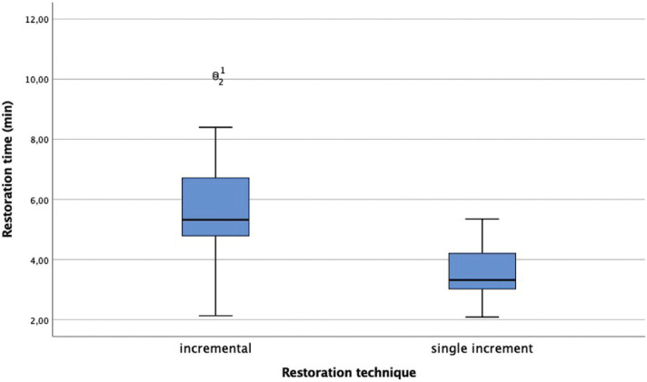
The descriptive analysis of the time according to the restoration technique is indicated in Table 2. The single-increment technique used with the Bulk fill resins obtains an average of 3.52 minutes with significant differences relative to the incremental technique (p <0.05) (Fig. 1).
Table 2. Restoration time according the restoration technique descriptive analysis: mean value, standard deviation, mínimum and máximum value. (µm) Statistical analysis: T test.
Figure 1.
Restoration time (min) according the restoration technique.
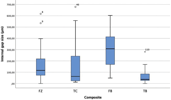
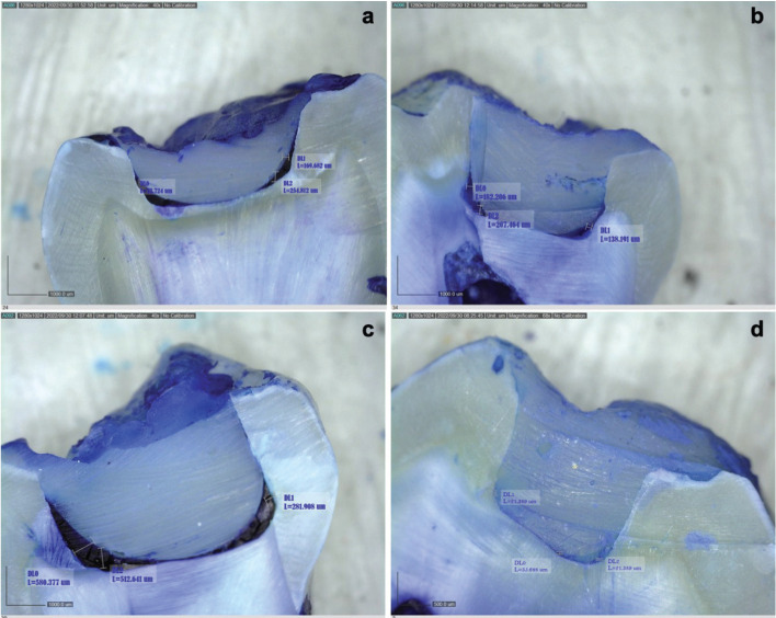
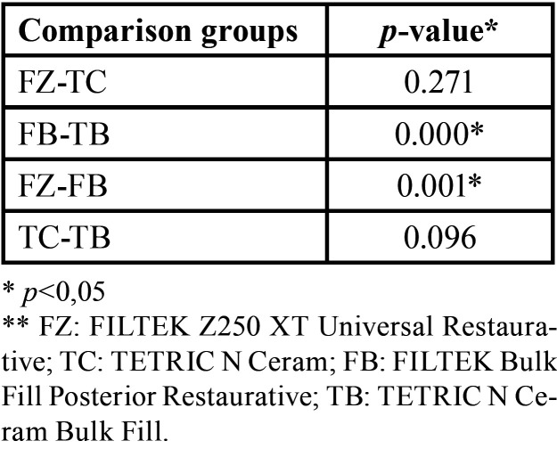
Figures 2 and 3 show the size of the internal gap of the restoration of the four resins considered for the present study. Table 3 shows very different behaviors between the two bulk fill resins (p <0.05), with the TB resin having the smallest internal gap size at an average value of 63.31 µm; the FB resin have the highest value of all the resins, with an average value of 313.14 µm. The two conventional resins (FZ and TC) present very similar average values (p> 0.05).
Figure 2.
Dental restoration internal gap size (µm). * FZ: FILTEK Z250 XT Universal Restaurative; TC: TETRIC N Ceram; FB: FILTEK Bulk Fill Posterior Restaurative; TB: TETRIC N Ceram Bulk Fill.
Figure 3.
Dental restoration internal gap size (µm) microscopical view. * a: FZ (FILTEK Z250 XT Universal Restaurative); b: TC (TETRIC N Ceram FB); c: FB (FILTEK Bulk Fill Posterior Restaurative); d: TB (TETRIC N Ceram Bulk Fill).
Table 3. Dental restoration internal gap size descriptive analysis (mesial, distal and floor levels of the cavity of each sample): mean value, standard deviation, mínimum and máximum value, (µm).
According to a comparison between groups (Table 4), the FB and FZ resins of the same commercial brand present significant differences between them (p <0.05).
Table 4. Dental restoration internal gap size statistical analysis: Mann-Whitney.
Finally, relative to the size of the internal gap according to the location, the TC, FB and TB resins present high values at the restoration floor level, while the FZ resin presents high values at the mesial level (Table 5).
Table 5. Dental restoration internal gap size mean (µm).
Discussion
In the present study, the internal gap formed between the restoration–tooth interface and the restoration operative time between bulk fill resins and conventional resins are observed; this analysis is crucial due to the importance of improving the clinical experience of a patient during restorations.
Composite resin is the most commonly used resin in direct adhesive restorations due to its aesthetic characteristics and mechanical properties (4,17,18). One of the great advantages of this material is that it does not require the removal of healthy tissue for its retention and that it has good mechanical behavior in the posterior sector (4,18,19). However, the disadvantages include the formation of secondary caries, fracture of the restoration, microleakage and marginal discoloration, which are all caused by polymerization shrinkage (4).
This contraction is responsible for the presence of marginal leaks, a high coefficient of thermal expansion and the absorption of water, impoverishing the internal adaptation and forming gaps between the restoration material and the dental structure, which allows the filtration of fluids, thus affecting long-term retention; however, the success of restorations depends on shrinkage stress and various other factors that impoverish the marginal seal (18,20,21).
The results of this study show that bulk fill resins present smaller internal gaps than conventional resins, even from the same commercial company, in cavitary preparations with 4-mm depth; the Tetric N-Ceram Bulk Fill presents the best results (63.31 µm), with other studies that corroborate these findings (15,17).
There are several factors to consider that can generate failures in the restoration–tooth interface, forming an internal gap that causes microfiltration that is responsible for postoperative sensitivity and secondary caries (21,22), such as cavitary preparation, restoration, adhesion, light irradiation, exposure time and the properties of the composite resin.
Bulk fill composite resins have small inorganic particle sizes, increasing the depth of light curing and allowing the passage of light in a better manner, reducing volumetric contraction and internal gap formation (23-27).
Regarding the restoration technique, it is important to indicate that the restoration time is measured from the moment the resin is introduced into the cavity preparation; this indication avoids confusion with the time necessary for the clinical preparation of the patient and for the placement of the adhesive system in each restoration. It is possible to determine that the single-increment technique used in Bulk Fill resins compared to the incremental technique improves clinical restoration times with significant differences, avoiding the gaps that the interfaces between layers may present when the incremental technique is conducted, improving marginal sealing. Notably, class I cavity preparations are used to improve Factor C (conFiguration factor), which can strongly affect the polymerization stress of the composite resin (28).
The FZ resin presents the greatest restoration–tooth interface with a value of 166.95 µm. The depth of the cavity preparation may affect the polymerization stress forming the internal gap because, despite using the incremental technique, the depth exceeds the specifications of conventional composite resins (1,5,6).
A two-step adhesive is used because studies indicate that etch-and-wash adhesives generate a thick hybrid layer with long, wide and dense resin layers; at this point, the filtration in dentin is not significant because the dentin tubules are sealed after the application of the adhesive, thus avoiding microfiltration because greater control is maintained in the time of application of the phosphoric acid and adhesive. In addition, chlorhexidine is used, which has been shown to improve the resistance of the hybrid layer (29). In this manner, it is possible to control errors in the adhesive sealing, improving the clinical restoration protocol (30,31).
It is important to remember that polymerization contraction affects the entire restoration–tooth interface. At this level, adding a poor-quality material can become more damaging to the dental structure.
Conclusions
By considering the limitations of the present in vitro study, it can be concluded that bulk fill resins present smaller internal gaps between restoration–tooth interfaces than conventional resins in 4 mm deep class I cavity preparations. In addition, single-increment restoration technique improves clinical restoration times relative to the oblique incremental technique.
Acknowledgments
Ackcnowledgements None declared.
${Availability of data and materials} None declared.
Authors contributions PMS raised the research idea, designed the methodology, supervised and excecuted the experimental part, processed the statistics, carried out the bibliographic search and wrote it. GO executed the experimental part (restorations). JA made the microscope readings and collected the sample. PO executed the experimental part, collect the sample and participated in the calibration. MR y RV sample sectioning, obteined the materials, collected the sample. All authors participated on the project administration, review, editing and approving the final draft.
Ethics We inform that we have followed the ethical guidelines set out in the Declaration of Helsinki on the use of human organs and tissue. The execution of this research project was approved by a ethical committee/IRB of the Universidad Católica de Cuenca with registration No. CEISH-UASB-O-CEISH-2022-001. All teeth were extracted for therapeutic reasons unrelated to the study at the Teaching Clinic of the Faculty of Stomatology of the Universidad Católica de Cuenca. Such teeth are collected, stored under special conditions and used for further research studies, with prior informed consent of the patients.
Conflicts of interest The authors declare that they have no conflict of interest with the development and publication of this research.
Funding The study was supported by the Universidad Católica de Cuenca (PICVII19-26).
References
- 1.Martins LC, Oliveira LRS, Braga SSL, Soares CJ, Versluis A, Borges GA. Effect of Composite Resin and Restorative Technique on Polymerization Shrinkage Stress, Cuspal Strain and Fracture Load of Weakened Premolars. J Adhes Dent. 2021;22:503–514. doi: 10.3290/j.jad.a45180. [DOI] [PubMed] [Google Scholar]
- 2.Ajaj RA, Farsi NJ, Alzain L, Nuwaylati N, Ghurab R, Nassar HM. Dental bulk-fill resin composites polymerization efficiency: A systematic review and meta-analysis. J Compos Sci. 2021;5:149. [Google Scholar]
- 3.Duarte JCL, Costa AR, Veríssimo C, Duarte RW, Filho SC, Spohr AM. Interfacial Stress and Bond Strength of Bulk-Fill or Conventional Composite Resins to Dentin in Class II Restorations. Braz Dent J. 2020;31:532–539. doi: 10.1590/0103-6440202003338. [DOI] [PubMed] [Google Scholar]
- 4.Kruly PC, Giannini M, Pascotto RC, Tokubo LM, Suga USG, Marques ACR. Meta-analysis of the clinical behavior of posterior direct resin restorations: Low polymerization shrinkage resin in comparison to methacrylate composite resin. PLOS ONE. 2018;13:e0191942. doi: 10.1371/journal.pone.0191942. [DOI] [PMC free article] [PubMed] [Google Scholar]
- 5.Aggarwal N, Jain A, Gupta H, Abrol A, Singh C, Rapgay T. The comparative evaluation of depth of cure of bulk-fill composites - An in vitro study. JCD. 2020;5:371–375. doi: 10.4103/JCD.JCD_453_18. [DOI] [PMC free article] [PubMed] [Google Scholar]
- 6.Alqudaihi FS, Cook NB, Diefenderfer KE, Bottino MC, Platt JA. Comparison of Internal Adaptation of Bulk-fill and Increment-fill Resin Composite Materials. Oper Dent. 2019;44:E32–E44. doi: 10.2341/17-269-L. [DOI] [PubMed] [Google Scholar]
- 7.Peskersoy C, Recen D, Kemaloğlu H. The effect of composite placement technique on the internal adaptation, gap formation and microshear bond strength. Eur Oral Res. 2022;56:10–16. doi: 10.26650/eor.2022897456. [DOI] [PMC free article] [PubMed] [Google Scholar]
- 8.Peutzfeldt A, Mühlebach S, Lussi A, Flury S. Marginal Gap Formation in Approximal "Bulk Fill" Resin Composite Restorations After Artificial Ageing. Oper Dent. 2018;43:180–189. doi: 10.2341/17-068-L. [DOI] [PubMed] [Google Scholar]
- 9.Pinto G, Prieto L, Pierote JJ, Ferraz L, Câmara JV, Aguiar FH. Effect of different adhesive strategies on the microtensile bond strength of dentin to indirect resin-based composite. J Clin Exp Dent. 2020;12:e1066–e1070. doi: 10.4317/jced.57094. [DOI] [PMC free article] [PubMed] [Google Scholar]
- 10.Zotti F, Falavigna E, Capocasale G, De Santis D, Albanese M. Microleakage of Direct Restorations-Comparisonbetween Bulk-Fill and Traditional Composite Resins: Systematic Review and Meta-Analysis. Eur J Dent. 2021;15:755–767. doi: 10.1055/s-0041-1724155. [DOI] [PMC free article] [PubMed] [Google Scholar]
- 11.Radwa A, Ahmed H. Fracture resistance of mod cavities in maxillary premolar teeth restored with different restorative protocols: an in-vitro study. E.D. J. 2022;68:1907–1916. [Google Scholar]
- 12.Iwasaki T, Kamiya N, Hirayama S, Tanimoto Y. Evaluation of the mechanical behavior of bulk-fill and conventional flowable resin composites using dynamic micro-indentation. Dent Mater J. 2022;41:87–94. doi: 10.4012/dmj.2021-116. [DOI] [PubMed] [Google Scholar]
- 13.Soto-Montero J, Giannini M, Sebold M, de Castro EF, Abreu JLB, Hirata R. Comparison of the operative time and presence of voids of incremental and bulk-filling techniques on Class II composite restorations. Quintessence Int. 2022;53:200–208. doi: 10.3290/j.qi.b2218737. [DOI] [PubMed] [Google Scholar]
- 14.Duarte JCL, Costa AR, Veríssimo C, Duarte RW, Filho SC, Spohr AM. Interfacial Stress and Bond Strength of Bulk-Fill or Conventional Composite Resins to Dentin in Class II Restorations. Braz Dent J. 2020;31:532–539. doi: 10.1590/0103-6440202003338. [DOI] [PubMed] [Google Scholar]
- 15.Han SH, Park SH. Incremental and Bulk-fill Techniques With Bulk-fill Resin Composite in Different Cavity Configurations. Oper Dent. 2018;43:631–641. doi: 10.2341/17-279-LR. [DOI] [PubMed] [Google Scholar]
- 16.Gale MS, Darvell BW. Thermal cycling procedures for laboratory testing of dental restorations. J Dent. 1999;27:89–99. doi: 10.1016/s0300-5712(98)00037-2. [DOI] [PubMed] [Google Scholar]
- 17.Cayo-Rojas CF, Hernández-Caba KK, Aliaga-Mariñas AS, Ladera-Castañeda MI, Cervantes-Ganoza LA. Microleakage in class II restorations of two bulk fill resin composites and a conventional nanohybrid resin composite: an in vitro study at 10,000 thermocycles. BMC oral health. 2021;21:619. doi: 10.1186/s12903-021-01942-0. [DOI] [PMC free article] [PubMed] [Google Scholar]
- 18.Liu J, Zhang H, Sun H, Liu Y, Liu W, Su B. The Development of Filler Morphology in Dental Resin Composites: A Review." Materials (Basel, Switzerland) Materials (Basel) 2021;14:5612. doi: 10.3390/ma14195612. [DOI] [PMC free article] [PubMed] [Google Scholar]
- 19.Rachmia J, Fauziyah S. Dental Composite Resin: A Review. AIP Conf Proc. 2019;2193 [Google Scholar]
- 20.Gamarra VSS, Borges GA, Júnior LHB, Spohr AM. Marginal adaptation and microleakage of a bulk-fill composite resin photopolymerized with different techniques. Odontology. 2018;106:56–63. doi: 10.1007/s10266-017-0294-5. [DOI] [PubMed] [Google Scholar]
- 21.Papagiannoulis L, Kakaboura A, Eliades G. In vivo vs in vitro anticariogenic behavior of glass-ionomer and resin composite restorative materials. Dent Mater. 2002;18:561–9. doi: 10.1016/s0109-5641(01)00090-2. [DOI] [PubMed] [Google Scholar]
- 22.Thomas RZ, Ruben JL, ten Bosch JJ, Fidler V, Huysmans MC. Approximal secondary caries lesion progression, a 20-week in situ study. Caries Res. 2007;41:399–405. doi: 10.1159/000104799. [DOI] [PubMed] [Google Scholar]
- 23.Guéders AM, Charpentier JF, Albert AI, Geerts SO. Microleakage After Thermocycling of 4 Etch and Rinse and 3 Self-etch Adhesives with and Without a Flowable Composite Lining. Oper Dent. 2006;31:450–455. doi: 10.2341/05-55. [DOI] [PubMed] [Google Scholar]
- 24.Hayashi J, Espigares J, Takagaki T, Shimada Y, Tagami J, Numata T. Real-time in-depth imaging of gap formation in bulk-fill resin composites. Dental materials: official publication of the Academy of Dental Materials. 2019;35:585–596. doi: 10.1016/j.dental.2019.01.020. [DOI] [PubMed] [Google Scholar]
- 25.Alsagob E, Bardwell D, Ali A, Dhayat S, Stark P. Comparison of microleakage between bulk-fill flowable and nanofilled resin-based composites. Interventional medicine & applied science. 2018;10:102–109. doi: 10.1556/1646.10.2018.07. [DOI] [PMC free article] [PubMed] [Google Scholar]
- 26.Poggio C, Chiesa M, Scribante A, Mekler J, Colombo M. Microleakage in Class II composite restorations with margins below the CEJ: In vitro evaluation of different restorative techniques. Med Oral Patol Oral Cir Bucal. 2013;1:793–8. doi: 10.4317/medoral.18344. [DOI] [PMC free article] [PubMed] [Google Scholar]
- 27.Benetti AR, Havndrup-Pedersen C, Honoré C, Pedersen MK, Pallesen U. Bulk-fill resin composites: polymerization contraction, depth of cure, and gap formation. Oper Dent. 2015;40:190–200. doi: 10.2341/13-324-L. [DOI] [PubMed] [Google Scholar]
- 28.Han SH, Sadr A, Shimada Y, Tagami J, Park SH. Adaptation Internal of composite restorations with or without intermediate an layer effect of polymerization shrinkage parameters of the layer material. JDent. 2019;80:41–8. doi: 10.1016/j.jdent.2018.10.013. [DOI] [PubMed] [Google Scholar]
- 29.Breschi L, Maravic T, Comba A, Cunha SR, Loguercio AD, Reis A. Chlorhexidine preserves the hybrid layer in vitro after 10-years aging. Dent Mater. 2020;36:672–680. doi: 10.1016/j.dental.2020.03.009. [DOI] [PubMed] [Google Scholar]
- 30.Tosco V, Vitiello F, Furlani M, Gatto ML, Monterubbianesi R, Giuliani A. Microleakage analysis of Different Bulk-Filling Techniques for Class II Restorations: CT, SEM and EDS Evaluations. Materials. 2021;14:31. doi: 10.3390/ma14010031. [DOI] [PMC free article] [PubMed] [Google Scholar]
- 31.Tran XV, Quang K. Microleakage and characteristics of resin-tooth tissues interface of a self-etch and an etch-and-rinse adhesive systems. " Restorative dentistry & endodontics. 2021;46:30. doi: 10.5395/rde.2021.46.e30. [DOI] [PMC free article] [PubMed] [Google Scholar]


