Correction: J Exp Clin Cancer Res 37, 13 (2018)
10.1186/s13046-018-0684-8
Following publication of the original article [1], an error was identified in Figs. 3b and 4d/e, and Fig. 7c.
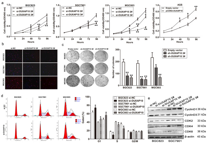
Fig. 3.
DUXAP10 promotes GC cells growth and cell cycle progression. a MTT assays were used to determine the cell viability for si-DUXAP10 or si-NC transfected BGC823, SGC7901 and MGC803 cells, and DUXAP10 vector or empty vector transfected AGS cells. Values represented the mean ± s.d. from three independent experiments. b Edu staining analysis showing significant decrease of cell viability in si-DUXAP10 transfected BGC823, SGC7901 and MGC803 cells. c Colon formation assays showing significant decrease of cloning viability in si-DUXAP10 transfected GC cells. d FACS analysis shows significant increases or decreases of cells in G1or S phase, respectively, in si-DUXAP10 transfected GC cells. e Cyclin D1, Cyclin D3, CDK2, CDK4, and CDK6 protein levels were detected by western blot analysis after DUXAP10 knockdown. *P < 0.05, **P < 0.01
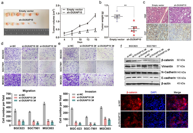
Fig. 4.
DUXAP10 down-regulation inhibits GC cells tumor growth in vivo, and invasion in vitro. a Representative images of tumors formed in nude mice injected subcutaneously with DUXAP10 knockdown BGC823 cells, and the tumor growth curves of DUXAP10 down-regulation and control groups. b Tumors induced by DUXAP10 knockdown in BGC823 cells showed markedly lower weight than control cells. c Tumors developed from sh-DUXAP10 transfected BGC823 cells showed lower ki67 protein levels than tumors developed by control cells. Up: H & E staining; Down: immunostaining. d,e Transwell assays were used to investigate the changes in migratory and invasive abilities of DUXAP10 knockdown cells. f E-cadherin, N-cadherin, Vimentin and β-catenin protein levels were detected by western blot and Immunofluorescence analysis after DUXAP10 knockdown in BGC823 cells. *P < 0.05, **P < 0.01
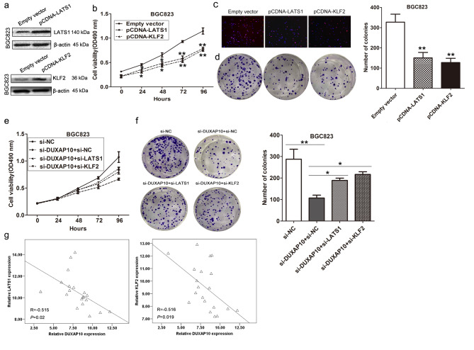
Fig. 7.
DUXAP10 promotes GC cell proliferation partly via regulating LATS1 and KLF2. a KLF2 and LATS1 protein levels were detected by western blot in BGC823 cells transfected with KLF2 or LATS1 vector. b MTT assays were used to determine the cell viability for LATS1 and KLF2 vector or empty vector transfected BGC823 and SGC7901 cells. c,d Edu staining and colony formation assays were used to determine the cell viability for LATS1, KLF2 vector or empty vector transfected cells. e,f MTT and colony formation assays showed that cell proliferation was partly rescued by KLF2 and LATS1 knockdown in DUXAP10 siRNA transfected cells. g The correlation between DUXAP10 and KLF2, or LATS1 expression was detected in 20 pairs of GC and corresponding noncancerous tissues by qRT-PCR
The corrected figures are given below. The corrections do not affect the conclusions of the article.
Footnotes
The online version of the original article can be found at 10.1186/s13046-018-0684-8.
Publisher’s Note
Springer Nature remains neutral with regard to jurisdictional claims in published maps and institutional affiliations.
Contributor Information
Fengqi Nie, Email: NieFengqi@njmu.edu.cn.
Mingde Huang, Email: 2471843860@qq.com.
Ming Sun, Email: msun7@mdanderson.org.
References
- 1.Xu Y, Yu X, Wei C, et al. Over-expression of oncigenic pesudogene DUXAP10 promotes cell proliferation and invasion by regulating LATS1 and β-catenin in gastric cancer. J Exp Clin Cancer Res. 2018;37:13. doi: 10.1186/s13046-018-0684-8. [DOI] [PMC free article] [PubMed] [Google Scholar]


