

Abstract
Background:
Rarely, chronic tophaceous gout can result in lumbar spinal stenosis and neural compression.
Case Description:
A 67-year-old male presented with the radiographic and magnetic resonance findings of gout involving and causing compression of the lumbar spine that responded to surgical decompression.
Conclusion:
It is difficult to diagnose lumbar spinal stenosis secondary to tophaceous gout. Notably, the treatment, based on the clinical presentation, may include both medication and surgical decompression.
Keywords: Joint inflammation, Lumbar spinal stenosis, Multidisciplinary approach, Tophaceous gouty arthritis, Tophi, Urate crystals

INTRODUCTION
Tophaceous gouty arthritis is characterized by the accumulation of urate crystals in and around joints. It constitutes a chronic and debilitating form of arthritis that primarily affects the peripheral joints (i.e., toes, fingers, and knees), but can also involve other structures (i.e., tendons, bursa, and spine).[2] In rare cases, tophaceous gouty arthritis may coexist with and exacerbate the symptoms/signs of lumbar spinal stenosis. The management of these two conditions may require a multidisciplinary approach involving rheumatologists, pain management specialists, and spine surgeons. In addition to the medical management with anti-gout medications, it may require surgical intervention. Here, a 67-year-old male presented with both tophaceous gouty arthritis and lumbar spinal stenosis warranting both medication and decompressive surgery.
CASE REPORT
A 67-year-old male presented with bilateral radicular lower back pain and neurogenic claudication (i.e., occurring within <5 min of ambulation) for 20 years. He also had pain in both his hands and feet. On physical examination, he had multiple hard, nodular swellings consistent with tophi in his hands and feet, including 0.5 cm tophi involving both big toes and the right little toe, a large 1.4 cm tophus involving the right middle finger, a 1 cm tophus on the right ring, and a 0.8 cm tophus at the right wrist [Figures 1 a-c]. There were also numerous 0.5 cm tophi noted involving the inter-phalangeal joints of the left hand [Figure 1b]. On examination, he had midline/paracentral tenderness over the L4 and L5 vertebrae, bilaterally decreased ankle reflexes and diminished L4-S1 sensation to pin appreciation. Laboratory studies showed hyperuricemia (i.e., initial uric acid level of 10.6 mg/dL), normal antinuclear antibody profile, and slightly elevated rheumatoid factor level at 16.17 IU/mL. (i.e., Reference range <14 IU/mL).
Figure 1:

Clinical photographs of patient’s bilateral feet (a), bilateral hands (b), and front view of the right hand (c) demonstrating multiple tophaceous swellings involving various joints of hands and feet.
Magnetic resonance (MR) studies
Axial and sagittal MR imaging views showed disc desiccation at multiple levels with disc space narrowing and Type II endplate changes at the L3/4 and L4/5 levels. At L3/4, L4/5, and L5/S1, there was significant bilateral lateral recess stenosis attributed to central disc bulges, and marked facet hypertrophy/reactive arthrosis and thickening of the yellow ligament. Added hyperintensities were also noted bilaterally adjacent to the spinous processes of L3 through L5 [Figures 2 and 3]. The patient was referred for surgery and to rheumatology for additional medical management with anti-gout medications.
Figure 2:

Sagittal views of magnetic resonance imaging lumbosacral spine of the patient showing right sagittal cut (a), midsagittal cut (b), and left sagittal cut (c).
Figure 3:

Axial views of the magnetic resonance imaging lumbosacral spine at (a) L3-4, (b) L4-5, and (c) L5-S1 levels of the patient. The green arrow in (a) is demonstrating the thickening of the spinous process noted at the L3-4 level. The yellow arrow in (b) demonstrates a hyper-intense signal in the left paraspinal region.
Surgery
He underwent a decompressive laminectomy from L3 to L5 with medial facetectomy/foraminotomy and bilateral excision of tophi involving the spinous process and around the bilateral facet joints under spinal anesthesia. Intraoperatively, thickened yellow ligament and hypertrophied facet joints and spinous processes were removed along with multiple tophaceous nodules [Figure 4]. The patient was discharged on the same day, and later the bilateral leg pain and claudication improved significantly. He was then referred to rheumatology.
Figure 4:
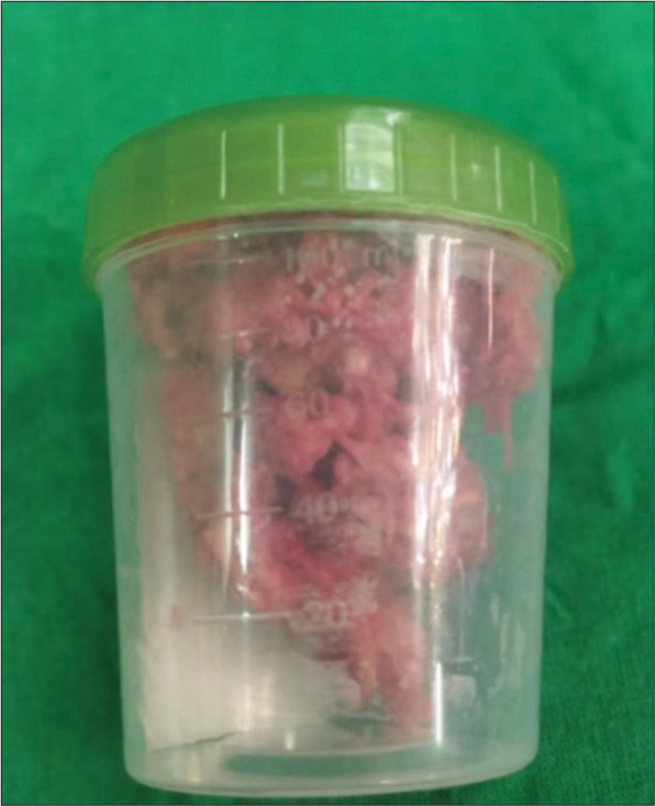
Photograph of the specimen excised intraoperatively demonstrating tophi along with bony tissue.
DISCUSSION
Definition of tophaceous spinal gout
Gout is a crystal arthropathy characterized by the deposition of uric acid within joints. The lumbar region is particularly susceptible to gout,[3,6] with various parts of the vertebral body, including the epidural space, ligamentum flavum, pedicle, and facet joint, being potential sites of manifestation.[5] Studies have indicated that axial involvement may occur in approximately 14–35% of cases.[3,6,8] Tophi deposits in the axial elements of the spine are typically found at the facet articulation, but other structures such as the ligamentum flavum, pedicle, lamina, epidural soft tissue, vertebral body, and disc space can also be affected.[10]
Symptoms of spinal tophaceous gout
Patients may present with back pain, myelopathy, and/or radiculopathy (i.e., radiculopathy is the most prevalent symptom in 34.5% of cases)[3] Spinal gout tends to occur more frequently in men between 35 and 75 years of age, particularly in those with chronic polyarticular tophaceous gout that is not optimally controlled.[4-6] There also appears to be a positive correlation between the onset of symptoms and elevated serum uric acid levels.[5]
MR diagnosis of tophaceous spinal gout
Tophi, characteristic of gout, in the spine may produce a homogenous intermediate to hypointense signal on T1-weighted images, while intensity is variable on T2-weighted sequences.[8] Gouty tophi may produce either homogeneous or peripheral heterogeneous patterns attributed to variations in the vascularization of the inflammatory tissue induced by the deposition of monosodium urate crystals.[9,10]
Medical management
Pharmacologic urate acid-lowering therapies form the mainstay of treatment for spinal gout and tophi. Gout is typically managed using colchicine, nonsteroidal anti-inflammatory drugs, or a combination of both to stabilize acute attacks. Urate-lowering therapy is achieved through the use of xanthine oxidase inhibitors (XOIs), which minimize uric acid synthesis and reduce serum urate levels.[7] Allopurinol and febuxostat are the two XOIs approved by the US food and drug administration. Uricosuric agents like probenecid may be used in combination with an XOI or as monotherapy for refractory gout.[10]
Surgery
When tophaceous gout contributes to spinal stenosis and root/cord compression, root surgical decompression often resulting in favorable outcomes may be warranted.[1]
CONCLUSION
Here, a 67-year-old male presented with lumbar radiculopathy and neurogenic claudication attributed to lumbar stenosis complicated by superimposed tophaceous gouty arthritis involving the yellow ligament, lumbar facet joints, and spinous processes. Notably, the patient markedly improved following the lumbar decompression surgery.
Footnotes
How to cite this article: Jazaib Ali M, Hussain M. Case of lumbar spinal stenosis and chronic tophaceous gout. Surg Neurol Int 2023;14:294.
Contributor Information
Muhammad Yassar Jazaib Ali, Email: yasirkhebar@yahoo.com.
Manzar Hussain, Email: manzar.hoseyn@gmail.com.
Declaration of patient consent
The authors certify that they have obtained all appropriate patient consent.
Financial support and sponsorship
Nil.
Conflicts of interest
There are no conflicts of interest.
Disclaimer
The views and opinions expressed in this article are those of the authors and do not necessarily reflect the official policy or position of the Journal or its management. The information contained in this article should not be considered to be medical advice; patients should consult their own physicians for advice as to their specific medical needs.
REFERENCES
- 1.Elgafy H, Liu X, Herron J. Spinal gout: A review with case illustration. World J Orthop. 2016;7:766–75. doi: 10.5312/wjo.v7.i11.766. [DOI] [PMC free article] [PubMed] [Google Scholar]
- 2.Golenbiewski J, Keenan RT. Moving the needle: Improving the care of the gout patient. Rheumatol Ther. 2019;6:179–93. doi: 10.1007/s40744-019-0147-5. [DOI] [PMC free article] [PubMed] [Google Scholar]
- 3.Hasegawa EM, de Mello FM, Goldenstein-Schainberg C, Fuller R. Gout in the spine. Rev Bras Reumatol. 2013;53:296–302. [PubMed] [Google Scholar]
- 4.Hasturk AE, Basmaci M, Canbay S, Vural C, Erten F. Spinal gout tophus: A very rare cause of radiculopathy. Eur Spine J. 2012;21(Suppl 4):S400–3. doi: 10.1007/s00586-011-1847-x. [DOI] [PMC free article] [PubMed] [Google Scholar]
- 5.King JC, Nicholas C. Gouty arthropathy of the lumbar spine: A case report and review of the literature. Spine (Phila Pa 1976) 1997;22:2309–12. doi: 10.1097/00007632-199710010-00023. [DOI] [PubMed] [Google Scholar]
- 6.Konatalapalli RM, Lumezanu E, Jelinek JS, Murphey MD, Wang H, Weinstein A. Correlates of axial gout: A cross-sectional study. J Rheumatol. 2012;39:1445–9. doi: 10.3899/jrheum.111517. [DOI] [PubMed] [Google Scholar]
- 7.Richette P, Doherty M, Pascual E, Barskova V, Becce F, Castañeda-Sanabria J, et al. 2016 updated EULAR evidence-based recommendations for the management of gout. Ann Rheum Dis. 2017;76:29–42. doi: 10.1136/annrheumdis-2016-209707. [DOI] [PubMed] [Google Scholar]
- 8.Wendling D, Prati C, Hoen B, Godard J, Vidon C, GodfrinValnet M, et al. When gout involves the spine: Five patients including two inaugural cases. Joint Bone Spine. 2013;80:656–9. doi: 10.1016/j.jbspin.2013.06.002. [DOI] [PubMed] [Google Scholar]
- 9.Yen PS, Lin JF, Chen SY, Lin SZ. Tophaceous gout of the lumbar spine mimicking infectious spondylodiscitis and epidural abscess: MR imaging findings. J Clin Neurosci. 2005;12:44–6. doi: 10.1016/j.jocn.2004.03.020. [DOI] [PubMed] [Google Scholar]
- 10.Zheng ZF, Shi HL, Xing Y, Li D, Jia JY, Lin S. Thoracic cord compression due to ligamentum flavum gouty tophus: A case report and literature review. Spinal Cord. 2015;53:881–6. doi: 10.1038/sc.2015.93. [DOI] [PMC free article] [PubMed] [Google Scholar]