Abstract
Since the emergence of cardiac computed tomography (Cardiac CT) at the turn of the 21st century, there has been an exponential growth in research and clinical development of the technique, with contributions from investigators and clinicians from varied backgrounds: physics and engineering, informatics, cardiology, and radiology. However, terminology for the field is not unified. As a consequence, there are multiple abbreviations for some terms, multiple terms for some concepts, and some concepts that lack clear definitions and/or usage. In an effort to aid the work of all those who seek to contribute to the literature, clinical practice, and investigation of the field, the Society of Cardiovascular Computed Tomography updates a standard set of medical terms commonly used in clinical and research activities related to cardiac CT.
Keywords: Cardiac, CT, Medical Terminology
Supplemental material is available for this article.
This article is published synchronously in Radiology: Cardiothoracic Imaging and Journal of Cardiovascular Computed Tomography.
©2023 Society of Cardiovascular Computed Tomography. Published by RSNA with permission.
Keywords: Cardiac, CT, Medical Terminology
See commentary by Roberts and Hanneman in this issue.
Introduction
Cardiac computed tomography (Cardiac CT) continues to expand with increasing applications and evolving technology. The terminology used in the field was previously unified by consensus agreement of cardiologists, medical physicists and radiologists in 2006 (1). A representative writing group with key stakeholders was reconvened to provide an update to the nomenclature of terms commonly used in clinical and research applications of cardiac CT. The purpose of this work is to consolidate multiple terms and to provide clear definitions for terms applicable to cardiac CT.
The writing group focused on terms most relevant to cardiac CT. Not included within the scope of this document were more general terms related to vascular imaging interpretation and analysis such as cross-sectional area or percent diameter stenosis. These were thought to be well understood or have been clearly defined in the literature of their respective fields. The one set of exceptions was terms used to describe plaque composition. New inclusions for version 2.0 include addition of expanding CT technology and radiology lexicon standard terminology. Radlex® (which is short for radiology lexicon) is developed and maintained by the Radiological Society of North America (RSNA) to provide common terminology for radiology exams and reports including the LOINC/RSNA Radiology Playbook, RadElement Common Data Elements and RadReport radiology reporting templates. The website www.radlex.org provides a search engine to support adoption of preferred terminology, with preferred name, RadLex ID and preferred user radiology lexicon (PURL) defined for each term (2).
The document underwent organization review by the SCCT Board of Directors and American Association of Physicists in Medicine's Board, the American College of Radiology's Board, North American Society for Cardiovascular Imaging's Board and by the Radiological Society of North America's Board and external peer review. Disclosures of potential conflicts of interest for the writing group and external peer reviewers may be found in Appendix 1. Affiliations of the external peer reviewers may be found in Appendix 2.
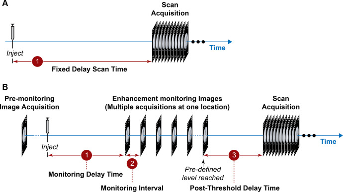
Contrast administration (A) fixed scan delay, (B) monitoring delay time, monitoring interval, post threshold delay time. Used with permission of Mayo Foundation for Medical Education and Research, all rights reserved.
Explanation of Tables
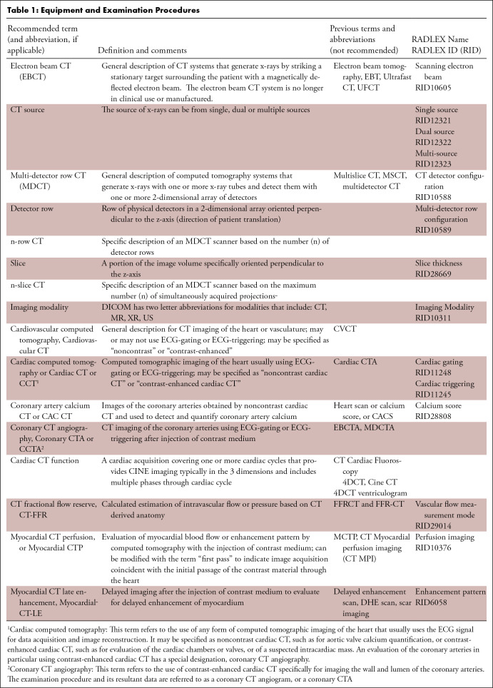
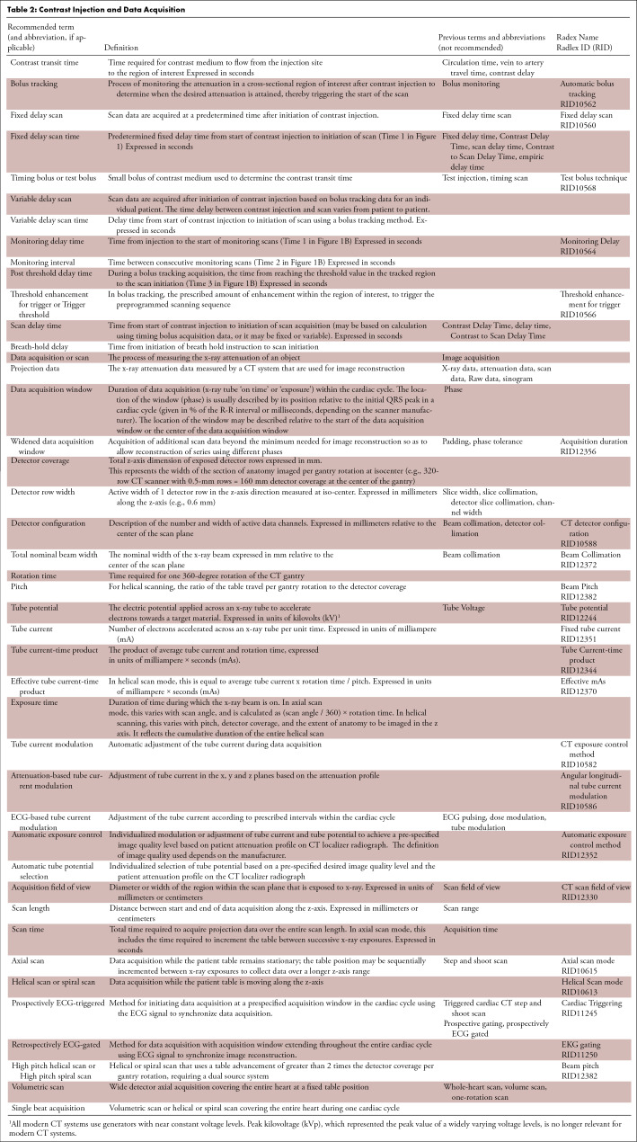
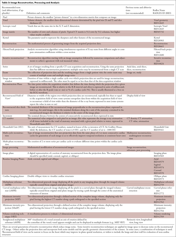
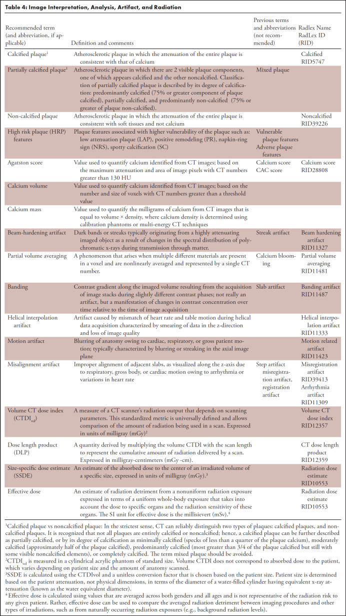
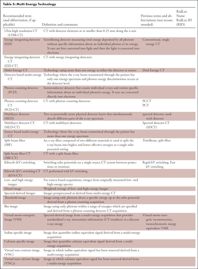
This document provides tables of standardized medical terminology for cardiac CT applying to Equipment and Examination Procedures (Table 1), Contrast Injection and Data Acquisition (Table 2), Image Reconstruction, Processing, and Analysis (Table 3), Image Interpretation, Analysis, Artifacts, and Radiation (Table 4) and Multi-energy Technology (Table 5) (3–7). In each table, the recommended terms are listed in the first column, with any recommended abbreviations in parentheses. Definitions and any comments regarding the terms or their usage, including occasional cases of other acceptable terms, are listed in the next column. Previous terms and abbreviations that are not recommended and hence to be avoided are in the third column. The RADLEX name and ID are given in the fourth column. Decisions about terms were made by consensus (majority opinion).
Table 1:
Equipment and Examination Procedures
Table 2:
Contrast Injection and Data Acquisition
Table 3:
Image Reconstruction, Processing and Analysis
Table 4:
Image Interpretation, Analysis, Artifact, and Radiation
Table 5:
Multi-Energy Technology
Footnotes
Writing group co-chairs;
Representative of American Association of Physicists in Medicine;
Representative of American College of Radiology;
Representative of North American Society for Cardiovascular Imaging;
Representative of Radiological Society of North America.
Disclosures of conflicts of interest: All Conflicts of interest are noted per SCCT guidelines committee reporting. See Appendix 1 and 2.
References
- 1. Weigold WG , Abbara S , Achenbach S , Arbab-Zadeh A , Berman D , Carr JJ , Cury RC , Halliburton SS , McCollough CH , Taylor AJ ; >Society of Cardiovascular Computed Tomography . Standardized medical terminology for cardiac computed tomography: a report of the Society of Cardiovascular Computed Tomography . J Cardiovasc Comput Tomogr 2011. May-Jun ; 5 ( 3 ): 136 – 144 . doi: 10.1016/j.jcct.2011.04.004 . [DOI] [PubMed] [Google Scholar]
- 2. RadLex Radiology Lexicon . https://www.rsna.org/practice-tools/data-tools-and-standards/radlex-radiology-lexicon. Last accessed 7/26/2022.
- 3. Shaw LJ , Blankstein R , Bax JJ , Ferencik M , Bittencourt MS , Min JK , Berman DS , Leipsic J , Villines TC , Dey D , Al'Aref S , Williams MC , Lin F , Baskaran L , Litt H , Litmanovich D , Cury R , Gianni U , van den Hoogen I , R van Rosendael A , Budoff M , Chang HJ , E Hecht H , Feuchtner G , Ahmadi A , Ghoshajra BB , Newby D , Chandrashekhar YS , Narula J . Society of Cardiovascular Computed Tomography / North American Society of Cardiovascular Imaging - Expert Consensus Document on Coronary CT Imaging of Atherosclerotic Plaque . J Cardiovasc Comput Tomogr 2021. Mar-Apr ; 15 ( 2 ): 93 – 109 . doi: 10.1016/j.jcct.2020.11.002 . [DOI] [PubMed] [Google Scholar]
- 4. Satharasinghe DM , Jeyasugiththan J , Wanninayake WMNMB , Pallewatte AS . Size-specific dose estimates (SSDEs) for computed tomography and influencing factors on it: a systematic review . J Radiol Prot 2021. Dec 6 ; 41 ( 4 ). doi: 10.1088/1361-6498/ac20b0 . [DOI] [PubMed] [Google Scholar]
- 5. DICOM Standards Committee, Working Groups 21 . Digital Imaging and Communications in Medicine (DICOM) Supplement 188: Multi-energy CT Images . ( 2018. ) https://www.dicomstandard.org/news-dir/progress/docs/sups/sup188.pdf. Last accessed 7/21/2022.
- 6. McCollough , Cynthia H. , Kirsten Boedeker , Dianna Cody , Xinhui Duan , Thomas Flohr , Sandra S. Halliburton , Jiang Hsieh , Rick R. Layman , and Norbert J. Pelc . “Principles and applications of multienergy CT: Report of AAPM Task Group 291.” Medical physics 47 , no. 7 ( 2020. ): e881 – e912 . [DOI] [PubMed] [Google Scholar]
- 7. Leng , S. , Bruesewitz , M. , Tao , S. , Rajendran , K. , Halaweish , A.F. , Campeau , N.G. , Fletcher , J.G. and McCollough , C.H. , 2019. . Photon-counting detector CT: system design and clinical applications of an emerging technology . RadioGraphics 39 ( 3 ), 729 . [DOI] [PMC free article] [PubMed] [Google Scholar]


