Abstract
We report on the production and characterization of Ehrlichia risticii, the agent of Potomac horse fever (PHF), from snails (Pleuroceridae: Juga spp.) maintained in aquarium culture and compare it genetically to equine strains. Snails were collected from stream waters on a pasture in Siskiyou County, Calif., where PHF is enzootic and were maintained for several weeks in freshwater aquaria in the laboratory. Upon exposure to temperatures above 22°C the snails released trematode cercariae tentatively identified as virgulate cercariae. Fragments of three different genes (genes for 16S rRNA, the groESL heat shock operon, and the 51-kDa major antigen) were amplified from cercaria lysates by PCR and sequenced. Genetic information was also obtained from E. risticii strains from horses with PHF. The PCR positivity of snail secretions was associated with the presence of trematode cercariae. Sequence analysis of the three genes indicated that the source organism closely resembled E. risticii, and the sequences of all three genes were virtually identical to those of the genes of an equine E. risticii strain from a property near the snail collection site. Phylogenetic analyses of the three genes indicated the presence of geographical E. risticii strain clusters.
Potomac horse fever (PHF), also called equine monocytic ehrlichiosis, is an important disease of horses caused by a monocytotropic rickettsia, Ehrlichia risticii (17–21, 24, 29). It was first observed in 1979 on pastures along the Potomac River in Maryland (18) and has since been identified in a number of other states and in Europe (24). Clinical signs include anorexia, lethargy, variable fever, and diarrhea; laminitis is a complication in a significant percentage of animals. Fatalities may result if severely affected horses are not treated promptly with fluid and antibiotic therapy.
The means by which horses become infected with E. risticii has remained a mystery (17). There is no evidence for the spread of the disease by arthropod vectors such as ticks (5, 15, 24–26, 33). It appears, rather, that cases of E. risticii infection are associated with riverine and other aquatic habitats and hence with potential aquatic vectors. E. risticii-caused diarrhea in horses is enzootic in northern California and southern Oregon and is known locally as the “Shasta River crud” (SRC) or “ditch fever,” owing to its association with pastures bordering rivers and irrigation ditches (5, 19). The hypothesis associating E. risticii with an aquatic environment is supported by recent phylogenetic studies that demonstrated a close phylogenetic relationship between E. risticii and three other rickettsiae associated with aquatic habitats: Neorickettsia helminthoeca, the agent of “salmon poisoning,” a frequently fatal enteric disease of canids; the SF agent, isolated in Japan from trematode metacercariae parasitic on gray mullet fish; and Ehrlichia sennetsu, the agent of human sennetsu ehrlichiosis in Japan and Malaysia (13, 14, 27, 28, 39). Together these four agents form a distinct cluster or genogroup separate from the other rickettsiae (9, 28, 30, 39).
This phylogenetic relationship and the association between PHF and aquatic habitats focused our attention on potential aquatic vectors that might be involved in the epizootiology of E. risticii in northern California and southern Oregon. We have since amplified fragments of three different E. risticii genes from operculate snails of the genus Juga (4). The fragments were virtually identical to the homologous genes of the SRC agent, an E. risticii strain isolated from a horse residing only a few miles from the snail collection site (19).
Here we describe the PCR-based detection of E. risticii genes in trematode cercariae released by operculate snails of the genus Juga. The snails were collected from stream waters on a pasture in Siskiyou County, Calif., when PHF is enzootic and were maintained for several weeks in freshwater aquaria in our laboratory. We hypothesize that trematodes that use operculate snails as intermediate hosts may be involved in the life cycle of E. risticii in northern California. We also compare genetic information obtained from snail-derived E. risticii with that from E. risticii from horses with PHF. We conclude that certain E. risticii strains obtained from horses are identical to those obtained from snail secretions and that geographical clusters of genetically polymorphic E. risticii strains are present. This information may have practical implications for current vaccination strategies.
MATERIALS AND METHODS
Snail collection.
Freshwater snails were collected in August 1997 from a pasture in Weed, Siskiyou County, Calif.; some horses residing on that pasture had a history of PHF. Snails were collected by hand or with a net from the clear, shallow margins of a stream that flows through the pasture and that is accessible to horses for drinking. The stream waters are fed by the nearby Shasta River. A total of about 400 pleurocerid snails of the genus Juga (6) were collected and transported in chilled stream water (10 to 12°C) to our laboratory at the University of California, Davis. On the basis of the current nomenclature derived from classical shell characters, the majority of the individuals resembled the species Juga hemphilli hemphilli, having ribs (costae) for the most part limited to the apical whorl of the shell. Recognizing, however, that the systematics of the family Pleuroceridae are in need of thorough revision (6, 8), identification of these snails to the species level remains presumptive.
Aquarium culture.
In the laboratory, the snails were rinsed briefly with tap water and were distributed into three freshwater aquaria (10 gal each). The floor of each tank was covered with washed sand pebbles and the tanks were filled with tap water (pH 8.1) treated with 5 ml of water conditioner (Amquel; Kordon, Hayward, Calif.) per 10 gal to eliminate possible ammonia, chloramine, and chlorine contamination. The water tested negative for copper, which is known to be lethal for snails. The lids of the tanks were fitted with daylight lamps that were used for about 8 to 10 h each day. Two tanks with 75 snails each were kept at room temperature (RT; about 22 to 24°C), and one tank with about 250 snails was kept at 8°C. The larger snails (about 2.0 to 2.5 cm) were kept at RT. The sizes of snails maintained at 8°C ranged from about 0.5 to 1.5 cm. Snails were fed alga pellets and fish flakes ad libitum. The tank water was continually filtered through activated carbon filters (Whisper Power Filter; Tetra Sales, Blacksburg, Va.) and was replaced weekly with fresh tap water. The filters were replaced weekly as well.
Source and preparation of E. risticii strains used for phylogenetic comparison.
Peripheral blood leukocytes (PBLs) were obtained from 10 horses clinically diagnosed with PHF and were used as the source of template DNA for PCR. Five horses (Buck, Bunn, Danny, Tate, and Thorenberg) were from Klamath Falls, Oreg. Three horses (Doc, Dr Pepper, and Ms Annie) were from three locations (Horse Creek, Weed, and Montague, respectively) in northern California. One horse (Eclipse) was from Elizabethville, Pa., and one horse (Mostly Memories) was from Richland, Mich. One additional horse (Shotgun), from Mount Shasta City, Calif., had previously been reported to be a source of the SRC agent (19). PBL lysates were prepared as described elsewhere (3, 5). Briefly, 10 ml of venous blood collected into tubes containing acid citrate dextrose were centrifuged at 700 × g for 5 min, and the buffy coat cells were removed and frozen overnight at −20°C. Erythrocytes were lysed with 0.2% NaCl, and the buffy coat cells were washed three times with phosphate-buffered saline. The buffy coat cells were pelleted and lysed for 3 h at 56°C in 100 μl of lysis buffer (10 mM Tris hydrochloride [pH 8.3], 0.45% Nonidet P-40, 0.45% Tween 20, 100 μg of proteinase K per ml). The proteinase K was subsequently heat inactivated for 15 min at 95°C. Three microliters of the cell lysate was used as template for the PCR. Three E. risticii strains obtained from snails were also used for phylogenetic comparison. Two strains (the Shasta snail-1 [SHSN-1] and Shasta snail-2 [SHSN-2] strains) have been characterized previously (4). One strain (the Klamath Falls snail [KLSN] strain) originated from a pool of lymnaeid snails, genus Stagnicola, collected in August 1996 near Klamath Falls, Oreg. DNA was extracted from the snails as described below.
Processing of snails, snail secretions, and tank water for PCR.
For further investigation, three separate experiments were performed. In experiment 1, snail secretions and tank water samples were obtained at different time points and were examined for the presence of E. risticii DNA by PCR. Snails were sampled on days 2, 6, 7, 13, 14, 17, and 21 after snail collection. Secretions were collected with sterile pipet tips from the anterior shell aperture of individual snails in the holding tanks, placed onto glass microscope slides, and examined under a light microscope at ×200 to ×400 magnification for the presence of trematode cercariae. Thereafter, the secretions were transferred from the slides into 2-ml microcentrifuge tubes and spun at top speed in a microcentrifuge for 1 min, and the supernatant was removed. Five hundred microliters of DNA lysis buffer was added, and the pellets were lysed as described above. Three microliters of the lysate was used for PCR without further DNA extraction. Tank water was prepared at different time points (days 2, 8, and 13 after the beginning of the experiment) for PCR examination by centrifuging various volumes (15, 70, 400 ml) at 1,500 × g for 20 min and subsequent lysis of the resulting pellet as described above.
In experiment 2, selected snails were removed from the holding tanks, rinsed for 2 min in running tap water, assigned to three groups of five snails each, and kept in petri dishes (8-cm-diameter petri dishes filled with 20 ml of tap water) for 3 days at different temperatures (8, 22, and 37°C). At the end of the experiment, the snails and water were frozen inside the petri dishes at −20°C for several days and were then thawed at RT. The snails and water were separated and processed as follows. The snails were dissected from their shells with sterile scissors and placed into 2-ml microcentrifuge tubes, and the tissues were mechanically disrupted for 1 min on a BeadBeater (Biospec Products, Bartlesville, Okla.). The tubes were spun at top speed in a microcentrifuge for 1 min. Excess crude extract was removed until approximately 1 ml remained in each microcentrifuge tube. The tubes were filled with 1.0 ml of DNA extraction buffer (10 mM Tris [pH 8.0], 2 mM EDTA, 0.1% sodium dodecyl sulfate [SDS], 500 μg of proteinase K per ml), vortexed well, placed in a heating block (56°C) for 3 h, vortexed again, and then heated to 97°C for 15 min to inactivate the proteinase K. The water from the petri dish was centrifuged at 1,500 × g for 30 min, and the pellet was resuspended in 1.5 ml of DNA extraction buffer and treated as described above for the snail tissues. DNA was then extracted from these samples by standard procedures (31). The DNA content was checked with a UV spectrophotometer, and each sample was adjusted so that it contained approximately 300 ng of DNA per μl. One microliter was used per PCR mixture.
In experiment 3, a group of 60 snails was removed from the holding tank (8°C) 10 weeks after collection from the field. The snails were rinsed for 2 min in running tap water, placed in a beaker with 50 ml of Amquel-treated tap water, and kept for 24 h at 29°C in a water bath. Snail secretions were sampled at 0, 1, 2, 3, 4, 5, 6, 7, 8, and 24 h after the start of the experiment and were microscopically examined for the presence of cercariae. One milliliter of the secretions was collected at each time point for PCR examination and was processed as described above.
Scanning electron microscopy.
Snail secretions were obtained as described above and centrifuged at 1,500 × g, and the supernatant was removed. The remaining pellet was fixed overnight at 4°C in modified Karnovsky’s fixative (2.0% paraformaldehyde and 2.5% glutaraldehyde in 0.06 M Sorensen’s phosphate buffer [pH 7.2]) and was attached to a 12-mm coverslip with 0.1% polylysine. The sample was rinsed briefly in 0.1 M Sorensen’s phosphate buffer and dehydrated in a graded acetone series (50 to 100%), 10 min per step; the 100% acetone step was repeated three times. The critical dry point was achieved with bone-dry-grade liquid carbon dioxide. The specimen was mounted on support stubs with silver suspension paste, coated with 5-nm gold particles in a Polaron E5000 sputter coater, and viewed and photographed in a Philips PSEM501 scanning electron microscope at 10 to 15 kV.
Nested PCR assays.
A nested PCR that amplifies a 5′ segment (527 bp) of the 16S rRNA gene of E. risticii was used as an initial screen for the presence of E. risticii DNA in snail secretions and in PBLs of horses. The components and conditions of this PCR have been described in detail elsewhere (5). Current cycling parameters were preheating at 94°C for 5 min and then 35 cycles of 94°C for 1 min, 60°C for 2 min, and 72°C for 1.5 min, followed by a final extension at 72°C for 7 min. The PCR products were visualized in ethidium bromide-stained 1.5% agarose minigels.
Segments of two additional ehrlichial genes were amplified by PCR. Sets of nested primers were designed to detect portions of the E. risticii homolog of the Escherichia coli groESL heat shock operon gene (35) and the E. risticii 51-kDa major antigen gene (10, 11, 37). Primer sequences for amplifying the heat shock operon were 5′-ACCAGGCTACCTCACAGGC-3′ and 5′-TTGACCCTCGCATCAATG-3′ (outer primers) and 5′-CACAAGTTGGTTCAATTTCTGC-3′ and 5′-CCGAGATCTTCAACAGTAAGGC-3′ (inner primers). The amplified sequence was entirely contained within the groEL portion of the operon. Primer sequences for amplifying the 51-kDa major antigen gene were 5′-GGATCGATAACTGCGATGCT-3′ and 5′-ACCGGCCTGACCACTAAAG-3′ for the outer primers and 5′-TCCTATAATGGCACCACTAGCG-3′ and 5′-CCATCCGCAGTAGAGTTTGAG-3′ for the inner primers.
Predicted molecular sizes for the groESL fragment were 823 bp (first-round product) and 526 bp (nested product). The nested product comprised nucleotides 500 through 1025 of the operon (numbering relative to that for GenBank accession no. U96732). The predicted sizes for the 51-kDa major antigen gene fragment were 818 bp (first-round product) and 569 bp (nested product). The nested product comprised nucleotides 1303 through 1871 of the gene (numbering relative to that for GenBank accession no. U85784). Components and conditions of the PCR assays for groESL and the 51-kDa major antigen genes were similar to those for the standard 16S rRNA gene amplification, except that the annealing temperatures varied from 45 to 55°C.
Cloning and sequencing of amplified PCR products.
For the majority of the equine E. risticii strains and for the strain obtained from the Klamath Falls snails, a 5′ segment of the 16S rRNA gene was amplified by nested PCR with primers ER-3 (5′-ATTTGAGAGTTTGATCCTGG-3′) (5, 7) and PC-5 (5′-TACCTTGTTACGACTT-3′) (40) for the first round and primers ER-3 and ER-2 (5′-GTTTTAAATGCAGTTCTTGG-3′) (5) for the second round. Nearly complete sequences of the 16S rRNA gene were obtained from Juga snail secretions and from two of the equine E. risticii strains (the strains from horses Bunn and Eclipse) by amplifying and cloning the gene in three overlapping fragments. The majority of the gene (ca. 1,440 bp) was amplified in the first round with primers ER-3 and PC-5. In the nested round the 5′ segment of the gene was amplified with primers ER-3 and ER-2; the middle segment was amplified with primers ER-2a(R) (5′-CCCGTAAGTTAGGTGTG-3′) and ER-X (5′-CATCTCACGACACGAGC-3′), and the 3′ segment was amplified with primers ER-Y (5′-CCAACACAGGTGTTGC-3′) and ER-Z2 (5′-ACCCCAGTCACCCACCCC-3′). Cycling conditions were as described above for the standard nested PCR, except that annealing was performed at 52°C and the 72°C extension was lengthened to 2 min.
The PCR products of the 16S rRNA, groESL, and 51-kDa major antigen genes were purified by spin chromatography (PCR SELECT-II spin columns; 5′→3′ Inc., Boulder, Colo.) and cloned with the pNoTA/T7 shuttle vector and competent E. coli (Prime PCR Cloner Cloning System; 5′→3′ Inc.). Double-stranded DNA was isolated with a PERFECTprep Plasmid DNA Preparation Kit (5′→3′ Inc.). One microgram of plasmid DNA was used for restriction endonuclease digestion with BamHI (New England Biolabs, Beverly, Mass.) to verify insert sizes. Inserts were sequenced with the universal M13 forward and reverse sequencing primers present in the vector. Sequencing was performed with a fluorescence-based automated sequencing system (Applied Biosystems, Foster City, Calif.). The sequences of both DNA strands were determined for all inserts.
Analysis of sequence data.
The sequences were subjected to BLAST analysis (1) of GenBank nucleic acid sequences for similarity rank, percent identity, and deduced amino acid sequences (the last one for the groESL and 51-kDa genes only). Multiple sequence alignments were made with ClustalW (36) and in some cases with GeneWorks (IntelliGenetics Inc., Mountain View, Calif.). Phylogenetic analyses were performed by the DNA maximum likelihood method (DNAML) of PHYLIP (12), which allows unequal expected frequencies of the four nucleotides, with the frequencies determined empirically from those present in the sequences analyzed, and unequal rates of transitions and tranversions. A single rate of change was assumed for all sites. Phylogenetic trees based on these analyses were generated by Treeview (PHYLIP) (12).
Nucleotide sequence accession numbers.
Most 16S rRNA gene sequences were obtained from the GenBank database and have the following accession numbers: E. risticii Illinois (type strain), M21290; E. sennetsu, M73225; E. canis, M73221; E. chaffeensis, U23503; E. equi, M73223; human granulocytic ehrichiosis agent, U02521; SHSN-1, AF037210; and SHSN-2, AF037211. The sequences for the Kentucky, Ohio 081, and SRC strains of E. risticii were derived from published sources (19, 38). The groESL (35) and 51-kDa major antigen (11) gene sequences of E. risticii (the strain from a horse in Maryland) (16) had GenBank accession nos. U96732 and U85784, respectively. The GenBank accession numbers of other relevant groESL sequences were as follows: E. risticii, U24396; E. sennetsu, U88092; E. chaffeensis, L10917; SHSN-1, AF037212; SHSN-2, AF037213; and the SRC agent, AF037214. The GenBank accession numbers for the 51-kDa major antigen gene sequences of SHSN-1, SHSN-2, and the SRC agent were AF037215, AF037216, and AF037217, respectively.
The sequences of strains from horses and snails newly reported in this paper have been assigned the following GenBank accession numbers: for the 16S rRNA gene fragments, Buck, AF036648; Bunn, AF036649; Danny, AF036650; Doc, AF036651; Dr Pepper, AF036652; Eclipse, AF036653; Juga, AF036654; KLSN, AF036655; Mostly Memories, AF036656; Ms Annie, AF036657; Tate, AF036658; and Thorenberg, AF036659; for groESL heat shock operon fragments, Bunn, AF036660; Danny, AF036661; Doc, AF036662; Dr Pepper, AF036663; Eclipse, AF036664; Juga, AF036665; KLSN, AF036666; Mostly Memories, AF036667; Ms Annie, AF036668; Tate, AF036669; and Thorenberg, AF036670; and for 51-kDa major antigen gene fragments, Doc, AF036671; Dr Pepper, AF036672; Eclipse, AF036673; Juga, AF036674; Ms Annie, AF036675; and Thorenberg, AF036676.
RESULTS
Aquarium culture.
Snails collected from stream waters readily adapted to the artificial situation in the aquarium. Within a few hours many of the snails attached with their foot to the glass walls of the aquarium as well as to rocks and pebbles. The two tentacles arising from the base of the head (proboscis) were slowly undulating, and both proboscis and foot protruded from the shell. The majority of the snails kept in water at a temperature of 8°C remained alive for more than 10 weeks after collection. In RT water most snails survived for as long as 3 weeks, with only a few surviving up to 5 weeks. Metabolism of snails, as measured by the amount of feces and other secretions produced, was visibly higher at RT than at 8°C. The carbon-activated filters in the tanks kept at RT clogged more easily and had to be replaced weekly to prevent clouding of the water, whereas the filter in the tank kept at 8°C had to be replaced at 3-week intervals.
Light microscopic examination.
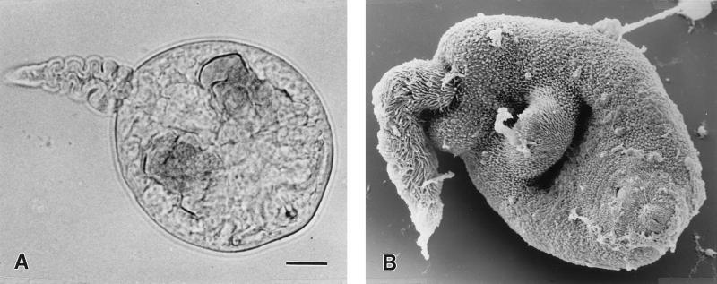
After several hours in RT water, snails frequently released tubiform feces and other cloudy, white secretions from their orifices. Microscopic examination of both types of secretions showed various numbers of rapidly motile, sperm-like organisms of about 0.1 to 0.15 mm in length (Fig. 1A). The organisms had a characteristic tail with a dorsoventral finfold that was shorter than the body and that was used at high revolutions for locomotion. The body had a bilobed virgula organ located in the region of the oral sucker. A smaller ventral sucker was also visible. On the basis of morphology and behavior, the cercariae were tentatively identified as virgulate cercariae (32). Cercariae were readily seen for about 7 days in secretions from snails maintained in water at RT and in concentrated and nonconcentrated RT water samples (experiment 1). Some cercariae were also observed in water secretions of experiment 2 when the snails were kept at RT. No cercariae were observed in experiment 2 in the secretions of snails kept at 8 and 37°C or in experiment 3 in any of the samples. Cercariae were also not observed in secretions of snails kept in the holding tank at 8°C. After about 7 days, cercariae were no longer observed in either secretions or water samples from tanks kept at RT.
FIG. 1.
Light-microscopic (A) and scanning electron microscopic (B) pictures of a virgulate cercaria released into aquarium water by pleurocerid snails of the genus Juga. Bar, 0.01 mm.
Scanning electron microscopy.
A scanning electron micrograph of a cercaria is shown in Fig. 1B. The tail with the dorsoventral finfold and the oral sucker containing a so-called stylet are clearly visible.
PCR.
The results of the nested PCRs for the E. risticii 16S rRNA, groESL, and 51-kDa major antigen genes are summarized in Table 1. The 5′ end of the 16S rRNA gene was readily amplified from all horse and snail samples (15 of 15). With the exception of the sample from one horse (Buck from Oregon), groESL gene fragments also were obtained from all samples. The 51-kDa major antigen gene was amplified from 9 of 15 samples. No amplification of this gene fragment was obtained from the Klamath Falls snail sample, samples from four horses from Oregon (Bunn, Buck, Danny and Tate), and a sample from a horse from Michigan (Mostly Memories).
TABLE 1.
Results of nested PCRs for the 16S rRNA, groESL, and 51-kDa major antigen genes of E. risticii with equine PBL- and snail-derived DNA as templates
| Name of DNA source (reference) | Species from which DNA was derived | Host origin | PCR result
|
||
|---|---|---|---|---|---|
| 16s rRNA gene | groESL gene | 51-kDa major antigen gene | |||
| SHSN-1 (4) | Snail | Weed, Calif. | + | + | + |
| SHSN-2 (4) | Snail | Weed, Calif. | + | + | + |
| JUGA | Snail | Weed, Calif. | + | + | + |
| KLSN | Snail | Klamath Falls, Oreg. | + | + | nega |
| Buck | Horse | Klamath Falls, Oreg. | + | neg | neg |
| Bunn | Horse | Klamath Falls, Oreg. | + | + | neg |
| Danny | Horse | Klamath Falls, Oreg. | + | + | neg |
| Tate | Horse | Klamath Falls, Oreg. | + | + | neg |
| Thorenberg | Horse | Klamath Falls, Oreg. | + | + | + |
| Doc | Horse | Horse Creek, Calif. | + | + | + |
| Dr Pepper | Horse | Weed, Calif. | + | + | + |
| Ms Annie | Horse | Montague, Calif. | + | + | + |
| Shotgunb | Horse | Mt. Shasta City, Calif. | + | + | + |
| Mostly Memories | Horse | Richland, Mich. | + | + | neg |
| Eclipse | Horse | Elizabethville, Pa. | + | + | + |
neg, negative.
Source of SRC agent (19).
In snail experiment 1, the secretions from and the tank water of snails maintained at RT were positive for all three genes at days 2 (Fig. 2), 6, and 7 after the start of the experiment and were negative on days 8, 13, 14, 17, and 21 (data not shown). The snail tissues derived from experiment 2 were PCR negative for the E. risticii 16S rRNA gene. Secretions obtained from snails kept at RT and at 37°C were faintly PCR positive for the 16S rRNA gene; secretions obtained from snails kept at 8°C were negative (experiment 2; data not shown). Secretions serially taken from snails kept at RT in experiment 3 were all PCR negative.
FIG. 2.
Nested PCR of E. risticii for the 16S rRNA (527 bp), groESL heat shock operon (526 bp), and 51-kDa major antigen (569 bp) genes with DNA from Juga secretions (lanes 1), concentrated tank water (lanes 2), and positive (lanes +) and negative (lanes −) E. risticii DNA controls. φ, φX174 replicative-form DNA HaeIII digest (molecular size marker).
16S rRNA sequences.
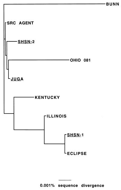
BLAST searches with the 16S rRNA gene sequences amplified from horse and snail samples consistently resulted in 98 to 100% homology with corresponding fragments of the 16S rRNA genes of E. risticii Illinois (data not shown). E. sennetsu was the next closest organism (about 1% less identity than E. risticii Illinois), followed by N. helminthoeca with about 94% identity (data not shown). For the Juga snail secretions and for samples from two horses (Eclipse from Pennsylvania and Bunn from Oregon), the majority (1,251 bp) of the 16S rRNA gene was available for sequence comparison. The 16S rRNA gene fragment amplified from Juga snail secretions was identical to the homologous fragment of the SRC agent. It had one nucleotide change each compared to the two sequences obtained for operculate snails from Siskiyou County (4). Compared to the type strain of E. risticii, strain Illinois, it had the same four nucleotide changes as the SRC agent. The 16S rRNA gene fragment amplified from a sample from Eclipse (Pennsylvania) was closely related to the Illinois type strain of E. risticii (two nucleotide changes), whereas that from a sample from Bunn (Oregon) was genotypically highly polymorphic (14 nucleotide changes). With the exception of the fragment from a sample from Eclipse all other sequences had the same four nucleotides changes (at positions 956, 1221, 1230, and 1246) in comparison to the sequence of the Illinois type strain of E. risticii. The data are summarized in Table 2.
TABLE 2.
Nucleotide differences in the 16S rRNA genes of six equine and three snail E. risticii strains
| DNA source (reference) | Nucleotide at the following positiona:
|
|||||||||||||||||||||||||
|---|---|---|---|---|---|---|---|---|---|---|---|---|---|---|---|---|---|---|---|---|---|---|---|---|---|---|
| 40 | 76 | 77 | 90 | 92 | 97 | 105 | 131 | 142 | 229 | 281 | 309 | 319 | 336 | 365 | 382 | 619 | 769 | 775 | 828 | 956 | 971 | 1221 | 1226 | 1230 | 1246 | |
| E. risticii Illinois | G | G | G | C | T | C | G | G | G | G | G | A | G | G | A | C | G | G | G | A | T | G | C | G | C | G |
| Eclipseb | . | . | . | . | . | . | . | . | . | . | . | . | . | . | . | T | . | . | . | . | . | . | . | A | . | . |
| E. risticii Ohio 081 | . | A | A | T | C | . | . | . | . | . | . | . | . | . | . | . | T | . | . | . | C | A | T | . | T | A |
| E. risticii Kentucky | . | . | . | . | . | A | . | A | . | . | . | . | . | . | . | . | . | . | . | . | C | . | T | . | C | A |
| SRCc | . | . | . | . | . | . | . | . | . | . | . | . | . | . | . | . | . | . | . | . | C | . | T | . | T | A |
| Jugad | . | . | . | . | . | . | . | . | . | . | . | . | . | . | . | . | . | . | . | . | C | . | T | . | T | A |
| SHSN-1 (4) | . | . | . | . | . | . | . | . | . | . | . | G | . | . | . | . | . | . | . | . | C | . | T | . | T | A |
| SHSN-2 (4) | . | . | . | . | . | . | . | . | . | . | . | . | . | . | . | . | . | . | . | G | C | . | T | . | T | A |
| Bunne | A | . | . | . | . | . | C | . | A | T | C | . | C | T | T | . | . | A | A | . | C | . | T | . | T | A |
Differences in the nucleotide sequence are relative to the sequence of the type strain E. risticii Illinois (GenBank accession no. U21290); identical nucleotides are indicated by periods, and nucleotide differences are indicated by capital letters. The fragment length used for the alignment was 1,251 bp (positions 36 through 1287). Position numbers refer to positions in the 16S rRNA gene of the type strain E. risticii Illinois.
Source of strain from Pennsylvania.
SRC agent from California (19).
Underlining indicates snails.
Source of strain from Oregon.
For isolates from nine horses (Buck, Bunn, Danny, Tate, and Thorenberg from Oregon; Doc, Dr Pepper and Ms Annie from California; and Mostly Memories from Michigan) and one snail pool (KLSN from Oregon), partial sequences (527 bp) from the 5′ end of the 16S rRNA gene were available for comparison. The sequence of this genomic region for isolates from two horses in California (Ms Annie and Dr Pepper) and one horse in Oregon (Thorenberg) were identical to that for the Illinois type strain of E. risticii; isolates from two other horses in Oregon (Danny and Tate) had one nucleotide change, the isolate from one horse in California (Doc) had four nucleotide changes; the isolate from a horse in Michigan (Mostly Memories) had three nucleotide changes. The E. risticii strains obtained from the Oregon snail sample (KLSN) and from a horse (Buck) residing in this area had four and five nucleotide changes, respectively, relative to the sequence of the type strain, and these changes were identical to those seen in E. risticii Ohio 081. The data are summarized in Table 3.
TABLE 3.
Nucleotide differences at the 5′ end of the 16S rRNA genes of 11 equine and one snail E. risticii strains
| DNA source | Nucleotide at the following positiona:
|
|||||||||||||||||||||
|---|---|---|---|---|---|---|---|---|---|---|---|---|---|---|---|---|---|---|---|---|---|---|
| 40 | 76 | 77 | 90 | 92 | 94 | 97 | 105 | 131 | 142 | 189 | 229 | 281 | 297 | 309 | 319 | 336 | 347 | 365 | 382 | 515 | 541 | |
| E. risticii Illinois | G | G | G | C | T | G | C | G | G | G | G | G | G | A | A | G | G | T | A | C | G | G |
| Dr Pepperb | . | . | . | . | . | . | . | . | . | . | . | . | . | . | . | . | . | . | . | . | . | . |
| Ms Annieb | . | . | . | . | . | . | . | . | . | . | . | . | . | . | . | . | . | . | . | . | . | . |
| Thorenbergc | . | . | . | . | . | . | . | . | . | . | . | . | . | . | . | . | . | . | . | . | . | . |
| Dannyc | . | . | . | . | . | . | . | . | . | . | . | . | . | . | . | . | . | . | . | . | A | . |
| Tatec | . | . | . | . | . | . | . | . | . | . | . | . | . | . | . | . | . | . | . | . | . | A |
| Mostly Memoriesd | . | . | . | . | . | . | . | . | . | . | A | . | . | . | . | . | . | C | G | . | . | . |
| E. risticii Kentucky | . | . | . | . | . | . | A | . | A | . | . | . | . | . | . | . | . | . | . | . | . | . |
| Docb | . | A | . | . | . | A | A | . | A | . | . | . | . | . | . | . | . | . | . | . | . | . |
| E. risticii Ohio 081 | . | A | A | T | C | . | . | . | . | . | . | . | . | . | . | . | . | . | . | . | . | . |
| Buckc | . | A | A | T | C | . | . | . | . | . | . | . | . | G | . | . | . | . | . | . | . | . |
| Klamath Falls snailc | . | A | A | T | C | . | . | . | . | . | . | . | . | . | . | . | . | . | . | . | . | . |
Differences in the nucleotide sequence are relative to the sequence of the type strain E. risticii Illinois (GenBank accession no. U21290); identical nucleotides are indicated by periods, and nucleotide differences are indicated by capital letters. The fragment length used for the alignment was 527 bp (positions 36 through 563). Position numbers refer to positions in the 16S rRNA gene of the type strain E. risticii Illinois.
Sources of strains from California.
Sources of strains from Oregon.
Source of strain from Michigan.
groESL heat shock operon sequences.
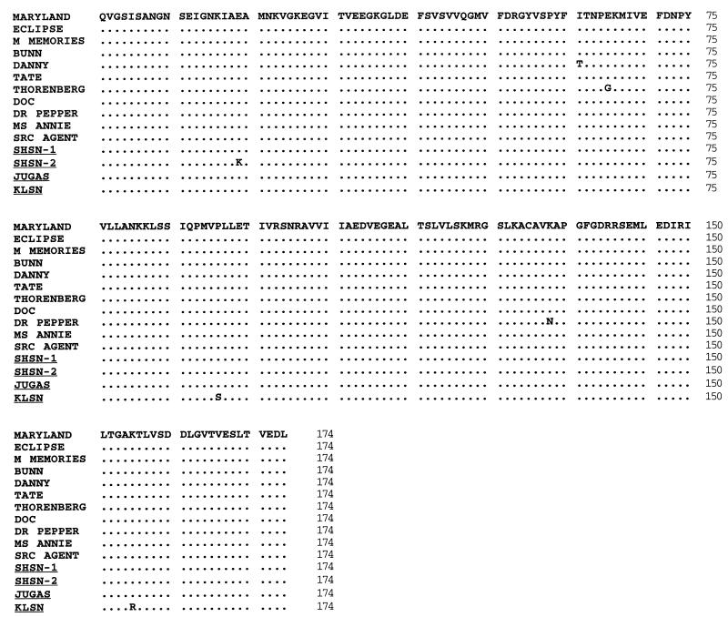
Partial nucleotide sequences of the groESL heat shock operon gene were obtained from nine horse and two snail samples and were used for BLAST searches. The groESL gene nucleotide sequences had between 91 and 100% identity to the corresponding groESL sequences of a Maryland strain of E. risticii (35). The next closest related groESL sequence was that from E. sennetsu (ranging between 89 and 96% identity), followed by E. chaffeensis (about 65% identity) (Table 4). The deduced amino acid sequences of groESL were compared to those for a reference equine E. risticii strain (Maryland), the SRC agent (19), and two previously characterized snail E. risticii strains (4) (Fig. 3). Most of the nucleotide mutations were silent changes which resulted in very similar amino acid sequences for all strains. The isolates from horses Danny, Doc, and Thorenberg had one amino acid change each relative to the sequence of the Maryland strain, while the amino acid sequence obtained from the Klamath Falls snail had two changes.
TABLE 4.
Percent nucleotide identities of E. risticii groESL heat shock operon nucleotide sequences (526 bp) from nine horses and two snails
| Name of DNA source | Species from which DNA was derived | Origin | % Nucleotide identity to the followinga:
|
||
|---|---|---|---|---|---|
| E. risticiib | E. sennetsuc | E. chaffeensisd | |||
| JUGA | Snail | Weed, Calif. | 98 | 96 | 65 |
| KLSN | Snail | Klamath Falls, Oreg. | 91 | 89 | 64 |
| Bunn | Horse | Klamath Falls, Oreg. | 98 | 96 | 65 |
| Danny | Horse | Klamath Falls, Oreg. | 98 | 95 | 65 |
| Tate | Horse | Klamath Falls, Oreg. | 98 | 96 | 65 |
| Thorenberg | Horse | Klamath Falls, Oreg. | 98 | 96 | 65 |
| Doc | Horse | Horse Creek, Calif. | 98 | 95 | 66 |
| Dr Pepper | Horse | Weed, Calif. | 98 | 96 | 65 |
| Ms Annie | Horse | Montague, Calif. | 98 | 96 | 65 |
| Mostly Memories | Horse | Richland, Mich. | 99 | 96 | 65 |
| Eclipse | Horse | Elizabethville, Pa. | 100 | 96 | 65 |
FIG. 3.
Alignment of the deduced amino acid sequences of groESL heat shock operon gene fragments amplified from E. risticii strains from 11 horses and four snails (the snail designations are underlined). The sequences of an equine E. risticii strain (from a horse in Maryland; GenBank accession no. U24396), the SRC agent (GenBank accession no. AF037214), and two snail-derived E. risticii strains (SHSN-1 and SHSN-2; GenBank accession nos. AF037212 and AF037213, respectively) are included for comparison. Identical amino acids are represented by periods; differences in sequence relative to the sequence of the strain from a horse in Maryland are indicated by capital letters.
Sequences of the 51-kDa major antigen genes.
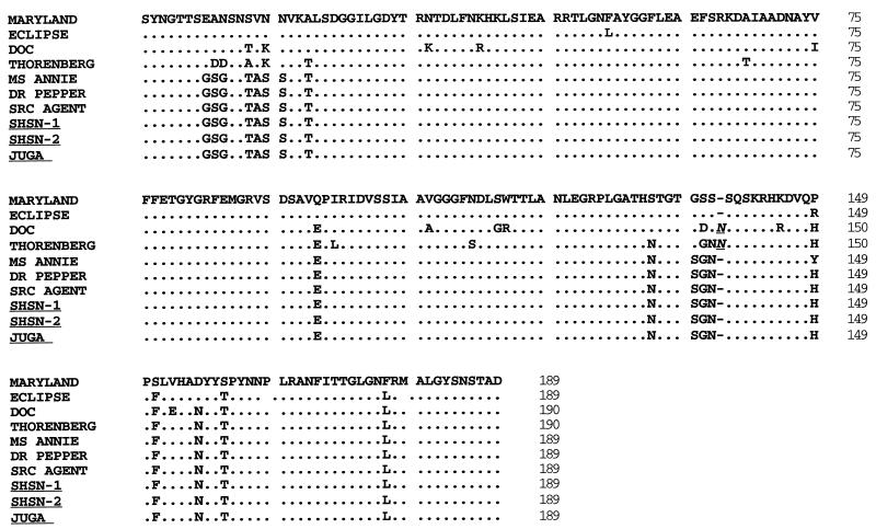
The nucleotide sequences of the 51-kDa major antigen genes were more diverse than those of the groESL heat shock operon and ranged from 92 to 99% nucleotide sequence identity and 90 to 97% amino acid sequence identity (Table 5). The sequence of the strain obtained from the Juga secretions was virtually identical to those of two previously characterized operculate snail strains collected in Siskiyou County (4), a strain from a horse in California (Dr Pepper), and the equine SRC agent (19) (Fig. 4). The sequence of another strain from a horse in California (Ms Annie) was closely related to that of the strain from Juga secretions (one amino acid change at position 149). The 51-kDa major antigen gene sequences of the Maryland reference strain, the strain from a horse in Pennsylvania (Eclipse), and two strains from horses in Oregon (Doc and Thorenberg) differed from those of the rest of the strains. Two hot spots of amino acid changes were apparent. The first was located at the 5′ end of the examined sequence and revealed up to eight amino acid changes compared to the sequences of strains from California. The second hot spot was located around amino acid position 135 and revealed up to seven amino acid changes. With five exceptions, the sequences of the strains from horses in Maryland and Pennsylvania were identical. The sequences of the strains from the horses Doc (California) and Thorenberg (Oregon) appeared to be more closely related to each other than to those of the other strains from horses in California and Oregon. Common to both strains were several amino acid changes that were not seen in the other strains. They also shared at position 139 one unique amino acid insertion which was not present in any of the other strains. The strain from Doc was the most diverse strain, with 18 amino acid changes relative to the Maryland strain of E. risticii.
TABLE 5.
Nucleotide and amino acid sequence identitiesa of E. risticii 51-kDa major antigen gene sequences (569 bp) from five horses and one snail in comparison to the sequences of a Maryland equine strainb of E. risticii
| Name of DNA source | Species from which DNA was derived | Origin | % Nucleotide identity | % Amino acid identity |
|---|---|---|---|---|
| JUGA | Snail | Weed, Calif. | 92 | 90 |
| Thorenberg | Horse | Klamath Falls, Oreg. | 92 | 90c |
| Doc | Horse | Horse Creek, Calif. | 94 | 91c |
| Dr Pepper | Horse | Weed, Calif. | 92 | 90 |
| Ms Annie | Horse | Montague, Calif. | 92 | 90 |
| Eclipse | Horse | Elizabethville, Pa. | 99 | 97 |
Sequence identities were calculated from GeneWorks alignments.
GenBank accession no. U85784.
Includes one amino acid insertion at position 146.
FIG. 4.
Alignment of the deduced amino acid sequences of 51-kDa major antigen gene fragments amplified from E. risticii strains from seven horses and three snails (the snail designations are underlined). The sequences of an equine E. risticii strain (from a horse in Maryland; GenBank accession no. U24396), the SRC agent (GenBank accession no. AF037217), and two snail-derived E. risticii strains (SHSN-1 and SHSN-2; GenBank accession nos. AF037215 and AF037216, respectively) are included for comparison. Identical amino acids are represented by periods, differences in sequence relative to the sequence of the strain from the horse in Maryland are indicated by capital letters; insertions are shown by underlined italics.
Phylogenetic analysis of the 16S rRNA gene.
The nucleotide changes observed in the multiple alignments were reflected in the outcome of the phylogenetic analysis. The phylogenetic relationship among equine and snail E. risticii strains inferred from the 5′ end sequences of the 16S rRNA genes was characterized by the presence of two main clusters and one clear outlier. The sequence divergence was generally very low, but a cluster consisting of an Oregon snail strain (strain KLSN), an Oregon equine strain (from the horse Buck), and the E. risticii Ohio 081 equine reference strain was observed. This cluster of strains was clearly remote from the majority of the other strains (Fig. 5). The strain from the horse Bunn (Oregon) showed the highest sequence divergence.
FIG. 5.
DNA maximum-likelihood phylogram inferred from the nucleotide sequences of 18 16S rRNA gene fragments (527 bp at the 5′ end) originating from E. risticii strains from 14 horses and four snails (the snail designations are underlined). Four of the sequences are for equine E. risticii reference strains: E. risticii Illinois (type strain; GenBank accession no. M21290), E. risticii Kentucky (38), E. risticii Ohio 081 (38), and the E. risticii SRC agent (19). Two snail-derived E. risticii strains (SHSN-1 and SHSN-2; GenBank accession nos. AF037210 and AF037211, respectively) are also included.
A similar picture was seen for those equine and snail E. risticii strains for which the majority of the 16S rRNA genes were available for analysis (Fig. 6). The sequence divergence was again very low, but the strain from horse Bunn (Oregon) and the E. risticii Ohio 081 strain were clearly separated from the remainder of the strains.
FIG. 6.
DNA maximum-likelihood phylogram inferred from the nucleotide sequences of nine 16S rRNA gene fragments (1,251 bp) originating from E. risticii strains from six horses and three snails (the snail designations are underlined). The divergence of 16S rRNA gene fragments from the strains from aquarium Juga snails is compared to the divergence of two newly obtained equine E. risticii strains from Oregon (from Bunn) and Pennsylvania (from Eclipse) and to four equine E. risticii reference strains (E. risticii Illinois [GenBank accession no. M21290], E. risticii Kentucky [38], E. risticii Ohio 081 [38], and the E. risticii SRC agent [19]). Two snail-derived E. risticii strains (SHSN-1 and SHSN-2; GenBank accession nos. AF037210 and AF037211, respectively) are also included.
Phylogenetic analysis of the groESL heat shock operon.
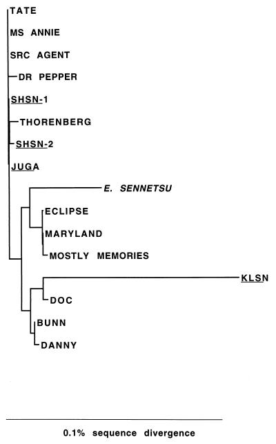
The close similarity in the deduced amino acid sequences among all E. risticii strains was reflected in a generally tight phylogenetic clustering (Fig. 7). However, three strains from horses in the eastern states (Eclipse from Pennsylvania, the strain from a horse in Maryland, and Mostly Memories from Michigan) formed a separate cluster. The strain from the Oregon snail sample showed the highest sequence divergence and was, with E. sennetsu, farther apart from the rest of the strains.
FIG. 7.
DNA maximum-likelihood phylogram inferred from the nucleotide sequences of 16 groESL heat shock operon gene fragments originating from E. risticii strains from 11 horses and four snails (the snail designations are underlined) and from an E. sennetsu reference strain (GenBank accession no. U88092). groESL heat shock operon genes of a Maryland strain of E. risticii (GenBank accession no. U24396), the SRC agent (GenBank accession no. AF037214), and two snail-derived E. risticii strains (SHSN-1 and SHSN-2; GenBank accession nos. AF037212 and AF037213, respectively) are included for comparison.
Phylogenetic analysis of the 51-kDa major antigen gene.
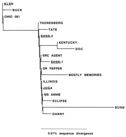
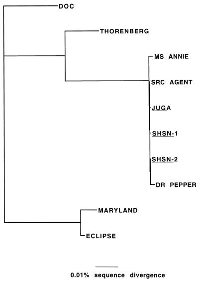
The fairly high nucleotide and amino acid sequence diversity among the 51-kDa major antigen genes was mirrored in their phylogenetic relationships (Fig. 8). Two strains from horses in the eastern states (Eclipse from Pennsylvania and the strain from a horse in Maryland) formed one group and the majority of the California equine and snail strains formed another. Two strains from horses (Doc from California and Thorenberg from Oregon) were separate from the rest of the strains and were on individual branches.
FIG. 8.
DNA maximum-likelihood phylogram inferred from the nucleotide sequences of 10 51-kDa major antigen gene fragments originating from E. risticii strains from seven horses and three snails (the snail designations are underlined). An equine E. risticii strain (from a horse in Maryland; GenBank accession no. U24396), the SRC agent (GenBank accession no. AF037217), and two snail-derived E. risticii strains (SHSN-1 and SHSN-2; GenBank accession nos. AF037215 and AF037216, respectively) are included for comparison.
DISCUSSION
Here we report the detection of E. risticii DNA in secretions from snails that were collected in the field and that were kept for several weeks in freshwater aquaria in the laboratory. For the first time it was possible to examine snails as potential vectors for the transmission of E. risticii and possibly other infectious organisms in a defined laboratory setting. The presence of trematode virgulate cercariae in snail secretions was surprising but not completely unexpected since many snail species are known to harbor trematodes (32). Virgulate cercariae, for example, can develop in operculate snails and are produced by trematodes of the family Lecithodendriidae (32). However, the association between the presence of cercariae and PCR positivity for three E. risticii gene sequences in snail secretions is the first description of a possible link between snails, trematodes, and the epizootiology of PHF in horses.
For more than 15 years it has remained a mystery as to how E. risticii infection is spread among horses (17). Transmission of the organism through arthropod vectors has been widely considered but never experimentally proved (5, 15, 23–25, 32). The disease has, however, been associated from the very beginning with riverine habitats (18), and it has frequently been noted by veterinary clinicians that horses kept on dry lot in areas where the disease is enzootic usually do not develop the disease. The hypothesis that freshwater snails are involved in the life cycle and transmission of E. risticii would provide an explanation for these observations. The field-collected snails in our study readily released cercariae upon exposure to water temperatures above 22°C. This would help to explain the seasonality of the disease, with cases often occurring on irrigated pastures after heat spells. Many trematodes have life cycles with several intermediate hosts (32). Among these is the trematode Nanophyetus salmincola, the vector for N. helminthoeca, which causes “salmon poisoning” disease in dogs (22). Transmission of E. risticii might similarly be achieved with other intermediate hosts, perhaps arthropod vectors such as ants or beetles that feed on snail secretions. In this regard the observed presence of darkling beetles and their larvae on farms where PHF is enzootic might be noteworthy (24).
The ease by which E. risticii DNA was detected in secretions from snails in aquaria is in contrast to our previous attempts to amplify E. risticii DNA from snail tissues, which were tedious and time-consuming and which resulted in the detection of only a few positive snails (4). By focusing on cercariae rather than whole snail tissues, the sensitivity of the PCR is likely to be increased. This could explain why we were able to detect E. risticii DNA in secretions at a much higher rate than in snail pools or individual whole snails. Because the DNA from entire snails is extracted, it is likely that dilution of rickettsial DNA occurs. The E. risticii load in the cercariae was obviously high enough for its DNA to be detectable by PCR with lysed secretions without the need for DNA extraction.
The fact that cercariae were not observed in secretions of snails maintained for several weeks at 8°C can be attributed to two facts. First, most of the snails kept at this temperature were smaller than those releasing cercariae. It is known for other trematodes that only large snails (those with a size of 2 cm or larger) will harbor cercariae (22). Second, the larger snails in the holding tank might have released cercariae after the water was replaced with fresh tap water (temperature, about 22°C). The fresh tap water may have temporarily increased the water temperature to a level inducing an unnoticed shedding of cercariae. We hypothesize that once cercariae are released from the snails, new cercariae are no longer produced and a new trematode life cycle (i.e., infection of the snail by miracidia) must start before the production of cercariae can begin again.
A substantial portion of this study was concerned with analyses of genome sequences obtained for strains from snail cercariae and strains from horses with PHF. The E. risticii sequences from the Juga snail secretions were nearly identical to those obtained from operculate snails previously collected at the same site as the aquarium snails (4) and to sequences derived from strain from a horse with PHF residing in close vicinity to the snail collection site (19). We feel that the presence of such highly similar genotypes in both species is sufficient evidence to conclude that horses and snails harbor the same or very similar strains of E. risticii. This conclusion is supported by the fact that strains from horses from different geographical areas such as the eastern United States appeared to have different genotypes. It is well documented that the antigenic profiles or restriction enzyme patterns of E. risticii strains originating from different geographical areas are quite diverse (7, 37, 38).
Previous studies of the genetic diversity of E. risticii strains used solely the 16S rRNA gene sequences for analysis (7, 37, 38). The sequences of this gene are known to vary in an orderly manner throughout the phylogenetic tree and hence represent desirable targets for PCR and phylogenetic analyses (40). Differences in the 16S rRNA gene sequences of different strains of the same species of rickettsial organisms are very small (0 to 0.1%) (2, 34), which can sometimes make conclusive phylogenetic analysis difficult. We conclude from the analysis of two additional E. risticii gene sequences (the groESL heat shock operon gene and the 51-kDa major antigen gene) that genetic diversity is more extensive in genes coding for antigenic determinants that serve as targets for antibody- or cell-mediated immune selection. Further studies of the molecular epidemiology of E. risticii should therefore focus on these or similarly diverse genes. Similar suggestions have recently been made for other members of the rickettsiae, such as Ehrlichia equi and Ehrlichia phagocytophila (35).
The presence of geographical clusters of E. risticii strains was evident from phylogenetic analyses of all three examined gene fragments. However, the most profound differences were observed in the 51-kDa major antigen genes. The 51-kDa major antigen sequences of strains from the Shasta and Juga snails, the SRC agent, and strains from the horses Dr Pepper and Ms Annie were virtually identical. These strains all originated in the same geographical area with the same river drainage (Shasta) and the same ecosystem. The strains from horses in Pennsylvania and Maryland are geographically associated as well and share significant genotypic homology to each other. A close association between the strains from the horses Thorenberg and Doc was noticed; both strains came from the same geographical area (the drainage here is the Klamath River). We were not able to amplify 51-kDa gene sequences from a few E. risticii strains that were more divergent (geographically clustered), even though PCR conditions (for example, the primer annealing stringency) were modified. We consider this to be additional evidence for the widespread genetic diversity of E. risticii, which may be a major factor in recognized vaccine failures (7, 23, 37).
The known geographical distribution of Juga spp. encompasses northern California, northern Nevada, Oregon, and Washington (6), an area similar to that of the pleurocerid host of N. salmincola. If snails have a role in the life cycle of E. risticii in other areas of the United States, different snails must certainly be involved. Preliminary evidence in this regard is the successful amplification of two E. risticii gene fragments from a pool of lymnaeid snails, genus Stagnicola. It is possible that strains of ehrlichiae causing PHF may eventually be found in a variety of snail genera, a host range that may be reflected in the genetic and antigenic variation observed among E. risticii isolates. Thus, the “agent” of PHF may in actuality represent an array of closely related ehrlichial strains differing in their particular snail hosts and vectors and perhaps in their virulence for horses as well.
The information derived from this study should aid future investigations of snails as vectors of disease-causing organisms and greatly simplify the detection of E. risticii in these mollusks. The cost-effectiveness of aquarium culture versus the collection of large numbers of snails with subsequent laborious DNA extraction is an important factor for consideration. Similar procedures might also be applied to other suspected cercaria-transmitted infections.
We conclude by emphasizing that cercarial transmission of E. risticii infection remains a hypothesis worthy of further testing. Because of a ubiquitous bacterial flora, the isolation of E. risticii directly from snail secretions appears to be difficult. An alternative method would be to attempt experimental transmission of E. risticii to horses through snail-derived cercariae, with subsequent isolation of the organism from peripheral blood cells. It will be interesting to see whether results similar to ours are obtained with snail species from other areas of the country where PHF is known to occur.
ACKNOWLEDGMENTS
We thank Stacia Hoover and Eric Bowman for nucleotide sequencing; Mike Ronne, Jon Goodell, Paul Miller, Tom Sampson, Amy Finken, and Hal Schott for providing equine blood specimens; and Larisa Vredevoe, Elfriede DeRock, and Inderpal Kaur W. Singh for generous help in collecting snails. We thank Bob Munn for electron microscopy, Carlos Munos for valuable information regarding snail husbandry, Stewart Schell for advice on cercarial identification, Steve Holloway for help with phylogenetic analyses, and Yasuko Rikihisa for valuable discussions and inspiration. The support of Nancy East is greatly appreciated.
This work was supported by grants from the Center for Equine Health, University of California, Davis, with funds provided by the Oak Tree Racing Association, the State of California satellite wagering fund, and contributions from private donors, and by discretionary funds from the Department of Medicine and Epidemiology, School of Veterinary Medicine, University of California, Davis.
REFERENCES
- 1.Altschul S F, Gish W, Miller W, Myers E W, Lipman D J. Basic local alignment search tool. J Mol Biol. 1990;215:403–410. doi: 10.1016/S0022-2836(05)80360-2. [DOI] [PubMed] [Google Scholar]
- 2.Anderson B E, Dawson J E, Jones D C, Wilson K H. Ehrlichia chaffeensis, a new species associated with human ehrlichiosis. J Clin Microbiol. 1991;29:2838–2842. doi: 10.1128/jcm.29.12.2838-2842.1991. [DOI] [PMC free article] [PubMed] [Google Scholar]
- 3.Barlough J E, Madigan J E, DeRock E, Bigornia L. Nested polymerase chain reaction for detection of Ehrlichia equi genomic DNA in horses and ticks (Ixodes pacificus) Vet Parasitol. 1996;63:319–329. doi: 10.1016/0304-4017(95)00904-3. [DOI] [PubMed] [Google Scholar]
- 4.Barlough, J. E., G. H. Reubel, J. E. Madigan, L. K. Vredevoe, P. E. Miller, and Y. Rikihisa. Detection of Ehrlichia risticii, the agent of Potomac horse fever, in freshwater stream snails (Pleuroceridae: Juga spp.) of northern California. Submitted for publication. [DOI] [PMC free article] [PubMed]
- 5.Barlough J E, Rikihisa Y, Madigan J E. Nested polymerase chain reaction for detection of Ehrlichia risticii genomic DNA in infected horses. Vet Parasitol. 1997;68:367–373. doi: 10.1016/s0304-4017(96)01083-7. [DOI] [PubMed] [Google Scholar]
- 6.Burch J B. North American freshwater snails. Hamburg, Mich: Malacological Publications; 1989. [Google Scholar]
- 7.Chaichanasiriwithaya W, Rikihisa Y, Yamamoto S, Reed S, Crawford T B, Perryman L E, Palmer G H. Antigenic, morphologic, and molecular characterization of new Ehrlichia risticii isolates. J Clin Microbiol. 1994;32:3026–3033. doi: 10.1128/jcm.32.12.3026-3033.1994. [DOI] [PMC free article] [PubMed] [Google Scholar]
- 8.Dillon R T. Karyotypic evolution in pleurocerid snails. II. Pleurocera, Goniobasis, and Juga. Malacologia. 1991;33:339–344. [Google Scholar]
- 9.Dumler J S, Bakken J S. Ehrlichial diseases of humans: emerging tick-borne infections. Clin Infect Dis. 1995;20:1102–1110. doi: 10.1093/clinids/20.5.1102. [DOI] [PubMed] [Google Scholar]
- 10.Dutta S K, Shankarappa B, Mattingly-Napier B L. Molecular cloning and analysis of recombinant major antigens of Ehrlichia risticii. Infect Immun. 1991;59:1162–1169. doi: 10.1128/iai.59.3.1162-1169.1991. [DOI] [PMC free article] [PubMed] [Google Scholar]
- 11.Dutta S K, Shankarappa B, Thaker S R, Mattingly-Napier B L. DNA restriction endonuclease cleavage pattern and protein antigen profile of Ehrlichia risticii. Vet Microbiol. 1990;25:29–38. doi: 10.1016/0378-1135(90)90090-i. [DOI] [PubMed] [Google Scholar]
- 12.Felsenstein J. PHYLIP—Phylogeny Inference Package. Cladistics. 1989;5:164–166. [Google Scholar]
- 13.Fukuda T, Sasahara T, Kitao T. Studies on the causative agent of “Hyuganetsu” disease. X. Vector. J Jpn Assoc Infect Dis. 1972;36:235–241. [Google Scholar]
- 14.Fukuda T, Yamamoto S. Neorickettsia-like organism isolated from metacercaria of a fluke, Stellantchasmus falcatus. Jpn J Med Sci Biol. 1981;34:103–107. doi: 10.7883/yoken1952.34.103. [DOI] [PubMed] [Google Scholar]
- 15.Hahn N E, Fletcher M, Rice R M, Kocan K M, Hansen J W, Hair J A, Barker R W, Perry B D. Attempted transmission of Ehrlichia risticii, causative agent of Potomac horse fever, by the ticks, Dermacentor variabilis, Rhipicephalus sanguineus, Ixodes scapularis and Amblyomma americanum. Exp Appl Acarol. 1990;8:41–50. doi: 10.1007/BF01193380. [DOI] [PubMed] [Google Scholar]
- 16.Holland C J, Ristic M, Cole A I, Johnson P, Baker G, Goetz T. Isolation, experimental transmission, and characterization of causative agent of Potomac horse fever. Science. 1985;227:522–524. doi: 10.1126/science.3880925. [DOI] [PubMed] [Google Scholar]
- 17.Kahler S. Transmission is unsolved mystery of equine monocytic ehrlichiosis. J Am Vet Med Assoc. 1989;194:1681–1687. [PubMed] [Google Scholar]
- 18.Knowles R C, Anderson C W, Shipley W D, Whitlock R H, Perry B D. Acute equine diarrhea syndrome (AEDS): a preliminary report. Proc Am Assoc Eq Pract. 1983;29:353–357. [Google Scholar]
- 19.Madigan J E, Barlough J E, Rikihisa Y, Wen B, Miller P E, Sampson T J. Identification of an enzootic diarrhea (“Shasta River crud”) in northern California as Potomac horse fever. J Eq Vet Sci. 1997;17:270–272. [Google Scholar]
- 20.Madigan J E, Cohen M, Stabbe M T, True R G, Wohlford L A. Serologic evidence of Potomac horse fever (Ehrlichia risticii) in three California horses with enterocolitis and fever. Calif Vet. 1987;41:8–10. [Google Scholar]
- 21.Madigan J E, Rikihisa Y, Palmer J E, DeRock E, Mott J. Evidence for a high rate of false-positive results with the indirect fluorescent antibody test for Ehrlichia risticii antibody in horses. J Am Vet Med Assoc. 1995;207:1448–1453. [PubMed] [Google Scholar]
- 22.Millemann R E, Knapp S E. Biology of Nanophyetus salmincola and “salmon poisoning” disease. Adv Parasitol. 1970;8:1–41. [PubMed] [Google Scholar]
- 23.Palmer J E. Prevention of Potomac horse fever. Cornell Vet. 1989;79:201–205. . (Editorial.) [PubMed] [Google Scholar]
- 24.Palmer J E. Potomac horse fever. Vet Clin N Am (Eq Pract) 1993;9:399–410. doi: 10.1016/s0749-0739(17)30406-6. [DOI] [PubMed] [Google Scholar]
- 25.Perry B D, Rikihisa Y, Saunders G K. Intradermal transmission of Potomac horse fever. Vet Rec. 1985;116:246–247. doi: 10.1136/vr.116.9.246. . (Letter.) [DOI] [PubMed] [Google Scholar]
- 26.Perry B D, Schmidtmann E T, Rice R M, Hansen J W, Fletcher M, Turner E C, Robl M G, Hahn N E. Epidemiology of Potomac horse fever: an investigation into the possible role of non-equine mammals. Vet Rec. 1989;125:83–86. doi: 10.1136/vr.125.4.83. [DOI] [PubMed] [Google Scholar]
- 27.Pretzman C, Ralph D, Stothard D R, Fuerst P A, Rikihisa Y. 16S rRNA gene sequence of Neorickettsia helminthoeca and its phylogenetic alignment with members of the genus Ehrlichia. Int J Syst Bacteriol. 1995;45:207–211. doi: 10.1099/00207713-45-2-207. [DOI] [PubMed] [Google Scholar]
- 28.Rikihisa Y. Cross-reacting antigens between Neorickettsia helminthoeca and Ehrlichia species, shown by immunofluorescence and Western immunoblotting. J Clin Microbiol. 1991;29:2024–2029. doi: 10.1128/jcm.29.9.2024-2029.1991. [DOI] [PMC free article] [PubMed] [Google Scholar]
- 29.Rikihisa Y, Perry B D. Causative ehrlichial organisms in Potomac horse fever. Infect Immun. 1985;49:513–517. doi: 10.1128/iai.49.3.513-517.1985. [DOI] [PMC free article] [PubMed] [Google Scholar]
- 30.Rikihisa Y, Pretzman C I, Johnson G C, Reed S M, Yamamoto S, Andrews F. Clinical, histopathological, and immunological responses of ponies to Ehrlichia sennetsu and subsequent Ehrlichia risticii challenge. Infect Immun. 1988;56:2960–2966. doi: 10.1128/iai.56.11.2960-2966.1988. [DOI] [PMC free article] [PubMed] [Google Scholar]
- 31.Sambrook J, Fritsch E F, Maniatis T. Molecular cloning: a laboratory manual. 2nd ed. Cold Spring Harbor, N.Y: Cold Spring Harbor Laboratory Press; 1989. [Google Scholar]
- 32.Schell S C. How to know the trematodes. W. C. Dubuque, Iowa: Brown Company Publishers; 1970. [Google Scholar]
- 33.Schmidtmann E T, Robl M G, Carroll J F. Attempted transmission of Ehrlichia risticii by field-captured Dermacentor variabilis (Acari: Ixodidae) Am J Vet Res. 1986;47:2393–2395. [PubMed] [Google Scholar]
- 34.Stothard D R, Clark J B, Fuerst P A. Ancestral divergence of Rickettsia bellii from the spotted fever and typhus groups of Rickettsia and antiquity of the genus Rickettsia. Int J Syst Bacteriol. 1994;44:798–804. doi: 10.1099/00207713-44-4-798. [DOI] [PubMed] [Google Scholar]
- 35.Sumner J W, Nicholson W L, Massung R F. PCR amplification and comparison of nucleotide sequences from the groESL heat shock operon of Ehrlichia species. J Clin Microbiol. 1997;35:2087–2092. doi: 10.1128/jcm.35.8.2087-2092.1997. [DOI] [PMC free article] [PubMed] [Google Scholar]
- 36.Thompson J D, Higgins D G, Gibson T J. CLUSTAL W: improving the sensitivity of progressive multiple sequence alignment through sequence weighting, position-specific gap penalties and weight matrix choice. Nucleic Acids Res. 1994;22:4673–4680. doi: 10.1093/nar/22.22.4673. [DOI] [PMC free article] [PubMed] [Google Scholar]
- 37.Vemulapalli R, Biswas B, Dutta S K. Pathogenic, immunologic, and molecular differences between two Ehrlichia risticii strains. J Clin Microbiol. 1995;33:2987–2993. doi: 10.1128/jcm.33.11.2987-2993.1995. [DOI] [PMC free article] [PubMed] [Google Scholar]
- 38.Wen B, Rikihisa Y, Fuerst P A, Chaichanasiriwithaya W. Diversity of 16S rRNA genes of new ehrlichia strains isolated from horses with clinical signs of Potomac horse fever. Int J Syst Bacteriol. 1995;45:315–318. doi: 10.1099/00207713-45-2-315. [DOI] [PubMed] [Google Scholar]
- 39.Wen B, Rikihisa Y, Yamamoto S, Kawabata N, Fuerst P A. Characterization of the SF agent, an Ehrlichia sp. isolated from the fluke Stellantchasmus falcatus, by 16S rRNA base sequence, serological, and morphological analyses. Int J Syst Bacteriol. 1996;46:149–154. doi: 10.1099/00207713-46-1-149. [DOI] [PubMed] [Google Scholar]
- 40.Wilson K H, Blitchington R B, Greene R C. Amplification of bacterial 16S ribosomal DNA with polymerase chain reaction. J Clin Microbiol. 1990;28:1942–1946. doi: 10.1128/jcm.28.9.1942-1946.1990. . (Erratum, 29:333, 1991.) [DOI] [PMC free article] [PubMed] [Google Scholar]


