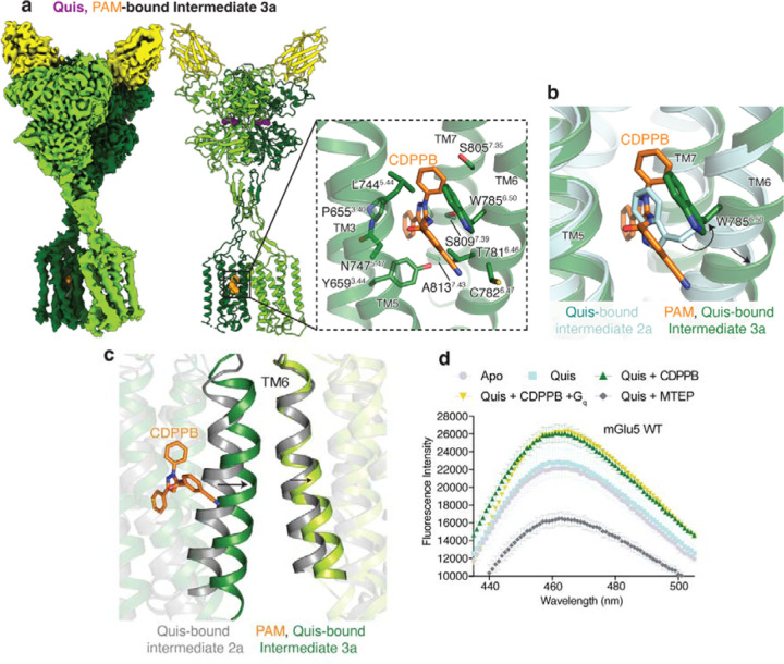
Fig 3: Structural changes of upon PAM binding to mGlu5.
a) Cryo-EM density and model of CDPPB (orange) and Quis-bound mGlu5 in a nanodisc. The structure represents the Intermediate 3a state with the CRDs and TMs in close proximity. Nb43 is shown in yellow. Insert: Binding pocket of CDPPB in the TM region showing residues within 4Å as sticks.
b) CDPPB binding to the TM causes the rearrangement of W7856.50 to accommodate the ligand.
c) Quis-bound Intermediate 2a and CDPPB, Quis-bound Intermediate 3a structures show differences in the conformation of TM6 at the protomer interface.
d) Bimane spectra of mGlu5 in nanodiscs labeled at positions C6914.30 (end of TM4) and C681ICL2. Adding Quis (cyan) results in no change in the spectra compared to Apo (grey). However, Quis and CDPPB increase the fluorescence (dark green), indicating a change in the ICL2 environment. Further addition of Gq does not result in a change in the bimane spectrum (yellow). The addition of Quis and MTEP causes a decrease in fluorescence. Data represented as mean ± SD, n = 3 individual.

