
Figure 3.
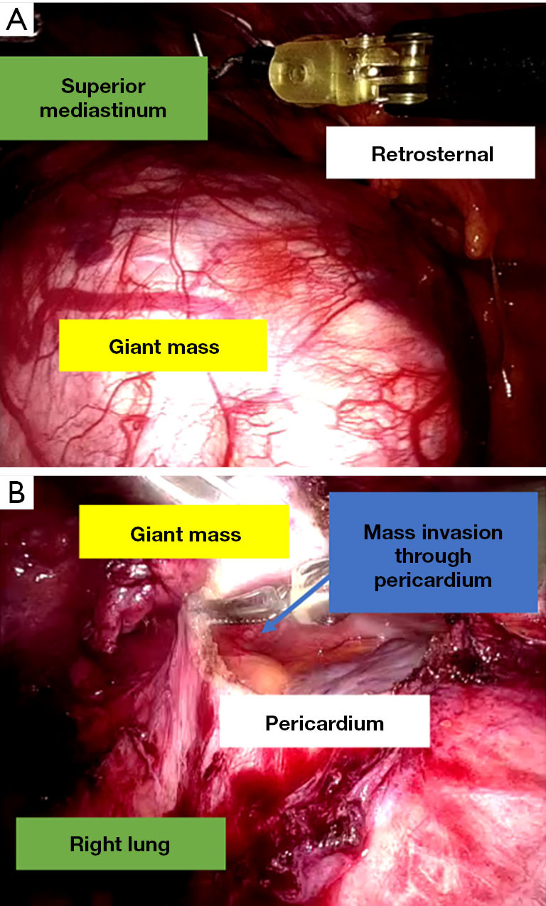
Pictures taken during surgery. (A) 15 cm anterior mediastinal mass. (B) Mass is pushed superiorly, the pericardium has been opened and lesions are found to be infiltrating through the pericardium.
Official websites use .gov
A
.gov website belongs to an official
government organization in the United States.
Secure .gov websites use HTTPS
A lock (
) or https:// means you've safely
connected to the .gov website. Share sensitive
information only on official, secure websites.
Pictures taken during surgery. (A) 15 cm anterior mediastinal mass. (B) Mass is pushed superiorly, the pericardium has been opened and lesions are found to be infiltrating through the pericardium.