Abstract
Background
Persistent increase in intraocular pressure (IOP) is often observed in eyes with thyroid-associated ophthalmopathy (TAO), which has irreversible effects on the visual function of patients. This retrospective cross-sectional study evaluated the value of magnetic resonance imaging (MRI) measurements of extraocular muscle (EOM) volume in prognosing IOP in patients with TAO.
Methods
This single-center study was conducted in Beijing Friendship Hospital (Beijing, China), a tertiary hospital. From 35 participants, 70 eyes (normal IOP group: 50 eyes; high IOP group: 20 eyes; random samples) were enrolled in this study. Basic data from patients were collected and compared using 2-sample t-tests and chi-squared tests. The volume of the EOM, orbit fat, and whole orbit were measured by orbital MRI. Moreover, proptosis, optic nerve (ON) sheath diameter, optic nerve angle (ONA), and gaze angle were additionally measured with MRI. These parameters were compared between the two groups using 2-sample t-tests and chi-squared tests. Before and after post-methylprednisolone therapy, the MRI data of 20 eyes were obtained and compared with a paired t-test. Peripapillary retinal nerve fiber layer (RNFL) thickness was obtained using an optical coherence tomography (OCT) scan. Spearman rank correlation analysis, logistic regression analysis, and receiver operating characteristic (ROC) curves were performed to evaluate the role of these factors in IOP changes.
Results
The EOM volume and axial and sagittal ONAs in the high-IOP group were significantly increased compared to the normal-IOP group (P<0.001, P=0.001, P=0.02, respectively). Logistic regression analysis indicate that the cutoff value for EOM volume for the diagnosis of high IOP was significantly larger than that of the other parameters except orbit volume (P=0.03, P=0.004, P=0.08, respectively). Spearman correlation analysis revealed a significant correlation between EOM volume and ON sheath diameter and average RNFL thickness (P=0.01, P=0.02, respectively). A paired t-test indicated a significant decrease of EOM volume and ON sheath diameter as well as a significant enlargement of the axial ONAs after methylprednisolone therapy (P=0.002, P=0.02, P=0.05, respectively).
Conclusions
EOM volume was an effective diagnostic factor for tracking IOP changes and ON patterns in patients with TAO. Methylprednisolone therapy is recommended for patients with TAO with secondary glaucoma to quickly reduce the EOM volume.
Keywords: Thyroid-associated ophthalmopathy (TAO), orbital MRI, intraocular pressure (IOP), extraocular muscle (EOM)
Introduction
Thyroid-associated ophthalmopathy (TAO) or Graves ophthalmopathy (GO) is the major extrathyroidal manifestation of Graves disease (GD), which is one of the most complex autoimmune disorders in human eyes, resulting in orbital disfigurement, double vision, and even vision loss (1). Patients with TAO have an inflammatory autoimmune condition caused by the change of thyrotropin receptor autoantibodies (TRAb) (2). The therapeutic strategies for patients with TAO are based on a precise assessment of the activity and severity of the disease. In most cases, TAO stabilizes after immunosuppressive therapy, which is the most common therapy in clinic (1,3). Several studies have evaluated the intraocular pressure (IOP) in patients with TAO using a cross-sectional study design, and a correlation between IOP and the different states of TAO has been established (4,5). In a recent study, 29.0% of patients with TAO under long-term evaluation of IOP were diagnosed with glaucoma compared with 6.0% of the non-TAO controls (6). Therefore, the mechanism and treatment of IOP in TAO orbits urgently need to be investigated.
IOP should be evaluated in patients due to its association with the increased risk of glaucoma and optic neuropathy (7), although very few studies have focused on the exact mechanism underlying the increased IOP in patients with TAO. IOP remains the disease’s primary modifiable risk factor from both a pathophysiological and treatment standpoint (8). TAO myopathy spans a wide spectrum of pathological changes, whose relevance to the evaluated IOP is poorly understood. Lymphocyte infiltration and glycosaminoglycan (GAG) deposits in the extraocular muscles (EOMs), connective tissue, and orbital fat (OF) are involved in TAO (9). Radiology, especially magnetic resonance imaging (MRI), is important for distinguishing the orbital contents and their alterations and has been considered as a potential solution for noninvasively classifying TAO (10). Proptosis, orbit enlargement, EOM proliferation, and OF expansion can be measured using orbital MRI and subsequently resolved (11). However, most of the existing studies have focused on the clinical features of orbital tissue in TAO, such as quantitation of EOM fibrosis by T1 mapping and T2 relaxation time (T2RT) (12,13), and the relationship between IOP and orbital tissue remains unexplored in comparison.
This study employed MRI to conduct an integrated assessment of orbital tissue and characterize the potential relationship between IOP and orbital tissue with the aim of evaluating their utility in assessing visual functional damage in patients with TAO. We present this article in accordance with the STROBE reporting checklist (available at https://qims.amegroups.com/article/view/10.21037/qims-23-44/rc).
Methods
Patients
This observational cross-sectional study was conducted in accordance with the Declaration of Helsinki (as revised in 2013) and was approved by the ethics committee of Beijing Friendship Hospital, Capital Medical University (approval No. 2022-P2-415). The requirement for written informed consent was waived due to the retrospective nature of the study. From January 2020 to September 2022, TAO inpatients aged 18 to 65 years were recruited from the Ophthalmology Department of the Beijing Friendship Hospital at Capital Medical University. Diagnoses were based on the Chinese guideline for the diagnosis and treatment of thyroid-associated ophthalmopathy [2022] (14) as follows: (I) if lid retraction is present, the diagnoses should include thyroid dysfunction, proptosis, and EOM involvement; (II) at least 1 of the 3 symptoms of lid retraction, proptosis, and effect of the EOMs should be present for a diagnosis of thyroid dysfunction. Additionally, all the recruited patients were required to have an IOP evaluation result from Goldmann applanation tonometry in the first position of the eyeball in their first hospitalization.
The exclusion criteria were as follows: (I) a history of orbital trauma, orbital tumors, orbital irradiation, or orbital surgery; (II) previous corticosteroid therapy, radiotherapy, or any other treatment for TAO; (III) intracranial mass lesions, hematencephalon, or other cranial disorders that could increase intracranial pressure; (IV) previous diagnosis of glaucoma, or a history of taking medicine for controlling IOP; (V) pathological myopia, anterior ischemic optic neuropathy, or a history of other retinal or optic nerve (ON) diseases; (VI) contraindications for MRI; and (VII) contraindications for glucocorticoid therapy.
In total, 70 eyes from patients who underwent MRI evaluation were enrolled, which were further classified into the normal IOP group (10< IOP ≤21) and the high IOP group (IOP >21) according to the IOP values.
Among all patients, most received methylprednisolone pulse therapy, which was administered at a dose of 500 mg of methylprednisolone for 6 successive weeks and was followed by administration of 250 mg of methylprednisolone for another 6 successive weeks. MRI data of 10 patients were collected, which was performed within 1 week after methylprednisolone pulse therapy at the ophthalmology clinic. The pre- and post-therapy MRI data of 20 eyes were obtained.
MRI
Orbital MRI was performed in all patients and evaluated using a 3.0 T MRI scanner (Philips Healthcare, Best, Netherlands). Axial, coronal, and sagittal MRI was obtained using T2-weighted spin echo (repetition time =660 ms; time to echo =11.1 ms, matrix size =256×256 mm, field of view =18×18 cm, slice thickness =3.0 mm). All patients were required to keep their eyes closed and stay motionless during the examinations. The MRI scans of patients were evaluated by two experienced ophthalmologists who both held positions of deputy chief physician.
Measurement of volumes
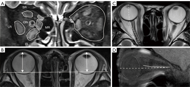
Orbital MRI data were anonymously and randomly measured by 2 using Dicom UniWeb Server (Radiont) which automatically provides the measurement. The two investigators were blinded to the information and clinical features of the patients. The cross-sectional area of whole orbit and the EOMs, including the superior rectus (SR), inferior rectus (IR), lateral rectus (LR), medial rectus (MR), superior oblique (SO), and ON, were measured in 4 continuous slices after the cut surface of the eyeball by tracing outlines of each tissue on T2-weighted coronal images. The levator palpebrae superioris was measured in complex with the SR because of its difficulty in being discerned. The volume of the whole orbit, EOMs, and ON was defined as the sum of traced areas multiplied by the slice thickness (3 mm). The volume of the OF was calculated by subtracting the volumes of the EOMs and ON from the whole orbit (Figure 1A).
Figure 1.
Measurements of orbital MRI data on T2-weighted images. (A) Measurements of the cross-sectional area of whole orbit and EOMs, including the SR, IR, LR, MR, SO, and ON. (B) Measurement of proptosis. The perpendicular distance (dual arrows) from the top of the cornea to the line connected to bilateral frontal processes in the zygomatic bones was measured. (C) Measurements of the axial ONA (solid line), horizontal gaze angle (dotted line), and the diameter of optic nerve sheath (I-shaped line). (D) Measurements of the sagittal ONA (solid line) and vertical gaze angle (dotted line). SR, superior rectus; IR, inferior rectus; LR, lateral rectus; MR, medial rectus; SO, superior oblique; ON, optic nerve; MRI, magnetic resonance imaging; ONA, optic nerve angle; EOM, extraocular muscle.
Measurement of proptosis and ON sheath diameter
The proptosis was measured on the axial T2-weighted images with Dicom UniWeb Server. A line was drawn between the bilateral frontal processes of the zygomatic bones, and a perpendicular line was drawn from the top of the cornea to this point. The length of the perpendicular line was measured automatically as the degree of proptosis (Figure 1B). The diameter of ON sheath was measured at 2 mm posterior to the ON insertion (Figure 1C).
Measurement of optic nerve angle (ONA) and gaze angle
The axial and sagittal ONAs were measured on T2-weighted images using Dicom UniWeb Server. The ONA is the angle of the greatest bend in the ON for each eye. Meanwhile, the angle formed by the long axis of the orbit and the long axis of the globe was also measured on T2-weighted axial and sagittal images to assess the direction of horizontal and vertical gaze (Figure 1C,1D).
Optical coherence tomography (OCT) measurements
The patients with TAO underwent an OCT scan using a spectral-domain OCT (Spectralis, Heidelberg Engineering). The peripapillary retinal nerve fiber layer (RNFL) thickness was obtained using the optic disc cube 200×200 protocol.
Statistical analysis
Statistical analyses were performed using SPSS statistical software (IBM Corp., New York, United States). The Kolmogorov-Smirnov test was used to identify the normality of distribution. Data are expressed as the mean ± standard deviation. The independent samples t-test and Mann-Whitney test were used to compare MRI parameters between different IOP groups. A paired t-test was used to compare the MRI parameters before and after methylprednisolone pulse therapy. Bivariate logistic regression was used to analyze the relationship between orbital MRI measurements and IOP. The area under the receiver operating characteristic (ROC) curve (AUC) was used to evaluate the accuracy of the regression model diagnosis. Multiple logistic regression was used to analyze the relationship between age, clinical activity score (CAS), orbital MRI measurements and IOP, area under curve (AUC), and confidence interval (CI) were calculated. Pearson product-moment correlation coefficient was used to analyze the correlation between the EOM volume and ON-related measurements. A P value <0.05 was considered statistically significant.
Results
Patient demographic and clinical characteristics
A total of 70 TAO eyes from 35 patients were enrolled in this study, including 50 in the normal IOP group and 20 in the high IOP group. Table 1 summarizes the patient demographics and clinical characteristics. Patients in the two groups showed no significant differences in age, gender, body mass index (BMI), or medical histories of hypertension, CAS, diabetes, or hyperlipidemia (P=0.30, P=0.13, P=0.68, P=0.08, P=0.20, P=0.79, P=0.50, respectively). Statistically significant increased IOPs were observed in the high IOP group (P<0.001) (Table 1).
Table 1. Patient demographic and clinical characteristics.
| Variables | Normal IOP group (n=50, eyes) | High IOP group (n=20, eyes) | P value |
|---|---|---|---|
| Age (years) | 47.78±11.13 | 50.45±8.90 | 0.30 |
| Gender (male/female) | 20/30 | 12/8 | 0.13 |
| BMI (kg/m2) | 24.90±3.36 | 25.34±4.90 | 0.68 |
| Hypertension (yes/no) | 0/50 | 3/17 | 0.08 |
| Hyperlipidemia (yes/no) | 27/23 | 9/11 | 0.50 |
| CAS | 3.30±0.84 | 3.75±1.12 | 0.20 |
| Diabetes/n% | 4/46 | 2/18 | 0.79 |
| IOP (mmHg) | 15.84±3.23 | 24.42±2.42 | <0.001 |
Data are presented as mean ± standard deviation. IOP, intraocular pressure; BMI, body mass index; CAS, clinical activity score.
Orbital MRI parameters
A significant proliferation of EOM volume was found in MRI of the high IOP group (P<0.001). Nevertheless, there were no significant differences in proptosis (P=0.76) or the volumes of OF (P=0.26) and the whole orbit (P=0.08). In terms of the angles in MRI measurement, we found a notable reduction in axial and sagittal ONA (P=0.001, P=0.02, respectively). As expected, the horizontal and vertical gaze angles did not change greatly (P=0.85, P=0.11, respectively) (Table 2).
Table 2. Comparison of orbital MRI parameters of the two groups.
| MRI parameters | Normal IOP group (n=50, eyes) | High IOP group (n=20, eyes) | P value |
|---|---|---|---|
| Proptosis (mm) | 21.07±3.10 | 21.36±4.38 | 0.76 |
| EOM volume (cm3) | 3.18±0.78 | 4.13±1.04 | <0.001 |
| OF volume (cm3) | 6.89 (5.81, 9.31) | 8.54 (6.22, 9.65) | 0.26 |
| Orbit volume (cm3) | 11.05±3.24 | 12.57±3.19 | 0.08 |
| Axial ONA (deg) | 168.80±6.30 | 162.35±5.81 | 0.001 |
| Sagittal ONA (deg) | 170.31±5.76 | 166.19±4.88 | 0.02 |
| Horizontal gaze (deg) | 158.30±10.53 | 158.90±10.80 | 0.85 |
| Vertical gaze (deg) | 166.12±10.36 | 171.23±10.01 | 0.11 |
Data are presented as mean ± standard deviation or median (quartile). MRI, magnetic resonance imaging; IOP, intraocular pressure; EOM, extraocular muscle; OF, orbital fat; ONA, optic nerve angle; deg, degree.
Logistic regression analysis and ROC of factors associated with high IOP
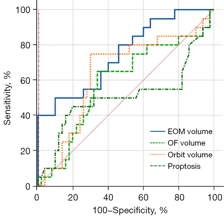
Bivariate logistic regression was used to examine the association between age, MRI parameters, and IOP (Table 3). ROC curve analysis showed that the cutoff value for proptosis for the diagnosis of high IOP was 23.2 mm (sensitivity: 45%; specificity: 80%), and the AUC was 0.509 (95% CI: 0.387–0.631). The cutoff value for EOM volume for the diagnosis of high IOP was 4.204 cm3 (sensitivity: 50%; specificity: 90%), and the AUC was 0.747 (95% CI: 0.629–0.843). The cutoff value for OF volume for the diagnosis of high IOP was 7.730 cm3 (sensitivity: 65%; specificity: 66%), and the AUC was 0.586 (95% CI: 0.461–0.702). The cutoff value for orbit volume for the diagnosis of high IOP was 12.045 cm3 (sensitivity: 75%; specificity: 60%), and the AUC was 0.641 (95% CI: 0.517–0.752). The result of pairwise comparison of ROC curves showed that the AUC of EOM volume was significantly larger than were the other parameters except orbit volume, which possessed high diagnostic efficacy (P=0.03, P=0.004, P=0.08, respectively) (Table 3, Figure 2). Multiple logistic regression was used to analyze the relationship between age, CAS, orbital MRI measurements, and IOP. The results showed that EOM volume (odds ratio =4.925; 95% CI: 1.946–12.464) were significantly associated with IOP changes, and the AUC of fit curve was 0.816 (95% CI: 0.705–0.899; P=0.06) (Table 4).
Table 3. Logistic regression, AUC, and cutoff values for the differential diagnosis of high IOP.
| MRI parameters | AUC | 95% CI | Cutoff point | Sensitivity | Specificity |
|---|---|---|---|---|---|
| EOM volume | 0.747 | 0.629–0.843 | 4.204 | 0.50 | 0.90 |
| OF volume | 0.586 | 0.461–0.702 | 7.730 | 0.65 | 0.66 |
| Orbit volume | 0.641 | 0.517–0.752 | 12.045 | 0.75 | 0.60 |
AUC, area under curve; IOP, intraocular pressure; CI, confidence interval; EOM, extraocular muscle; OF, orbital fat.
Figure 2.

ROC curves of factors associated with high IOP. Bivariate logistic regression was used to examine the association between MRI parameters and IOP, and ROC curve analysis indicated the cutoff value. EOM, extraocular muscle; OF, orbital fat; ROC, receiver operating characteristic; IOP, intraocular pressure; MRI, magnetic resonance imaging.
Table 4. Multiple logistic regression of age, CAS, and MRI parameters with IOP.
| Variables | Coefficient | Std. error | Wald | P | Odds ratio | 95% CI |
|---|---|---|---|---|---|---|
| Age | 0.0338 | 0.037 | 0.840 | 0.36 | 1.034 | 0.962–1.112 |
| CAS | 0.440 | 0.363 | 1.472 | 0.23 | 1.553 | 0.763–3.162 |
| EOM volume | 1.594 | 0.474 | 11.327 | <0.001 | 4.925 | 1.946–12.464 |
| OF volume | −0.095 | 0.150 | 0.403 | 0.53 | 0.909 | 0.678–1.220 |
| Proptosis | −0.136 | 0.115 | 1.408 | 0.24 | 0.873 | 0.697–1.093 |
| Constant | −6.335 | 3.592 | 3.111 | 0.08 | 1.034 | 0.962–1.112 |
CAS, clinical activity score; MRI, magnetic resonance imaging; IOP, intraocular pressure; Std., standard; CI, confidence interval; EOM, extraocular muscle; OF, orbital fat.
The relationship between ON patterns and EOM volume
Spearman correlation analysis was performed to explore the relationship between ON changes and EOM volume. The EOM volume was significantly related to the ON sheath diameter (P=0.01). As for the RNFL thickness, average RNFL thickness showed high correlation with EOM volume (P=0.02) (Table 5).
Table 5. The relationship between optic nerve patterns and EOM volume.
| Optic nerve patterns | Correlation coefficient | P value |
|---|---|---|
| Optic nerve sheath diameter | 0.450 | 0.01 |
| Average RNFL thickness | 0.408 | 0.02 |
| SN RNFL thickness | 0.322 | 0.08 |
| N RNFL thickness | 0.236 | 0.20 |
| IN RNFL thickness | 0.282 | 0.12 |
| ST RNFL thickness | 0.294 | 0.11 |
| T RNFL thickness | 0.204 | 0.27 |
| IT RNFL thickness | 0.191 | 0.30 |
EOM, extraocular muscle; RNFL, retinal nerve fiber layer; SN, superior nasal; N, nasal; IN, inferior nasal; ST, superior temporal; T, temporal; IT, inferior temporal.
The changes of orbital MRI parameters before and after methylprednisolone therapy
A paired t-test was used to compare the MRI measurements before and after methylprednisolone therapy in 20 eyes. A significant decrease of EOM volume was found after methylprednisolone therapy (P=0.002). Meanwhile, proptosis, orbit volume, and OF volume were not significantly changed (P=0.65, P=0.76, P=0.21, respectively). There was a significant reduction of ON sheath diameter and a notable enlargement of axial ONA (P=0.02, P=0.05, respectively), while the sagittal ONA showed no significant change (P=0.75) (Table 6).
Table 6. The changes of orbital MRI parameters within 1 week before and after methylprednisolone therapy.
| MRI parameters | Pretherapy | Post-therapy | P value |
|---|---|---|---|
| Proptosis (mm) | 23.03±2.66 | 23.18±2.97 | 0.65 |
| EOM volume (cm3) | 4.06±1.27 | 3.43±1.07 | 0.002 |
| OF volume (cm3) | 6.41±1.42 | 6.99±2.91 | 0.21 |
| Orbit volume (cm3) | 10.60±2.00 | 10.72±3.04 | 0.76 |
| Optic nerve sheath diameter (cm3) | 0.59±0.07 | 0.57±0.06 | 0.02 |
| Axial ONA (deg) | 165.35±7.73 | 169.16±5.89 | 0.05 |
| Sagittal ONA (deg) | 170.00±5.83 | 170.53±5.18 | 0.75 |
Data are presented as mean ± standard deviation. MRI, magnetic resonance imaging; EOM, extraocular muscle; OF, orbital fat; ONA, optic nerve angle; deg, degree.
Discussion
TAO, which is closely related to thyroid disease, is a disease of the orbit and is characterized by enlargement in the EOMs, OF, and orbital volume, as well as increased proptosis (1). In clinical practice, persistent increase in IOP is often observed, which has irreversible effects on the visual function of patients with TAO. The present study demonstrated that EOM volume is the most critical MRI parameter affecting IOP changes, as it had a higher AUC, sensitivity, and specificity in estimating IOP changes than did the other MRI parameters in TAO eyes. Moreover, EOM volume was correlated with ON changes, which reflect the visual function. Methylprednisolone therapy mainly causes reduction of EOM volume. This study used orbital MRI measurement to demonstrate the critical influence EOM volume has on IOP changes and visual function in patients with TAO.
IOP is correlated with the severity and activity of TAO, which was demonstrated by a significant increase of IOP in patients with severe TAO (15). It has been shown that patients with high IOP are also at a higher risk of developing chronic conditions requiring strabismus surgery and orbital decompression procedures (16). The underlying mechanism and key factors of increased IOP in TAO eyes remain unclear. A recent study found the EOMs to be significantly enlarged in patients with high IOP compared to those with normal IOP, while another study reported that compared to normal participants, patients with TAO who presented IR muscle thickening had a significantly larger increase in IOP, indicating there to be a correlation between EOM and IOP (17,18). Our study found greater EOM enlargement in patients with high IOP patients than in individuals with normal IOP, while the changes in OF and orbit volume, obtained through MRI measurements, were not significantly associated with increased IOP. Our study further demonstrated the importance of EOM volume as a key factor in estimating IOP changes over other MRI parameters. The current opinion is that IOP increase is mostly due to orbital edema, which consists of the increased production of GAGs by the orbital fibroblasts, and inflammation-activated fibroblasts differentiating into adipocytes (19). A recent study used T2RT, which reflects the edema changes caused by autoimmune inflammation and vascular congestion, and found that the high IOP in patients with TAO was positively correlated with EOM inflammation (13). This is consistent with our finding of a significant increase of EOM volume in the MRI T2 phase.
Methylprednisolone and mycophenolate sodium are recommended as first-line treatment according to the latest European Group on Graves’ Orbitopathy (EUGOGO) guidelines (1). We observed a significant decrease in EOM volume after methylprednisolone therapy. However, there was no significant change in proptosis, OF volume, or orbit volume after therapy, and some cases even showed worsening despite treatment. This result is consistent with a previous study by Higashiyama et al. who found that the mean volume of the OF tissue was not significantly decreased after methylprednisolone treatment (20). Wiersinga et al. also reported an earlier enlargement of the EOM as opposed to of OF during the disease’s progression and observed a more severe inflammatory reaction in EOM tissue (21). The possible reason for this may be that the differences in cytokine profiles and variable degrees of response to inflammation between OF and EOMs leads to an early reduction of EOM volume after methylprednisolone therapy. Proptosis changes were not found to be significantly in our study, which is consistent with a previous study. The value of proptosis depends on the ratio of the volume of the whole orbit to that of the bony orbital cavity, and the OF, which constitutes the highest proportion of the whole orbit, mainly determines the proptosis value (22). Moreover, we considered short-term methylprednisolone-related tissue edema is also an important reason. The long-term effect of methylprednisolone on the degree of proptosis is also a direction for future research.
The main site of retinal ganglion cell damage by abnormal IOP is the ON. Recent studies reported a more tortuous ON with primary gaze in patients with normal-tension and high-tension (IOP >21 mmHg) glaucoma than in controls (23,24). In this study, patients with high IOP had a significantly smaller ONA compared with patients with normal IOP, which indicated that the tortuosity of the ON was associated with high tension. Moreover, methylprednisolone therapy straightened the ON sheath and reduced its edema, which was reflected by a larger ONA and a reduction of ON sheath diameter. A previous study demonstrated increased ON tortuosity on MRI in patients with idiopathic intracranial hypertension (IIH), which means the ONA is significantly smaller in patients with IIH compared to controls (25). The mechanism underlying this relationship is likely to be that the relatively higher intracranial pressure increases the cerebrospinal fluid (CSF) distention of the perioptic subarachnoid space, leading to increased bending and edema of the ON sheath (26). Regarding the current study, we speculated that high IOP causes a relative increase in orbital pressure, which leads to an ON that is more susceptible to bending.
This study clarified the relationship between ON patterns and EOM volume. EOM enlargement has a strong correlation with an increased ON diameter and a decreased RNFL thickness. Hu et al. detected intraorbital ON changes using T2 mapping in patients with TAO and found that T2RT at the intraorbital ON was significantly correlated with the total thickness of EOMs and negatively correlated with visual acuity and visual field indices in patients with active TAO (27), which was in good agreement with our results. Hence, EOM volume is an important factor affecting ON patterns at an early stage and has a high affinity with the visual outcome of the disease.
The limitations of this study were its single-center, retrospective design and a relatively low number of high-tension patients. Moreover, relatively few patients were included in our study due to incomplete imaging and clinical data. Additionally, even when examined by an experienced imaging technician, a few patients had head position deviation, which also affected the results.
Conclusions
EOM volume was highly correlated with IOP and ON patterns, and this measurement represents a noninvasive, replicable, and diagnostically efficient means to tracking IOP and ON patterns in patients with TAO. Methylprednisolone therapy can significantly reduce the EOM volume. Therefore, methylprednisolone is recommended for patients with TAO with secondary glaucoma to quickly reduce the EOM volume.
Supplementary
The article’s supplementary files as
Acknowledgments
Funding: This work was funded by Capital’s Funds for Health Improvement and Research (No. 2022-2-20211 to Hongyang Li).
Ethical Statement: The authors are accountable for all aspects of the work in ensuring that questions related to the accuracy or integrity of any part of the work are appropriately investigated and resolved. This observational cross-sectional study was conducted in accordance with the Declaration of Helsinki (as revised in 2013) and was approved by the ethics committee of Beijing Friendship Hospital, Capital Medical University (No. 2022-P2-415). The requirement for written informed consent was waived due to the retrospective nature of the study.
Footnotes
Reporting Checklist: The authors have completed the STROBE reporting checklist. Available at https://qims.amegroups.com/article/view/10.21037/qims-23-44/rc
Conflicts of Interest: All authors have completed the ICMJE uniform disclosure form (available at https://qims.amegroups.com/article/view/10.21037/qims-23-44/coif). HL reports that this work was funded by Capital’s Funds for Health Improvement and Research (No. 2022-2-20211). The other authors have no conflicts of interest to declare.
References
- 1.Bartalena L, Kahaly GJ, Baldeschi L, Dayan CM, Eckstein A, Marcocci C, Marinò M, Vaidya B, Wiersinga WM; EUGOGO. The 2021 European Group on Graves' orbitopathy (EUGOGO) clinical practice guidelines for the medical management of Graves' orbitopathy. Eur J Endocrinol 2021;185:G43-67. 10.1530/EJE-21-0479 [DOI] [PubMed] [Google Scholar]
- 2.Taylor PN, Zhang L, Lee RWJ, Muller I, Ezra DG, Dayan CM, Kahaly GJ, Ludgate M. New insights into the pathogenesis and nonsurgical management of Graves orbitopathy. Nat Rev Endocrinol 2020;16:104-16. 10.1038/s41574-019-0305-4 [DOI] [PubMed] [Google Scholar]
- 3.Bartalena L, Piantanida E, Gallo D, Lai A, Tanda ML. Epidemiology, Natural History, Risk Factors, and Prevention of Graves' Orbitopathy. Front Endocrinol (Lausanne) 2020;11:615993. 10.3389/fendo.2020.615993 [DOI] [PMC free article] [PubMed] [Google Scholar]
- 4.Fan SX, Zeng P, Li ZJ, Wang J, Liang JQ, Liao YR, Hu YX, Xu MT, Wang M. The Clinical Features of Graves' Orbitopathy with Elevated Intraocular Pressure. J Ophthalmol 2021;2021:9879503. 10.1155/2021/9879503 [DOI] [PMC free article] [PubMed] [Google Scholar]
- 5.Eslami F, Borzouei S, Khanlarzadeh E, Seif S. Prevalence of increased intraocular pressure in patients with Graves' ophthalmopathy and association with ophthalmic signs and symptoms in the north-west of Iran. Clin Ophthalmol 2019;13:1353-9. 10.2147/OPTH.S205112 [DOI] [PMC free article] [PubMed] [Google Scholar]
- 6.Delavar A, Radha Saseendrakumar B, Lee TC, Topilow NJ, Ting MA, Liu CY, Korn BS, Weinreb RN, Kikkawa DO, Baxter SL. Associations Between Thyroid Eye Disease and Glaucoma Among Those Enrolled in the National Institutes of Health All of Us Research Program. Ophthalmic Plast Reconstr Surg 2023;39:336-40. 10.1097/IOP.0000000000002310 [DOI] [PMC free article] [PubMed] [Google Scholar]
- 7.Kang JM, Tanna AP. Glaucoma. Med Clin North Am 2021;105:493-510. 10.1016/j.mcna.2021.01.004 [DOI] [PubMed] [Google Scholar]
- 8.Fenwick EK, Man RE, Aung T, Ramulu P, Lamoureux EL. Beyond intraocular pressure: Optimizing patient-reported outcomes in glaucoma. Prog Retin Eye Res 2020;76:100801. 10.1016/j.preteyeres.2019.100801 [DOI] [PubMed] [Google Scholar]
- 9.Jain AP, Gellada N, Ugradar S, Kumar A, Kahaly G, Douglas R. Teprotumumab reduces extraocular muscle and orbital fat volume in thyroid eye disease. Br J Ophthalmol 2022;106:165-71. 10.1136/bjophthalmol-2020-317806 [DOI] [PubMed] [Google Scholar]
- 10.Song C, Luo Y, Yu G, Chen H, Shen J. Current insights of applying MRI in Graves' ophthalmopathy. Front Endocrinol (Lausanne) 2022;13:991588. 10.3389/fendo.2022.991588 [DOI] [PMC free article] [PubMed] [Google Scholar]
- 11.Cohen LM, Yoon MK. Update on Current Aspects of Orbital Imaging: CT, MRI, and Ultrasonography. Int Ophthalmol Clin 2019;59:69-79. 10.1097/IIO.0000000000000288 [DOI] [PubMed] [Google Scholar]
- 12.Ma R, Geng Y, Gan L, Peng Z, Cheng J, Guo J, Qian J. Quantitative T1 mapping MRI for the assessment of extraocular muscle fibrosis in thyroid-associated ophthalmopathy. Endocrine 2022;75:456-64. 10.1007/s12020-021-02873-0 [DOI] [PubMed] [Google Scholar]
- 13.Luo B, Wang W, Li X, Zhang H, Zhang Y, Hu W. Correlation Analysis between Intraocular Pressure and Extraocular Muscles Based on Orbital Magnetic Resonance T2 Mapping in Thyroid-Associated Ophthalmopathy Patients. J Clin Med 2022;11:3981. 10.3390/jcm11143981 [DOI] [PMC free article] [PubMed] [Google Scholar]
- 14.Oculoplastic and Orbital Disease Group of Chinese Ophthalmological Society of Chinese Medical Association. Thyroid Group of Chinese Society of Endocrinology of Chinese Medical Association . Chinese guideline on the diagnosis and treatment of thyroid-associated ophthalmopathy (2022). Zhonghua Yan Ke Za Zhi 2022;58:646-68. 10.3760/cma.j.cn112142-20220421-00201 [DOI] [PubMed] [Google Scholar]
- 15.Tu Y, Mao B, Li J, Liu W, Xu M, Chen Q, Wu W. Relationship between the 24-h variability of blood pressure, ocular perfusion pressure, intraocular pressure, and visual field defect in thyroid associated orbitopathy. Graefes Arch Clin Exp Ophthalmol 2020;258:2007-12. 10.1007/s00417-020-04733-5 [DOI] [PubMed] [Google Scholar]
- 16.Karhanová M, Kalitová J, Kovář R, Schovánek J, Karásek D, Čivrný J, Hübnerová P, Mlčák P, Šín M. Ocular hypertension in patients with active thyroid-associated orbitopathy: a predictor of disease severity, particularly of extraocular muscle enlargement. Graefes Arch Clin Exp Ophthalmol 2022;260:3977-84. 10.1007/s00417-022-05760-0 [DOI] [PubMed] [Google Scholar]
- 17.Stoyanova NS, Konareva-Kostianeva M, Mitkova-Hristova V, Angelova I. Correlation between intraocular pressure and thickness of extraocular muscles, the severity and activity of thyroid-associated orbitopathy. Folia Med (Plovdiv) 2019;61:90-6. 10.2478/folmed-2018-0050 [DOI] [PubMed] [Google Scholar]
- 18.Li X, Bai X, Liu Z, Cheng M, Li J, Tan N, Yuan H. The effect of inferior rectus muscle thickening on intraocular pressure in thyroid-associated ophthalmopathy. J Ophthalmol 2021;2021:9736247. 10.1155/2021/9736247 [DOI] [PMC free article] [PubMed] [Google Scholar]
- 19.Wang Y, Smith TJ. Current concepts in the molecular pathogenesis of thyroid-associated ophthalmopathy. Invest Ophthalmol Vis Sci 2014;55:1735-48. 10.1167/iovs.14-14002 [DOI] [PMC free article] [PubMed] [Google Scholar]
- 20.Higashiyama T, Nishida Y, Ohji M. Changes of orbital tissue volumes and proptosis in patients with thyroid extraocular muscle swelling after methylprednisolone pulse therapy. Jpn J Ophthalmol 2015;59:430-5. 10.1007/s10384-015-0410-4 [DOI] [PubMed] [Google Scholar]
- 21.Wiersinga WM, Regensburg NI, Mourits MP. Differential involvement of orbital fat and extraocular muscles in graves' ophthalmopathy. Eur Thyroid J 2013;2:14-21. 10.1159/000348246 [DOI] [PMC free article] [PubMed] [Google Scholar]
- 22.Ugradar S, Goldberg RA, Rootman DB. Bony Orbital Volume Expansion in Thyroid Eye Disease. Ophthalmic Plast Reconstr Surg 2019;35:434-7. 10.1097/IOP.0000000000001292 [DOI] [PubMed] [Google Scholar]
- 23.Demer JL, Clark RA, Suh SY, Giaconi JA, Nouri-Mahdavi K, Law SK, Bonelli L, Coleman AL, Caprioli J. Magnetic resonance imaging of optic nerve traction during adduction in primary open-angle glaucoma with normal intraocular pressure. Invest Ophthalmol Vis Sci 2017;58:4114-25. 10.1167/iovs.17-22093 [DOI] [PMC free article] [PubMed] [Google Scholar]
- 24.Wang X, Rumpel H, Baskaran M, Tun TA, Strouthidis N, Perera SA, Nongpiur ME, Lim WEH, Aung T, Milea D, Girard MJA. Optic nerve tortuosity and globe proptosis in normal and glaucoma subjects. J Glaucoma 2019;28:691-6. 10.1097/IJG.0000000000001270 [DOI] [PubMed] [Google Scholar]
- 25.Chen BS, Asnafi S, Lin MY, Bruce BB, Lock JH, Sharma RA, Newman NJ, Biousse V, Saindane AM. Optic Nerve Angle in Idiopathic Intracranial Hypertension. J Neuroophthalmol 2021;41:e464-9. 10.1097/WNO.0000000000000986 [DOI] [PubMed] [Google Scholar]
- 26.Hu R, Holbrook J, Newman NJ, Biousse V, Bruce BB, Qiu D, Oshinski J, Saindane AM. Cerebrospinal fluid pressure reduction results in dynamic changes in optic nerve angle on magnetic resonance imaging. J Neuroophthalmol 2019;39:35-40. 10.1097/WNO.0000000000000643 [DOI] [PubMed] [Google Scholar]
- 27.Hu H, Chen HH, Chen W, Wu Q, Chen L, Zhu H, Shi HB, Xu XQ, Wu FY. Thyroid-Associated Ophthalmopathy: Preliminary Study Using T2 Mapping to Characterize Intraorbital Optic Nerve Changes Before Dysthyroid Optic Neuropathy. Endocr Pract 2021;27:191-7. 10.1016/j.eprac.2020.09.006 [DOI] [PubMed] [Google Scholar]
Associated Data
This section collects any data citations, data availability statements, or supplementary materials included in this article.
Supplementary Materials
The article’s supplementary files as


