Abstract
Inappropriate and chronic activation of the cytosolic NOD‐, LRR‐, and pyrin domain‐containing 3 (NLRP3) inflammasome, a key component of innate immunity, likely underlies several inflammatory diseases, including coronary artery disease. This first‐in‐human phase I trial evaluated safety, pharmacokinetics (PKs), and pharmacodynamics (PDs) of oral, single (150–1800 mg) and multiple (300 or 900 mg twice daily for 7 days) ascending doses (SADs and MADs) of GDC‐2394, a small‐molecule inhibitor of NLRP3, versus placebo in healthy volunteers. The study also assessed the food effect on GDC‐2394 and its CYP3A4 induction potential in food‐effect (FE) and drug–drug interaction (DDI) stages, respectively. Although GDC‐2394 was adequately tolerated in the SAD, MAD, and FE cohorts, two participants in the DDI stage experienced grade 4 drug‐induced liver injury (DILI) deemed related to treatment, but unrelated to a PK drug interaction, leading to halting of the trial. Both participants experiencing severe DILI recovered within 3 months. Oral GDC‐2394 was rapidly absorbed; exposure increased in an approximately dose‐proportional manner with low‐to‐moderate intersubject variability. The mean terminal half‐life ranged from 4.1 to 8.6 h. Minimal accumulation was observed with multiple dosing. A high‐fat meal led to delays in time to maximum concentration and minor decreases in total exposure and maximum plasma concentration. GDC‐2394 had minimal CYP3A4 induction potential with the sensitive CYP3A4 substrate, midazolam. Exploratory ex vivo whole‐blood stimulation assays showed rapid, reversible, and near‐complete inhibition of the selected PD biomarkers, IL‐1β and IL‐18, across all tested doses. Despite favorable PK and target engagement PD, the GDC‐2394 safety profile precludes its further development.
Study Highlights.
WHAT IS THE CURRENT KNOWLEDGE ON THE TOPIC?
Inappropriate stimulation by endogenous ligands or chronic activation of the NLRP3 inflammasome may underlie several inflammatory diseases; therefore, inhibition of NLRP3 may be broadly therapeutic. GDC‐2394 is an orally available, small‐molecule inhibitor of NLRP3 that selectively blocks IL‐1β and IL‐18 secretion following activation of the NLRP3 pathway by various stimuli.
WHAT QUESTION DID THIS STUDY ADDRESS?
This first‐in‐human, dose‐escalation study aimed to evaluate the safety, tolerability, pharmacokinetics (PKs), and pharmacodynamics (PDs) of single and multiple oral doses of GDC‐2394 in healthy volunteers. The trial also assessed the effect of food on GDC‐2394 and, during a drug–drug interaction stage, examined the CYP3A4 induction potential of GDC‐2394.
WHAT DOES THIS STUDY ADD TO OUR KNOWLEDGE?
After oral administration, GDC‐2394 was rapidly absorbed with approximately dose‐proportional increases in exposure. A high‐fat meal had a minor effect on GDC‐2394 PKs, and GDC‐2394 had no apparent impact on midazolam PKs. GDC‐2394 also showed rapid, reversible, and near‐complete inhibition of IL‐1β and IL‐18 secretion in ex vivo stimulation of whole blood from trial participants. The study was halted because of serious adverse events that indicated possible drug‐induced hepatotoxicity. Overall, this study adds information on PK/PD target engagement of NLRP3 inhibitors, and translating this information into clinically meaningful responses may prove useful for evaluating other NLRP3 inhibitors in future studies.
HOW MIGHT THIS CHANGE CLINICAL PHARMACOLOGY OR TRANSLATIONAL SCIENCE?
Although GDC‐2394 had favorable PK and target engagement PD, the safety findings led to stopping the trial. Nevertheless, the PK/PD relationship and safety findings identified from this study may help guide the development of other NLRP3 inhibitors in coronary artery disease and other inflammatory diseases.
INTRODUCTION
Inflammasomes are cytosolic multiprotein complexes that are key components of the innate immune system and are responsible for the activation of inflammatory responses in myeloid cells. 1 Inflammatory ligands stimulate the assembly and activation of the inflammasome, which consists of a pattern‐recognition receptor that acts as a sensor, an adaptor protein, and pro‐caspase 1. 2
Nucleotide‐binding oligomerization domain, leucine‐rich repeat, and pyrin domain‐containing 3 (NLRP3) inflammasomes are triggered by a wide variety of endogenous and microbial stimuli. 2 Stimulation of the NLRP3 inflammasome activates caspase 1, leading to cleavage, maturation, and secretion of the cytokines interleukin (IL)‐1β and IL‐18, and cleavage of gasdermin D, which are responsible for inflammatory signaling and pyroptosis, respectively. 2 Because inappropriate stimulation by endogenous ligands or chronic inflammasome activation underlies multiple inflammatory diseases, including gout, chronic obstructive pulmonary disease, inflammatory bowel disease, and coronary artery disease (CAD), 3 inhibition of NLRP3 may have broad therapeutic potential.
Although current therapies for patients with CAD focus on cholesterol reduction, many patients still have chronic inflammation, which carries a risk of further cardiovascular events and is associated with poor clinical outcomes. 4 Atherogenic triggers, such as cholesterol crystals, activate NLRP3, leading to IL‐1β and IL‐18 release. 5 , 6 Models of atherosclerosis and atherosclerotic plaque formation have demonstrated the dependency of IL‐1β expression on NLRP3. 5 , 7 Although treatment with canakinumab, an anti‐IL‐1β antibody, in patients with atherosclerosis and residual inflammation significantly reduced the frequency of cardiovascular events, 8 this therapy is not currently approved for atherosclerosis, and additional anti‐inflammatory therapies are needed.
GDC‐2394 is an orally available, selective, and reversible small‐molecule inhibitor of NLRP3. 9 It selectively blocks IL‐1β and IL‐18 secretion following activation of the NLRP3 pathway by various stimuli, including cholesterol crystals, in biochemical and ex vivo cellular assays. Clinical development of GDC‐2394 was initiated based on nonclinical safety and pharmacology data targeting CAD as the lead indication. 9 This first‐in‐human, dose‐escalation study evaluated the safety, tolerability, pharmacokinetics (PKs), and pharmacodynamics (PDs) of single and multiple oral doses of GDC‐2394, and evaluated the effect of food and the CYP3A4 induction potential of GDC‐2394 in healthy volunteers.
METHODS
Study design
This first‐in‐human study of GDC‐2394 in healthy adult volunteers (https://www.isrctn.com/ISRCTN15492429) consisted of single‐ and multiple‐ascending dose (SAD, MAD), food‐effect (FE), and drug–drug interaction (DDI) stages (Figure S1a). The study was conducted at a single clinical research unit in New Zealand. The SAD, MAD, and FE stages were randomized, double‐blinded (to the participants and investigators), and placebo‐controlled, whereas the DDI stage was open‐label.
The study protocol was approved by an institutional review board and was conducted according to the International Conference on Harmonization E6 Guidelines for Good Clinical Practice and the laws of the country in which the trial took place. Each participant provided signed informed consent prior to study enrollment.
Study stages
The SAD stage consisted of four cohorts of eight participants each, randomized in a 6:2 ratio to receive a single oral dose of GDC‐2394 (150 mg, 450 mg, 900 mg, 1800 mg) or placebo on day 1. The MAD stage consisted of two cohorts of eight participants each, randomized in a 6:2 ratio to receive oral GDC‐2394 (300 mg twice daily [b.i.d.] or 900 mg b.i.d.) or placebo on days 1 through 6, followed by a single dose on the morning of day 7. In the FE stage, 10 participants were randomized in an 8:2 ratio to receive a single dose of 600 mg GDC‐2394 or placebo administered after fasting (period 1) and again after a high‐fat meal (period 2). Each dose period was separated by a washout of 4 days. The DDI stage evaluated the potential of GDC‐2394 for inducing cytochrome P4503A4 (CYP3A4) through co‐administration with midazolam, a sensitive CYP3A4 substrate, in one cohort of nine participants (15 participants were planned; see Results below). On the morning of day 1, participants received a single 5‐mg dose of midazolam (midazolam HCL syrup, 2 mg/mL). On days 3–9, participants received 900 mg b.i.d. GDC‐2394. On the morning of day 10, participants received a single 5‐mg dose of midazolam together with a final dose of GDC‐2394. Details on randomization and blinding procedures, and screening, treatment, and follow‐up periods for each stage are provided in Appendix S1.
Study drug administration
GDC‐2394 was provided by Genentech, Inc. (South San Francisco, CA) as a powder‐in‐capsule formulation in doses of 150 mg. Placebo capsules without the active ingredient were designed to match GDC‐2394 capsules. GDC‐2394 was administered under fasting conditions in the SAD, MAD, and DDI stages and in period 1 of the FE stage. Detailed fasting rules are described in Appendix S1.
Dose selection, escalation, and stopping criteria
The starting dose in the SAD (150 mg) was selected by considering the safety margins derived from the 4‐week toxicology study in cynomolgus monkeys. Using the maximum recommended starting dose (MRSD) approach, the human equivalent dose for the cynomolgus monkey no‐observed‐adverse‐effect level (NOAEL; 600 mg/kg/d) was 193 mg/kg, or 11,580 mg for a 60‐kg person. A default uncertainty factor of 10 was applied to derive an MRSD of 1158 mg. To account for uncertainty around the translatability of nonclinical studies to humans, an additional safety margin was added to the MRSD to arrive at the proposed starting dose of 150 mg. Based on preclinical predictions of human PKs, the safety margins for the 150‐mg dose were projected to be 15 and 51, based on the maximum plasma concentration (Cmax) of 98,600 ng/mL and the area under the concentration–time curve from time 0 to 24 h (AUC0–24 h) of 798,000 ng•h/mL, respectively, at the NOAEL in cynomolgus monkeys. The minimum anticipated biological effect level and pharmacologically active dose were not expected to be relevant in healthy volunteers, because the signaling pathways involving NLRP3 (i.e., inflammation) are not thought to be homeostatically active. 3
In the SAD and MAD stages, the sponsor safety monitoring committee (SMC) made dose‐escalation decisions in consultation with the investigator, based on a review of required available safety and PK data. Dose escalation steps were not to exceed the dose evaluated in the prior cohort by three‐fold, or doses of 1800 mg and 3000 mg daily in the SAD and MAD stages, respectively. The predicted exposure for the subsequent cohort was not to exceed a mean cohort Cmax or an AUC0–24 h equivalent to the NOAEL in cynomolgus monkeys. Detailed dose‐escalation stopping criteria are described in Appendix S1. Dose‐stopping criteria (Appendix S1) for individuals, cohorts, and for the study were prespecified in the protocol, and the SMC, together with the investigator, reviewed cases to determine if criteria were met.
Participants
Eligible participants were adult men and non‐pregnant, non‐lactating women who agreed to remain abstinent or use highly effective contraception, 18–65 years old, and in good health, as determined by medical history, 12‐lead electrocardiogram (ECG), vital signs, and clinical laboratory tests. Detailed exclusion criteria are provided in Appendix S1.
Safety end points and assessments
The primary objective of the study was to evaluate the safety of single and multiple doses of GDC‐2394 based on the incidence and severity of adverse events (AEs) and changes from baseline in targeted vital signs, clinical laboratory test results, and 12‐lead ECG findings. AEs were mapped to the Medical Dictionary for Regulatory Activities (version 25.0) thesaurus terms, and AE severity was graded according to the Division of AIDS Table for Grading the Severity of Adult and Pediatric Adverse Events (Corrected Version 2.1). 10 Statistical summaries provided were descriptive (e.g., incidence rates, means, and percentiles).
Pharmacokinetic end points and assessments
The PK objectives of the study were to evaluate the plasma PK of single and multiple doses of GDC‐2394 and the effect of food on the PK of GDC‐2394 based on plasma concentrations of GDC‐2394 and to evaluate the CYP3A induction potential of GDC‐2394 based on plasma concentrations of midazolam and 1‐hydroxymidazolam when midazolam was administered alone and in combination with GDC‐2394. Exploratory PK objectives included the assessment of renal elimination and preliminary metabolite profiling and characterization of GDC‐2394. Plasma concentrations of indacenyl amine (1,2,3,5,6,7‐hexahydro‐s‐indacen‐4‐amine), a GDC‐2394 metabolite, were quantified in all PK samples from GDC‐2394‐treated participants in the SAD, MAD, and DDI cohorts, and the metabolite:parent AUC and Cmax ratios (MRAUC and MRCmax) were calculated, accounting for the molecular weights of GDC‐2394 and indacenyl amine (431.51 g/mol and 173.25 g/mol, respectively).
Blood and urine samples were collected for PK analysis; detailed collection methods and timepoints are described in Appendix S1. GDC‐2394, indacenyl amine, midazolam, and 1‐hydroxymidazolam plasma concentrations were measured with validated high‐performance pressure liquid chromatography tandem mass spectrometry (HPLC‐MS/MS) by Labcorp Laboratory Bioanalytical Services (Indianapolis, IN; Madison, WI); lower limits of quantification (LLOQ) for GDC‐2394, indacenyl amine, midazolam, and 1‐hydroxymidazolam were 5 ng/mL, 0.1 ng/mL, 0.1 ng/mL, and 0.1 ng/mL, respectively. All study samples were run within the established stability window. GDC‐2394 urine concentrations were determined using a qualified HPLC‐MS/MS method (LLOQ: 10 ng/mL) by Labcorp Laboratory Bioanalytical Services (Indianapolis, IN).
Descriptive statistics were calculated for concentration data and PK parameters using standard noncompartmental analysis with Phoenix WinNonlin software, version 8.3 (Certara, Princeton, NJ). Comparison of exposures in the FE and DDI stages was conducted using a linear mixed‐effects model, including treatment as the fixed effect and participant as the random effect using SAS, version 9.4 (SAS Institute, Cary, NC). Log‐transformed Cmax, AUC from time 0 to last quantifiable concentration (AUC0–last), and AUC from time 0 to infinity (AUC0–inf) were evaluated to estimate geometric mean ratios (GMRs), and corresponding 90% confidence intervals (CIs) were used to quantify the uncertainty of point estimates.
Pharmacodynamic end points and assessments
The main PD end point was mature IL‐1β and IL‐18 production, assessed by ex vivo stimulation of whole blood from study participants with a cocktail of lipopolysaccharide (LPS), a known trigger, and adenosine triphosphate (ATP), an activator of the NLRP3 inflammasome. 11 Baseline (predose) and postdose blood sample collection, processing, and assay methods are described in Appendix S1.
Each measurement of IL‐1β and IL‐18 consisted of the mean of two technical replicates for each participant at each timepoint. Percent inhibition of IL‐1β production relative to baseline was calculated as [1‐(postdose stimulated IL‐1β level)/(predose stimulated IL‐1β level)]*100%. Percent inhibition of IL‐18 production relative to baseline was calculated in a similar manner but was adjusted for endogenous circulating levels of IL‐18 by deducting IL‐18 levels measured in unstimulated (‐LPS/ATP) samples from corresponding stimulated (+LPS/ATP) samples (as the endogenous circulating IL‐18 levels were above the LLOQ). Mean percent inhibition of IL‐1β and IL‐18 production relative to baseline was calculated and plotted against time; for exploratory PK/PD analyses, percent IL‐1β inhibition relative to baseline and percent IL‐18 inhibition relative to baseline (with values >100% removed) were plotted against time‐matched drug concentrations and fitted to a sigmoid maximum effect (Emax) model using R Statistical Software (version 4.0.1). 12
Statistical considerations
Tabulation and listing of descriptive statistics were performed using SAS. The safety analysis population consisted of all participants who received at least one dose of study drug. The PK population consisted of all participants who received at least one dose of GDC‐2394.
The sample size for this study was based on dose‐escalation rules rather than statistical considerations, and the sample size of six participants receiving GDC‐2394 per SAD or MAD cohort was consistent with similarly designed SAD/MAD studies. The planned sample size of 15 for the DDI cohort was calculated to provide 80% power yielding a 90% CI for the GMR to lie within the “no‐effect window” of 0.5–2 when the true GMR is 1.0, and assumed a dropout rate of 20%.
RESULTS
Participant disposition and demographics
The first participant enrolled in the study on April 20, 2021, and the last patient's last visit was on March 21, 2022. Of 124 participants screened, the study enrolled 67 participants (SAD, n = 32; MAD, n = 16; FE, n = 10; and DDI, n = 9; Figure S1b). Although the initial plan was to enroll 15 participants into the DDI stage, the study was halted after nine participants received GDC‐2394 due to two serious AEs (SAEs) of drug‐induced liver injury (DILI; see Safety results below). One participant in the SAD stage who received 450 mg GDC‐2394 withdrew on day 9. Fifty‐three participants received at least one dose of GDC‐2394.
The mean age across all cohorts and stages ranged from 30 to 37 years, and the mean weight ranged from 74.0 to 84.6 kg. The majority of participants were White, ranging from 78% to 91% of each stage (Table 1; Tables S1–S4), and not Hispanic or Latino. Women comprised 75%, 56%, 30%, and 33% of participants in the SAD, MAD, FE, and DDI stages, respectively.
TABLE 1.
Participant demographics and baseline characteristics.
| All SAD participants (n = 32) | All MAD participants (n = 16) | All FE participants (n = 10) | All DDI participants (n = 9) | |
|---|---|---|---|---|
| Age, year, mean (SD) | 31.1 (10.8) | 29.9 (9.8) | 37.4 (13.9) | 29.9 (12.3) |
| Sex | ||||
| Male | 8 (25.0%) | 7 (43.8%) | 7 (70.0%) | 6 (66.7%) |
| Female | 24 (75.0%) | 9 (56.2%) | 3 (30.0%) | 3 (33.3%) |
| Ethnicity | ||||
| Hispanic or Latino | 1 (3.1%) | 1 (6.2%) | 1 (10.0%) | 0 |
| Not Hispanic or Latino | 31 (96.9%) | 14 (87.5%) | 9 (90.0%) | 9 (100.0%) |
| Not stated | 0 | 1 (6.2%) | 0 | 0 |
| Race | ||||
| Asian | 1 (3.1%) | 1 (6.2%) | 1 (10.0%) | 0 |
| Black or African American | 1 (3.1%) | 1 (6.2%) | 0 | 0 |
| Native Hawaiian or other Pacific Islander | 1 (3.1%) | 1 (6.2%) | 1 (10.0%) | 0 |
| White | 29 (90.6%) | 13 (81.2%) | 8 (80.0%) | 7 (77.8%) |
| Multiple | 0 | 0 | 0 | 1 (11.1%) |
| Unknown | 0 | 0 | 0 | 1 (11.1%) |
| Weight, kg, mean (SD) | 75.9 (17.4) | 74.0 (23.0) | 81.8 (18.7) | 84.6 (19.2) |
Abbreviations: DDI, drug‐drug interaction; FE, food‐effect; MAD, multiple‐ascending dose; SAD, single‐ascending dose.
Safety
Across the SAD, MAD, and FE stages, the incidence of AEs was comparable between subjects receiving GDC‐2394 and placebo, and most AEs were mild in severity (Table 2). There were no deaths, SAEs, or AEs leading to dose modification/interruption or discontinuation, and there were no clinically meaningful, abnormal ECG or vital signs findings (Table 2; Supplementary Results; Tables S5–S7). No AEs affecting dose escalation in the SAD and MAD stages were reported. Among participants who received the highest dose of GDC‐2394 (900 mg b.i.d.) in the MAD stage, there were no laboratory abnormalities for aspartate aminotransferase (AST), bilirubin, or international normalized ratio. However, one subject had an asymptomatic, mild elevation of alanine aminotransferase (ALT) on day 7, the final dosing day, through day 21, with a peak elevation of 50 U/L (1.67× the upper limit of normal [ULN]) on day 14.
TABLE 2.
Overview of AEs.
| SAD | MAD | FE | DDI | ||||
|---|---|---|---|---|---|---|---|
| All GDC‐2394 (n = 24) | All placebo (n = 8) | All GDC‐2394 (n = 12) | All placebo (n = 4) | GDC‐2394 (n = 8) | Placebo (n = 2) | GDC‐2394 + midazolam (n = 9) | |
| Total number of subjects with at least one AE | 14 (58.3%) | 3 (37.5%) | 8 (66.7%) | 2 (50.0%) | 3 (37.5%) | 2 (100.0%) | 9 (100.0%) |
| Total number of AEs | 22 | 5 | 18 | 6 | 3 | 3 | 36 |
| Total number of deaths | 0 | 0 | 0 | 0 | 0 | 0 | 0 |
| Total number of subjects withdrawn from study due to an AE | 0 | 0 | 0 | 0 | 0 | 0 | 0 |
| Total number of subjects with at least one | |||||||
| AE with fatal outcome | 0 | 0 | 0 | 0 | 0 | 0 | 0 |
| Serious AE | 0 | 0 | 0 | 0 | 0 | 0 | 2 (22.2%) |
| Related serious AE | 0 | 0 | 0 | 0 | 0 | 0 | 2 (22.2%) |
| AE leading to withdrawal from treatment | 0 | 0 | 0 | 0 | 0 | 0 | 0 |
| AE leading to dose modification/interruption | 0 | 0 | 0 | 0 | 0 | 0 | 0 |
| Related AE | 6 (25.0%) | 1 (12.5%) | 4 (33.3%) | 1 (25.0%) | 1 (12.5%) | 0 | 7 (77.8%) |
| Grade 3–5 AE (at greatest intensity) | 0 | 0 | 0 | 1 | 0 | 0 | 2 (22.2%) |
Abbreviations: AEs, adverse events; DDI, drug‐drug interaction; FE, food‐effect; MAD, multiple‐ascending dose; SAD, single‐ascending dose.
In the DDI stage, all nine (100%) participants experienced at least one AE (Table 2; Table S8). There were no deaths, but there were two SAEs (see below). The most frequently reported (≥20%) AEs were fatigue, headache, and rash. Seven (78%) participants reported a total of 14 events that were considered related to study treatment; the most frequent were fatigue and DILI. Three participants experienced events consistent with hepatic injury after completion of GDC‐2394 dosing. One participant experienced a grade 2 ALT increase on day 11 that was considered related to study drug, and resolved to grade 1 by day 20. Two SAEs of grade 4 DILI occurred in two participants on days 18 and 20, respectively. The SAEs were considered to be related to study treatment, and both met criteria for Hy's law (AST or ALT >3× ULN and total bilirubin >2× ULN or clinical jaundice). 13 The sponsor halted all further enrollment. Both participants recovered, with one receiving treatment with oral ursodeoxycholic acid for DILI. See Appendix S1 Safety Narratives for detailed descriptions of these cases and the Appendix S2 Listing for a listing of all AEs by patient.
Pharmacokinetics
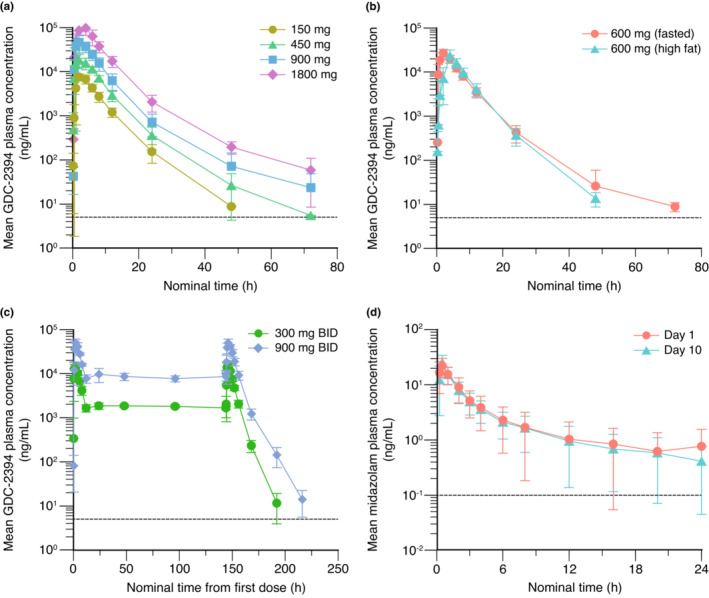
Following single oral doses across 150–1800 mg, plasma drug concentration‐time profiles showed rapid absorption of GDC‐2394, with a median time to maximum concentration (Tmax) ranging from 2 to 4 h (Table 3; Figure 1a); the mean terminal half‐life (t 1/2) ranged from 4.1 to 8.6 h, and apparent clearance was comparable (mean values ranging from 2590 to 3390 mL/h). The interindividual variability of exposure parameters (Cmax, AUC0–last, and AUC0–inf) was low to moderate; coefficients of variation (%CV) were less than 50%. Based on dose‐normalized Cmax and AUC0–last, increases in systemic exposure to GDC‐2394 were approximately dose proportional. A high‐fat meal with the 600‐mg dose led to a delay in Tmax (median, 4 h; range, 2–6 h) compared with dosing under fasted conditions (median: 2 h; range: 1–4 h; Table 3; Figure 1b). A high‐fat meal had a small negative effect on GDC‐2394 exposure, leading to decreases of ~16% for Cmax, 14% for AUC0–last, and 14% for AUC0–inf (Table 4).
TABLE 3.
GDC‐2394 plasma pharmacokinetic parameters in SAD, MAD, and FE stages.
| Treatment | Statistic | T max (h) a | C max (ng/mL) | AUC0–last (h*ng/mL) | AUC0–inf (h*ng/mL) | DN C max (ng/mL/mg) | DN AUC0–last (h*ng/mL/mg) | T 1/2 (h) | Vz/F (mL) | CL/F (mL/h) |
|---|---|---|---|---|---|---|---|---|---|---|
| SAD stage | ||||||||||
| 150 mg (n = 6) | Mean | 2.1 | 8110 | 53,500 | 54,000 | 54.1 | 356 | 4.1 | 16,700 | 2870 |
| SD | (1.0–4.0) | 1270 | 10,200 | 10,100 | 8.47 | 67.6 | 0.5 | 2450 | 558 | |
| CV% | 48.1 | 15.7 | 19.0 | 18.7 | 15.6 | 19.0 | 12.6 | 14.7 | 19.5 | |
| 450 mg (n = 6) | Mean | 2.0 | 22,900 | 138,000 | 138,000 | 50.8 | 307 | 5.8 | 30,200 | 3390 |
| SD | (1.0–4.0) | 10,800 | 29,200 | 29,200 | 24.1 | 64.5 | 2.7 | 20,700 | 779 | |
| CV% | 55.0 | 47.4 | 21.1 | 21.1 | 47.4 | 21.0 | 46.9 | 68.5 | 23.0 | |
| 900 mg (n = 6) | Mean | 2.0 | 50,400 | 317,000 | 317,000 | 56.0 | 352 | 6.8 | 34,900 | 3040 |
| SD | (1.0–4.0) | 15,200 | 77,400 | 77,100 | 16.9 | 86.4 | 4.6 | 39,000 | 1020 | |
| CV% | 45.3 | 30.2 | 24.5 | 24.3 | 30.1 | 24.6 | 66.8 | 111.8 | 33.4 | |
| 1800 mg (n = 6) | Mean | 4.0 | 101,000 | 738,000 | 739,000 | 56.3 | 410 | 8.6 | 32,100 | 2590 |
| SD | (2.0–4.2) | 22,200 | 188,000 | 188,000 | 12.4 | 104 | 1.9 | 11,300 | 731 | |
| CV% | 31.5 | 21.9 | 25.4 | 25.5 | 21.9 | 25.4 | 22.4 | 35.2 | 28.2 | |
| Treatment | Statistic | First dose (day 1) | Steady‐state (day 7) | |||||||
|---|---|---|---|---|---|---|---|---|---|---|
| C max (ng/mL) | AUC0–tau (h*ng/mL) | C min (ng/mL) | C max (ng/mL) | CLss/F (mL/h) | AUC0–tau (h*ng/mL) | T 1/2 (h) | AR (AUC0–tau) | AR (C max) | ||
| MAD stage | ||||||||||
| 300 mg b.i.d. (n = 6) | Mean | 15,300 | 82,000 | 1820 | 16,400 | 3290 | 92,200 | 4.2 | 1.1 | 1.1 |
| SD | 2620 | 8700 | 506 | 1840 | 335 | 10,000 | 0.9 | 0.02 | 0.14 | |
| CV% | 17.1 | 10.6 | 27.8 | 11.3 | 10.2 | 10.9 | 21.9 | 2.2 | 12.6 | |
| 900 mg b.i.d. (n = 6) | Mean | 52,700 | 301,000 | 9080 | 51,000 | 2780 | 327,000 | 6.7 | 1.1 | 1.0 |
| SD | 8450 | 31,900 | 2390 | 8290 | 288 | 36,200 | 1.3 | 0.08 | 0.13 | |
| CV% | 16.0 | 10.6 | 26.3 | 16.3 | 10.4 | 11.1 | 19.4 | 7.5 | 13.2 | |
| Statistic | Fasted period (n = 8) | High‐fat period (n = 8) | ||||||||
|---|---|---|---|---|---|---|---|---|---|---|
| T max (h) a | C max (ng/mL) | AUC0–last (h*ng/mL) | AUC0–inf (h*ng/mL) | T 1/2 (h) | Tmax (h) a | C max (ng/mL) | AUC0–last (h*ng/mL) | AUC0–inf (h*ng/mL) | T 1/2 (h) | |
| FE stage (600 mg) | ||||||||||
| Mean | 2.0 | 28,000 | 172,000 | 173,000 | 5.3 | 4.0 | 24,100 | 150,000 | 150,000 | 4.2 |
| SD | 1.0–4.0 | 5500 | 28,500 | 28,500 | 2.1 | 2.0–6.0 | 7730 | 29,900 | 30,100 | 0.4 |
| CV% | 46.2 | 19.6 | 16.5 | 16.5 | 40.4 | 26.7 | 32.1 | 20.0 | 20.1 | 10.0 |
Abbreviations: AR (AUC0–tau), accumulation ratio based on AUC0–tau; AR (Cmax), AR based on C max; AUC0–inf, area under the concentration–time curve from time 0 to infinity; AUC0–last, AUC from time 0 to last quantifiable concentration; AUC0–tau, AUC from beginning to end of a dosing interval, tau (12 h); CL/F, apparent clearance; CLss/F, apparent clearance at steady‐state; C max, maximum plasma concentration; C min, minimum plasma concentration; DN C max, dose‐normalized C max; DN AUC0‐last, dose‐normalized AUC0–last; FE, food‐effect; MAD, multiple‐ascending dose; SAD, single‐ascending dose; T 1/2, terminal half‐life; T max time to maximum plasma concentration; Vz/F, apparent volume of distribution.
T max is presented as median (range) instead of arithmetic mean (SD).
FIGURE 1.

Mean GDC‐2394 plasma concentration‐time profiles in the (a) single‐ascending dose (150 mg [n = 6], 450 mg [n = 6], 900 mg [n = 6], 1800 mg [n = 6]), (b) food‐effect (n = 8), and (c) multiple‐ascending dose stages (300 mg b.i.d. [n = 6], 900 mg b.i.d. [n = 6]), and (d) mean midazolam plasma concentration‐time profile in the drug‐drug interaction stage (n = 9). Dashed lines represent the lower limit of quantification. Error bars represent standard deviation.
TABLE 4.
Statistical analysis to assess (1) the effect of food on GDC‐2394 pharmacokinetics and (2) the effect of GDC‐2394 on the PKs of midazolam and 1‐hydroxymidazolam.
| Analyte | Parameter | Geometric LS means | GMR (test/reference) | ||||
|---|---|---|---|---|---|---|---|
| n | High‐fat (test) | n | Fasted (reference) | Estimate | 90% CI | ||
| (1) Effect of food on GDC‐2394 PKs | |||||||
| GDC‐2394 | C max (ng/mL) | 8 | 23,100 | 8 | 27,600 | 0.84 | 0.73–0.96 |
| AUC0–last (h*ng/mL) | 8 | 147,000 | 8 | 170,000 | 0.86 | 0.81–0.92 | |
| AUC0–inf (h*ng/mL) | 8 | 147,000 | 8 | 170,000 | 0.86 | 0.81–0.92 | |
| Analyte | Parameter | n | Midazolam + GDC‐2394 (test) | n | Midazolam (reference) | Estimate | 90% CI |
|---|---|---|---|---|---|---|---|
| (2) Effect of GDC‐2394 on midazolam and 1‐hydroxymidazolam PKs | |||||||
| Midazolam | C max (ng/mL) | 9 | 25.6 | 9 | 22.9 | 1.12 | 0.85–1.47 |
| AUC0–last (h*ng/mL) | 9 | 53.8 | 9 | 53.4 | 1.01 | 0.89–1.14 | |
| AUC0–inf (h*ng/mL) | 9 | 56 | 9 | 57.8 | 0.97 | 0.83–1.13 | |
| 1‐hydroxymidazolam | C max (ng/mL) | 9 | 8.48 | 9 | 9.25 | 0.92 | 0.63–1.33 |
| AUC0–last (h*ng/mL) | 9 | 16.4 | 9 | 18.8 | 0.88 | 0.78–0.98 | |
| AUC0–inf (h*ng/mL) | 9 | 18.3 | 9 | 20.3 | 0.9 | 0.81–1.00 | |
Abbreviations: AUC0–inf, area under the concentration‐time curve from time 0 to infinity; AUC0–last, AUC from time 0 to last quantifiable concentration; CI, confidence interval; C max, maximum plasma concentration; GMR, geometric mean ratio; LS, least‐squares; PKs, pharmacokinetics.
In the MAD, apparent clearance at steady state and the mean t 1/2 after the last dose were similar to the values observed during the SAD stage (Table 3). Interindividual variability of PK parameters was low to moderate; %CVs were less than 50%. Exposure parameters at steady‐state were comparable to those observed on day 1, with accumulation ratios of ~1 for Cmax and AUC0–tau (AUC over the dosing interval) for both cohorts. Across the two cohorts, similar trough concentrations observed on day 2 and at later timepoints indicated likely achievement of steady‐state by day 2 (Figure 1c), consistent with the relatively short half‐life. Steady‐state systemic exposure of GDC‐2394 increased with increasing b.i.d. doses in an approximately dose‐proportional manner.
In the DDI stage, 900 mg b.i.d. GDC‐2394 did not have an apparent impact on the systemic exposure of midazolam and its metabolite 1‐hydroxymidazolam. Consistent predose GDC‐2394 concentrations on day 7 through day 10 confirmed that steady‐state had been achieved (Table S9). Following co‐administration of GDC‐2394 and midazolam on day 10, mean midazolam Cmax, AUC0–last, and AUC0–inf were similar to those following administration of midazolam alone on day 1, with GMRs of 112%, 101%, and 97%, respectively (Table 4; Figure 1d). Mean 1‐hydroxymidazolam Cmax, AUC0–last, and AUC0–inf were also similar on day 10 versus on day 1, with GMRs of 92%, 88%, and 90%, respectively (Table 4). Of note, predose GDC‐2394 concentrations on days 7–10 in all DDI cohort participants (Table S9) were within the observed values of the day 7 predose GDC‐2394 concentrations in the MAD 900‐mg b.i.d. cohort (range: 6020–13,600 ng/mL).
Following single doses of GDC‐2394 ranging from 150 mg to 1800 mg, the mean fraction of drug excreted in urine through 48 h postdose ranged from 11.9% to 18.1%, with no apparent dose‐dependent trends (Table S10). Following multiple b.i.d. dosing, the mean fraction of drug excreted from 0 to 12 h postdose on day 7 was similar, ranging from 12.3% to 14.8% (Table S11).
GDC‐2394 may be metabolized to indacenyl amine, which contains an aniline substructure that poses some theoretical safety risks (e.g., mutagenicity and hepatotoxicity). 14 We therefore measured the plasma levels of this metabolite along with GDC‐2394 as an exploratory PK objective (Tables S12 and S13; Figures S2a,b). Across all SAD and MAD cohorts, mean MRAUC and MRCmax values were consistently less than or equal to 1%. Compared to GDC‐2394, indacenyl amine had a more prolonged terminal half‐life (~36–49 h), which translated to observed accumulation ratios of 4.3 and 6.1 for AUC0–tau in the 300‐mg b.i.d. and 900‐mg b.i.d. cohorts, respectively. In the DDI stage, predose indacenyl amine concentrations on days 7–10 in all participants (Table S9) were within the observed values of the day 7 predose indacenyl amine concentrations in the MAD 900‐mg b.i.d. cohort (range: 65.8–158 ng/mL).
Pharmacodynamic biomarkers
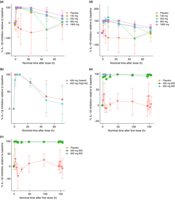
Single oral doses of GDC‐2394 inhibited production of IL‐1β after ex vivo stimulation of blood in a dose‐dependent manner, achieving significant inhibition (Figure 2a, Table S14). A maximum mean inhibition of greater than 97% (150‐mg and 450‐mg doses) and greater than 99% (900‐mg dose), relative to values using baseline samples, was achieved shortly after dosing and was sustained for ~8 h. With a single dose of 1800 mg GDC‐2394, a mean inhibition of greater than or equal to 99% was sustained for ~24 h. Single doses of 600 mg GDC‐2394 achieved greater than 99% mean inhibition, which was sustained for 8 h in both the fasted and high‐fat periods (Figure 2b), showing that food had little effect on the ability of GDC‐2394 to inhibit IL‐1β production. GDC‐2394 doses of 300 mg b.i.d. and 900 mg b.i.d. were able to achieve and sustain mean inhibition of IL‐1β production of greater than 95% and greater than 99% relative to baseline, respectively, over the 7‐day dosing period (Figure 2c, Table S15).
FIGURE 2.

Mean percent inhibition of IL‐1β and IL‐18 relative to baseline after LPS/ATP stimulation of whole blood samples collected from participants at the indicated timepoints in the SAD, FE, and MAD stages. Each measurement of secreted IL‐1β/IL‐18 consisted of the mean of two technical replicates. (a) IL‐1β: SAD stage (placebo [n = 8], 150 mg [n = 6], 450 mg [n = 6], 900 mg [n = 6], 1800 mg [n = 6]), (b) IL‐1β: FE stage (placebo [n = 2], 600 mg [n = 8]), (c) IL‐1β: dosing period of the MAD stage (placebo [n = 4], 300 mg b.i.d. [n = 6], 900 mg b.i.d. [n = 6]), (d) IL‐18: SAD stage (placebo [n = 8], 150 mg [n = 6], 450 mg [n = 6], 900 mg [n = 6], 1800 mg [n = 6]), and (e) IL‐18: dosing period of the MAD stage (placebo [n = 4], 300 mg b.i.d. [n = 6], 900 mg b.i.d. [n = 6]). Percent IL‐18 inhibition was adjusted for circulating levels of IL‐18. Error bars represent standard deviation. FE, food‐effect; MAD, multiple‐ascending dose; SAD, single‐ascending dose.
After adjusting for circulating levels of IL‐18, the extent and duration of IL‐18 inhibition in the ex vivo stimulation assay followed the same pattern as IL‐1β inhibition. Near‐complete or complete inhibition of IL‐18 production was observed in all SAD cohorts, an effect that was sustained for ~8 h following the 150‐mg, 450‐mg, and 900‐mg doses and for ~24 h following the 1800‐mg dose (Figure 2d, Table S14). Mean inhibition of IL‐18 production of greater than 93% and greater than 99% was observed across most of the 7‐day dosing period following the 300‐mg and 900‐mg b.i.d. doses, respectively (Figure 2e, Table S15). On average, across all SAD and MAD cohorts, participants receiving placebo showed variable levels of induced IL‐1β and IL‐18, but no significant inhibition of IL‐1β and IL‐18 production (Figure 2a–e). Inhibition of IL‐1β and IL‐18 production was reversible, with full recovery observed in many participants by 72 h after single doses of GDC‐2394, albeit with notable variability (Figure 2a,d).
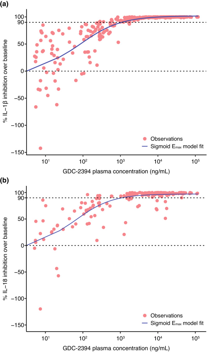
The onset of the PD response occurred shortly after dosing, which indicated that the PK/PD relationship was consistent with a PD effect that occurred in the systemic circulation, with no time delay between the plasma concentration and the response. Therefore, we explored the relationship between GDC‐2394 plasma concentrations and target engagement by plotting and fitting time‐matched GDC‐2394 plasma concentrations and the percent inhibition of IL‐1β release and percent inhibition of IL‐18 release (adjusted for circulating levels of IL‐18) to a sigmoid Emax model. Based on the model fit, we estimated that a plasma GDC‐2394 concentration of ~1100 ng/mL (~2.5 μM) or greater (correlating with ≥150 mg b.i.d.) could achieve at least 90% inhibition of both IL‐1β (Figure 3a) and IL‐18 (Figure 3b).
FIGURE 3.

Pharmacokinetic/pharmacodynamic modeling of individual data for (a) percent IL‐1β inhibition relative to baseline from all GDC‐2394‐treated participants in SAD, food‐effect, and MAD stages (n = 44) and (b) percent IL‐18 inhibition relative to baseline (adjusted for circulating levels of IL‐18) from all GDC‐2394‐treated participants in SAD and MAD stages (n = 36). Blue line represents the sigmoidal Emax model fit, and red circles represent individual observations. Dashed lines represent 0% and 90% inhibition relative to baseline. Emax, maximum effect; MAD, multiple‐ascending dose; SAD, single‐ascending dose.
DISCUSSION
GDC‐2394 is an investigational small‐molecule inhibitor of the NLRP3 inflammasome. The objective of this phase I trial was to characterize the safety, PKs, and PDs of GDC‐2394 in healthy participants to inform further development in the target population of patients with CAD.
Following oral dosing, GDC‐2394 was rapidly absorbed with approximately dose‐proportional increases in exposure and low‐to‐moderate intersubject PK variability. We expected that the relatively short half‐life of GDC‐2394 would likely require multiple daily doses to maintain target coverage. We therefore incorporated a FE stage into this first‐in‐human study to provide dosing guidance for future clinical studies in an outpatient setting, and observed that a high‐fat meal had only a minor impact on PKs. Urine PK data indicated that renal elimination made a minor contribution to the total clearance of GDC‐2394. In vitro CYP3A4 induction assays and preliminary physiologically based pharmacokinetic modeling suggested GDC‐2394 may have a mild‐to‐moderate CYP3A4 induction potential (unpublished data), an important consideration given that a large proportion of patients with CAD is expected to be taking statins. Specifically, simvastatin and lovastatin are sensitive CYP3A4 substrates, and atorvastatin is a moderately sensitive substrate. 15 Midazolam, a sensitive index CYP3A4 substrate, was used as the victim drug to investigate the CYP3A induction potential of GDC‐2394, in accordance with regulatory guidance specifying the use of index substrates in order to extrapolate to concomitant medications with similar DDI properties. 16 GDC‐2394 was dosed for 7 days in the DDI cohort; this duration of dosing was supported by trends from CYP3A clinical induction data indicating that 5–7 days of dosing were likely sufficient to achieve maximal CYP3A induction for drugs with a half‐life of less than 30 h. 17 GDC‐2394 had no apparent impact on midazolam PKs, with the 90% CI of the AUC GMR contained within the bioequivalence range of 80%–125%, indicating concomitant CYP3A4 substrates, such as statins, would not require dose adjustments.
To confirm GDC‐2394 target engagement and explore the PK/PD relationship to support dose selection, we used an ex vivo PD stimulation assay using whole blood collected at baseline and postdose. We used a cocktail of LPS and ATP to activate the NLRP3 inflammasome through the canonical (caspase‐1) pathway, 11 which led to high IL‐1β and IL‐18 levels. Through frequent sample collection, we observed a rapid, reversible, and near‐complete inhibition of IL‐1β production across all the doses tested. Similarly, across all the doses tested, GDC‐2394 showed near‐complete inhibition of IL‐18 production, which is also implicated in atherosclerosis. 18 , 19 Of note, the 150‐mg starting dose in this study was anticipated to be subtherapeutic based on the prediction of human PK from preclinical studies, but the observed AUC was approximately three‐fold higher than the preclinical prediction and thus explains the discrepancy between the predicted and observed target coverage. Based on PK/PD modeling, the estimated plasma 90% inhibitory concentration (IC90) value of ~1100 ng/mL (~2.5 μM) for IL‐1β and IL‐18 inhibition was in good agreement with the in vitro human whole blood IC90 value (639 ng/mL, 1.48 μM) after correcting for the blood:plasma ratio of 0.6. 9 Based on the observed PDs and the estimated clinical IC90, twice‐daily dosing of 150 mg GDC‐2394 or greater is expected to provide at least 90% target inhibition in peripheral blood throughout the dosing interval. However, the extent of IL‐1β and/or IL‐18 inhibition needed for clinical efficacy in patients with CAD with residual inflammation requires further investigation and determination in future trials.
GDC‐2394 was found to be adequately tolerated at all dose levels evaluated in the SAD and MAD stages. In the DDI stage, 900 mg b.i.d., the highest tested dose in the MAD, was selected to maximize the possibility of identifying a DDI. However, after the ninth participant in the DDI cohort completed dosing, two participants experienced SAEs of DILI that met criteria for Hy's law. 13 A third DDI participant experienced a non‐SAE of increased ALT. Based on these safety events, the sponsor halted GDC‐2394 dosing and enrollment. Follow‐up investigation showed similar steady‐state predose GDC‐2394 and indacenyl amine concentrations in the DDI cohort compared to the MAD 900‐mg b.i.d. cohort, which was expected because midazolam is not known to be a perpetrator of CYP enzyme‐mediated interactions.
In previous phase II trials, a similarly structured NLRP3 inhibitor, MCC950 (CP‐456,773) was tested, but further development was halted because of elevated liver enzyme levels. 3 In this clinical study, we observed hepatotoxicity despite preclinical toxicology studies in rats and cynomolgus monkeys indicating that the risk of DILI for GDC‐2394 was low (unpublished data). The occurrence of DILI at the highest dose tested and within a short time frame in our study may suggest contributions from a direct hepatotoxicity mechanism; however, we cannot rule out immune‐mediated or other mechanisms that are considered to be underlying idiosyncratic DILI. 20 Because the number of participants receiving multiple doses of GDC‐2394 was small, and follow‐up PK characterization of GDC‐2394 and indacenyl amine in participants with DILI showed levels that were similar to the previously cleared MAD cohort, we cannot establish the dose‐dependency of the DILI findings. Further clinical development of GDC‐2394 has stopped due to the lack of a clear explanation, unrelated to the study drug, for the observed severe hepatic events, despite favorable PK and target engagement PD. Nevertheless, the PK/PD target engagement relationship and safety findings identified in this study may help guide the development of other NLRP3 inhibitors in CAD and other inflammation‐related conditions.
AUTHOR CONTRIBUTIONS
F.T., T.C., A.H., A.N., U.O., and D.P. wrote the manuscript. F.T., R.K., A.H., A.N., J.M.M., Y.Z., S.S., M.W., J.L., N.L.‐K., R.O., and D.P. designed the research. A.N., A.T., L.C., and C.W. performed the research. F.T., T.C., A.N., J.M.M., J.L., A.M., N.L.‐K., and V.R. analyzed data.
FUNDING INFORMATION
Genentech, Inc. supported this study.
CONFLICT OF INTEREST STATEMENT
F.T., R.K., T.C., A.H., A.N., J.M.M., Y.Z., S.S., M.W., A.T., L.C., J.L., A.M., N.L.‐K., V.R., U.O., R.O., and D.P. are or were employees of Genentech, Inc., a member of the Roche Group, and are or were Roche stockholders at the time this work was performed. R.K. is currently an employee of IGM Biosciences. C.W. is an employee of the Christchurch Clinical Studies Trust Ltd.
Supporting information
Appendix S1
Appendix S2
ACKNOWLEDGMENTS
The authors thank the volunteers who participated in this trial and the site staff who assisted with the conduct of this trial. We would also like to thank Katie Maass and Dhruvit Sutaria for providing constructive feedback on the manuscript. Editing and writing assistance was provided by Deborah Solymar (Genentech, Inc.) and was funded by Genentech, Inc.
Tang F, Kunder R, Chu T, et al. First‐in‐human phase 1 trial evaluating safety, pharmacokinetics, and pharmacodynamics of NLRP3 inflammasome inhibitor, GDC‐2394, in healthy volunteers. Clin Transl Sci. 2023;16:1653‐1666. doi: 10.1111/cts.13576
DATA AVAILABILITY STATEMENT
For eligible studies, qualified researchers may request access to individual patient‐level clinical data through a data request platform. At the time of writing, this request platform is Vivli: https://vivli.org/ourmember/roche/. For up‐to‐date details on Roche's Global Policy on the Sharing of Clinical Information and how to request access to related clinical study documents, see here: https://go.roche.com/data_sharing. Anonymized records for individual patients across more than one data source external to Roche cannot, and should not, be linked due to a potential increase in risk of patient re‐identification.
REFERENCES
- 1. Deets KA, Vance RE. Inflammasomes and adaptive immune responses. Nat Immunol. 2021;22:412‐422. [DOI] [PubMed] [Google Scholar]
- 2. Broz P, Dixit VM. Inflammasomes: mechanism of assembly, regulation and signalling. Nat Rev Immunol. 2016;16:40720‐40420. [DOI] [PubMed] [Google Scholar]
- 3. Mangan M, Olhava E, Roush W, Seidel HM, Glick GD, Latz E. Targeting the NLRP3 inflammasome in inflammatory diseases. Nat Rev Drug Discov. 2018;17:588‐606. [DOI] [PubMed] [Google Scholar]
- 4. Ridker PM, Bhatt DL, Pradhan AD, et al. Inflammation and cholesterol as predictors of cardiovascular events among patients receiving statin therapy: a collaborative analysis of three randomised trials. Lancet. 2023;401:1293‐1301. [DOI] [PubMed] [Google Scholar]
- 5. Duewell P, Kono H, Rayner KJ, et al. NLRP3 inflammasomes are required for atherogenesis and activated by cholesterol crystals. Nature. 2010;464:1357‐1361. [DOI] [PMC free article] [PubMed] [Google Scholar]
- 6. Rajamäki K, Lappalainen J, Oörni K, et al. Cholesterol crystals activate the NLRP3 inflammasome in human macrophages: a novel link between cholesterol metabolism and inflammation. PLoS One. 2010;5:e11765. [DOI] [PMC free article] [PubMed] [Google Scholar]
- 7. van der Heijden T, Kritikou E, Venema W, et al. NLRP3 inflammasome inhibition by MCC950 reduces atherosclerotic lesion development in apolipoprotein E–deficient mice—brief report. Arterioscler Thromb Vasc Biol. 2017;37:1457‐1461. [DOI] [PubMed] [Google Scholar]
- 8. Ridker PM, Everett BM, Thuren T, et al. Antiinflammatory therapy with canakinumab for atherosclerotic disease. New Engl J Med. 2017;377:1119‐1131. [DOI] [PubMed] [Google Scholar]
- 9. McBride C, Trzoss L, Povero D, et al. Overcoming preclinical safety obstacles to discover (S)‐N‐((1,2,3,5,6,7‐hexahydro‐s‐indacen‐4‐yl)carbamoyl)‐6‐(methylamino)‐6,7‐dihydro‐5H‐pyrazolo[5,1‐b][1,3]oxazine‐3‐sulfonamide (GDC‐2394): a potent and selective NLRP3 inhibitor. J Med Chem. 2022;65:14721‐14739. [DOI] [PubMed] [Google Scholar]
- 10. U.S. Department of Health and Human Services, National Institutes of Health, National Institute of Allergy and Infectious Diseases, Division of AIDS . Division of AIDS (DAIDS) Table for Grading the Severity of Adult and Pediatric Adverse Events, Corrected Version 2.1. 2017. Accessed September 30, 2022. https://rsc.niaid.nih.gov/sites/default/files/daidsgradingcorrectedv21.pdf
- 11. Tran TAT, Grievink HW, Lipinska K, et al. Whole blood assay as a model for in vitro evaluation of inflammasome activation and subsequent caspase‐mediated interleukin‐1 beta release. PLoS One. 2019;14:e0214999. [DOI] [PMC free article] [PubMed] [Google Scholar]
- 12. R Core Team . R: A Language and Environment for Statistical Computing. R Foundation for Statistical Computing; 2022. [Google Scholar]
- 13. Temple R. Hy's law: predicting serious hepatotoxicity. Pharmacoepidemiol Drug Safe. 2006;15:241‐243. [DOI] [PubMed] [Google Scholar]
- 14. Liu R, Yu X, Wallqvist A. Data‐driven identification of structural alerts for mitigating the risk of drug‐induced human liver injuries. J Chem. 2015;7:4. [DOI] [PMC free article] [PubMed] [Google Scholar]
- 15. U.S. Food and Drug Administration . Drug development and drug interactions; table of substrates, inhibitors and inducers. Accessed September 30, 2022. https://www.fda.gov/drugs/drug‐interactions‐labeling/drug‐development‐and‐drug‐interactions‐table‐substrates‐inhibitors‐and‐inducers
- 16. U.S. Food and Drug Administration . FDA Center for drug evaluation and research. Clinical Drug Interaction Studies – Cytochrome P450 Enzyme and Transporter‐Mediated Drug Interactions Guidance for Industry. 2020. Accessed September 30, 2022. https://www.fda.gov/media/134581/download
- 17. Ramsden D, Fung C, Hariparsad N, et al. Perspectives from the innovation and quality consortium induction working group on factors impacting clinical drug‐drug interactions resulting from induction: focus on cytochrome 3A substrates. Drug Metab Dispos. 2019;47:1206‐1221. [DOI] [PubMed] [Google Scholar]
- 18. Blankenberg S, Tiret L, Bickel C, et al. Interleukin‐18 is a strong predictor of cardiovascular death in stable and unstable angina. Circulation. 2002;106:24‐30. [DOI] [PubMed] [Google Scholar]
- 19. Ridker PM, MacFadyen JG, Thuren T, Libby P. Residual inflammatory risk associated with interleukin‐18 and interleukin‐6 after successful interleukin‐1β inhibition with canakinumab: further rationale for the development of targeted anti‐cytokine therapies for the treatment of atherothrombosis. Eur Heart J. 2020;41:2153‐2163. [DOI] [PubMed] [Google Scholar]
- 20. Teschke R, Uetrecht J. Mechanism of idiosyncratic drug induced liver injury (DILI): unresolved basic issues. Ann Transl Med. 2021;9:730. [DOI] [PMC free article] [PubMed] [Google Scholar]
Associated Data
This section collects any data citations, data availability statements, or supplementary materials included in this article.
Supplementary Materials
Appendix S1
Appendix S2
Data Availability Statement
For eligible studies, qualified researchers may request access to individual patient‐level clinical data through a data request platform. At the time of writing, this request platform is Vivli: https://vivli.org/ourmember/roche/. For up‐to‐date details on Roche's Global Policy on the Sharing of Clinical Information and how to request access to related clinical study documents, see here: https://go.roche.com/data_sharing. Anonymized records for individual patients across more than one data source external to Roche cannot, and should not, be linked due to a potential increase in risk of patient re‐identification.


