Ikeda et al. highlight work from Su and colleagues that describes the mechanism by which NME3 and phosphatidic acid promote mitochondrial tethering prior to fusion.
Abstract
Mitochondrial fusion plays an important role in both their structure and function. In this issue, Su et al. (2023. J. Cell Biol. https://doi.org/10.1083/jcb.202301091) report that a nucleoside diphosphate kinase, NME3, facilitates mitochondrial tethering prior to fusion through its direct membrane-binding and hexamerization but not its kinase activity.
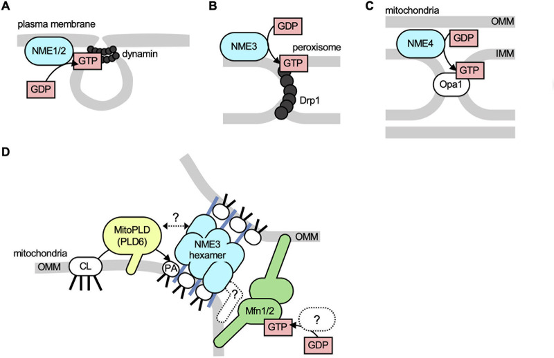
Nucleoside diphosphate kinases (NDPKs) catalyze the formation of nucleoside triphosphates, such as GTP, from nucleoside diphosphates such as GDP, using ATP as a phosphate donor (1, 2). The mammalian NDPK family consists of 10 members encoded by the NME1-10 genes. Some NDPK family members regulate membrane dynamics by supplying GTP to dynamin and dynamin-related GTPases locally. For instance, during endocytosis, NME1/2 provide GTP to dynamin, facilitating membrane scission at the coated pits and the subsequent release of endocytic vesicles into the cytosol (3; Fig. 1 A). NME3 regulates peroxisomal division (4; Fig. 1 B), while NME4 supplies GTP to Opa1, a dynamin-related GTPase in the mitochondrial inner membrane, promoting mitochondrial fusion (3; Fig. 1 C).
Figure 1.
Roles of nucleoside diphosphate kinases in membrane dynamics. (A) NME1/2 fuel dynamin at the plasma membrane during endocytosis. (B) NME3 facilitates peroxisomal division, a process that is mediated by the dynamin-related GTPase Drp1, which also plays a role in mitochondrial division. (C) NME4 channels GTP to Opa1 for the fusion of the inner mitochondrial membrane (IMM). (D) NME3 promotes the fusion of the outer mitochondrial membrane (OMM) by tethering mitochondria. The localization of NME3 at mitochondrial contact sites depends on the phosphatidic acid produced by MitoPLD.
A new paper by Su et al. (5) adds another thrilling layer to this understanding. The authors show that NME3 also regulates mitochondrial fusion at the outer membrane by mediating the connection between two mitochondria prior to fusion (Fig. 1 D). Strikingly, this function of NME3 is independent of its kinase activity. Instead, it requires NME3’s ability to directly bind to the membrane and to form homo-oligomers—more specifically, hexamers.
In previous work, the authors reported that a mutation in NME3 is linked to a fatal neurodegenerative disorder (6). They also discovered that NME3 is associated with the mitochondrial outer membrane and observed that patient cells lacking functional NME3 exhibit defects in mitochondrial fusion (6). Interestingly, NME3 was found to co-immunoprecipitate with MFN1 and MFN2, two homologous dynamin-related GTPases that mediate fusion of the mitochondrial outer membrane. The same study also revealed that the role of NME3 in mitochondrial fusion does not depend on its kinase activity but does require its hexamerization ability (6). This raises intriguing mechanistic questions about how NME3 regulates mitochondrial fusion. Since the kinase activity is not required, it seems unlikely that NME3 supplies MFNs with GTP.
In their current work, Su et al. explore further the mechanism by which NME3 regulates mitochondrial fusion. First, the authors discovered that the N-terminal region, consisting of 17 amino acids, is essential for targeting NME3 to the mitochondria. This region is predicted to form an amphipathic alpha helix, suggesting that it binds to lipid membranes. Confirming this, the authors found that this 17-amino-acid region binds to membranes likely via a phosphatidic acid (PA)-induced change in the lipid packing property in the membrane. Supporting this protein-membrane interaction mechanism, NME3 preferentially interacted with liposomes of smaller diameters, suggesting that NME3 recognizes membrane curvature. This 17-amino-acid region alone was sufficient for mitochondrial localization, as demonstrated by its GFP fusion being located in the mitochondria in cells. In contrast, neither the kinase nor the hexamerization activity of NME3 is necessary for its mitochondrial localization, as evidenced by mutations that inhibit these biochemical activities.
On the outer membrane, NME3 facilitates the connection of mitochondria through hexamerization. While a mutant form of NME3 defective in hexamerization is still associated with mitochondria, it cannot induce their clustering in cells. Interestingly, once the N-terminal region has been artificially brought to the mitochondria, it is not required for the subsequent clustering; NME3 lacking this region can still promote mitochondrial clustering when recruited through the rapamycin-induced chemical dimerization system. Remarkably, the authors revealed that NME3 is enriched at sites where mitochondria come into contact with each other, suggesting that it directly connects two mitochondria. Supporting this idea, in vitro purified NME3 was able to tether both PA-containing liposomes and isolated mitochondria.
How is PA generated on the mitochondrial outer membrane? Previous studies identified an enzyme located in the mitochondria, MitoPLD (also known as PLD6), which converts the mitochondrial phospholipid cardiolipin into PA and acts in mitochondrial tethering (7–9). Su et al. discovered that, while not strictly essential, MitoPLD influences the mitochondrial localization of the N-terminal region of NME3. Specifically, the knockdown of MitoPLD results in a partial reduction of this localization, whereas its overexpression enhances it. Remarkably, optogenetically recruiting bacterial PLD to the mitochondria, which artificially stimulates PA production, replaces the function of MitoPLD. This induced both the mitochondrial localization of NME3’s N-terminal region and mitochondrial clustering in cells. Furthermore, the PA produced by MitoPLD enhances the enrichment of NME3 at sites where mitochondria come into contact. In addition to fusion, previous research has shown that PA, generated by MitoPLD, negatively regulates Drp1-mediated mitochondrial division (9). In this mechanism, PA directly binds to Drp1, inhibiting its division activity on the outer membrane. As a result, PA significantly contributes to mitochondrial elongation by both suppressing division and promoting fusion.
Mitochondrial morphology is maintained by a delicate equilibrium between fusion and division processes (10). This balance is crucial not only for maintaining the structure of the mitochondria but also for regulating their function, distribution, and degradation via autophagy. Importantly, this dynamic balance adapts to various stress conditions. For instance, during periods of starvation, the mitochondria adjust the equilibrium to favor fusion over division (11, 12). This leads to elongated mitochondrial structures, which in turn affect both bioenergetics and mitophagy-based degradation. In this context, the authors discovered that glucose starvation increases the presence of NME3 at sites where mitochondria come into contact. This likely facilitates more efficient mitochondrial tethering, and consequently, more effective fusion under this stressful condition.
In summary, thanks to the work of Su et al., we now have compelling insights into an intricate mechanism by which NME3 brings two mitochondria into close proximity prior to membrane fusion (Fig. 1 D). This is facilitated through its membrane-binding activity and hexamerization. The study also sheds light on the role of MitoPLD in mitochondrial tethering, specifically in recruiting NME3 and enriching it at contact sites between mitochondria. The identification of NME3 opens up new, fascinating avenues for research aimed at better understanding the dynamics of mitochondrial tethering and fusion, as well as their alterations in neurodegenerative conditions. Several stimulating questions are raised by this research (Fig. 1 D). First, it would be exciting to understand how NME3 accumulates at mitochondrial contact sites. Could MitoPLD, or perhaps its activated form, create a unique domain on the outer membrane that serves as a clustering site for NME3? Regulation of membrane curvature could also play a critical role in this step. Given that mitochondria have a tubular morphology and that fusion can occur either tip-to-tip or tip-to-side, there may be a mechanism that targets MitoPLD to the tips of these tubules. Second, how are NME3 and MitoPLD regulated under glucose-starvation conditions to increase mitochondrial tethering? These proteins might undergo various post-translational modifications or interact with specific proteins under stress conditions. Third, while the authors showed that PA is important for localizing NME3 to mitochondria, it is not sufficient by itself. Identifying a specific NME3 receptor protein in the outer membrane could provide further insights. Fourth, does NME3 or another protein generate GTP locally in the vicinity of MFN1/2? Finally, from a human health perspective, it would be crucial to determine how defects in NME3 contribute to neurodegenerative diseases.
Acknowledgments
This work was supported by a National Institutes of Health grant to H. Sesaki (GM144103) and a Uehara Memorial Foundation postdoctoral fellowship to A. Ikeda.
References
- 1.Boissan, M., et al. 2018. Lab. Invest. 10.1038/labinvest.2017.137 [DOI] [Google Scholar]
- 2.Georgescauld, F., et al. 2020. Int. J. Mol. Sci. 10.3390/ijms21186779 [DOI] [PMC free article] [PubMed] [Google Scholar]
- 3.Boissan, M., et al. 2014. Science. 10.1126/science.1253768 [DOI] [Google Scholar]
- 4.Honsho, M., et al. 2020. Int. J. Mol. Sci. 10.3390/ijms21218040 [DOI] [Google Scholar]
- 5.Su, Y.A., et al. 2023. J. Cell Biol. 10.1083/jcb.202301091 [DOI] [Google Scholar]
- 6.Chen, C.W., et al. 2019. Proc. Natl. Acad. Sci. USA. 10.1073/pnas.1818629116 [DOI] [Google Scholar]
- 7.Choi, S.Y., et al. 2006. Nat. Cell Biol. 10.1038/ncb1487 [DOI] [Google Scholar]
- 8.Kameoka, S., et al. 2018. Trends Cell Biol. 10.1016/j.tcb.2017.08.011 [DOI] [PMC free article] [PubMed] [Google Scholar]
- 9.Adachi, Y., et al. 2016. Mol. Cell. 10.1016/j.molcel.2016.08.013 [DOI] [Google Scholar]
- 10.Murata, D., et al. 2020. J. Biochem. 10.1093/jb/mvz106 [DOI] [Google Scholar]
- 11.Gomes, L.C., et al. 2011. Nat. Cell Biol. 10.1038/ncb2220 [DOI] [PMC free article] [PubMed] [Google Scholar]
- 12.Rambold, A.S., et al. 2011. Proc. Natl. Acad. Sci. USA. 10.1073/pnas.1107402108 [DOI] [Google Scholar]


