Abstract
Objective
Renal involvement in anti-neutrophil cytoplasmic antibody (ANCA)-associated vasculitis (AAV) can lead to severe renal dysfunction requiring dialysis at diagnosis. We aimed to study the clinical and pathologic characteristics of patients with AAV dependent on dialysis at presentation and the long-term renal outcomes of patients who recovered from dialysis.
Methods
This retrospective study analyzed data of patients diagnosed with AAV who were on dialysis from July 2005 to May 2021 at a single tertiary center in Korea.
Results
Thirty-four patients were included in the study (median age 64.5 years, females 61.8%), of which 13 discontinued and 21 continued dialysis. The proportion of normal glomeruli (p<0.001) and interstitial fibrosis (p=0.024) showed significant differences between both groups. Multivariable analysis showed that the proportion of normal glomeruli was associated with dialysis discontinuation (odds ratio=1.29, 95% confidence interval 0.99~1.68, p=0.063), although without statistical significance. Treatment modalities, including plasmapheresis, did not show significance with dialysis discontinuation. In the follow-up analysis of 13 patients who had discontinued dialysis for a median of 81 months, 12 did not require dialysis, and their glomerular filtration rate values significantly increased at follow-up time compared to when they stopped dialysis (37.5 [28.5~45.5] vs. 24.0 [18.5~30.0] mL/min/1.73 m²; p=0.008).
Conclusion
Approximately 38% of AAV patients on dialysis discontinued dialysis, and the recovered patients had improved renal function without dialysis during longer follow-up. Patients with AAV on dialysis should be given the possibility of dialysis discontinuation and renal recovery, especially those with normal glomeruli in kidney pathology.
Keywords: Dialysis, Anti-neutrophil cytoplasmic antibody-associated vasculitis
INTRODUCTION
Anti-neutrophil cytoplasmic antibody (ANCA)-associated vasculitis (AAV) is a small vessel vasculitis commonly involving the kidneys and lungs [1]. Depending on the subtype, the prevalence of renal involvement is almost 100% for microscopic polyangiitis (MPA) and greater than 70% for granulomatosis with polyangiitis [2]. Renal involvement in AAV is important due to its high frequency and association with poor prognosis. Approximately 20% or more of AAV patients with renal involvement have been shown to have progressed to end-stage renal disease (ESRD) and were on dialysis during follow-up [3-6]. The risk of patients’ survival is significantly related to impaired renal function at diagnosis, with a hazard ratio of 2.63~4.45 [7-10].
A significant proportion of patients with AAV who have severe renal dysfunction require dialysis at presentation [11,12]. However, when treatment with immunosuppressants is effective, dialysis may be discontinued in 28% to 70% of patients with AAV within 6 to 12 months [13-15]. Kidney pathologies, such as the proportion of normal glomeruli and the extent of tubular atrophy or interstitial fibrosis, have been reported to be associated with dialysis discontinuation [15]. In terms of treatment, however, the effectiveness of rituximab, cyclophosphamide, or plasmapheresis for dialysis discontinuation remains controversial [13,16-18]. Furthermore, the long-term renal prognosis of patients after recovery from dialysis in AAV is not known. Therefore, we aimed to identify the clinicopathologic characteristics of patients who required dialysis at the time of AAV diagnosis and long-term renal prognosis, including whether they can remain dialysis-free after dialysis discontinuation.
MATERIALS AND METHODS
Patients and data collection
We retrospectively analyzed the data of patients diagnosed with AAV who were on dialysis due to renal involvement from July 2005 to May 2021 at a single tertiary center in Seoul, Korea. Diagnosis of AAV was based on the International Chapel Hill Consensus Conference Nomenclature of Vasculitides, and kidney involvement of AAV was clinically or histologically confirmed [19]. The patients on dialysis at the time of AAV diagnosis were included in our study. Patients who stopped dialysis during follow-up were classified into the 'discontinuation of dialysis' group, and those who continued dialysis were classified into the 'on dialysis' group. This study was performed in accordance with the Declaration of Helsinki and its later amendments. The study protocol was approved by the Institutional Review Board of Asan Medical Center (Seoul, South Korea) (2021-1357). The requirement for informed consent was waived, given the retrospective nature of the study.
The following data from electronic medical records were reviewed—age, sex, body mass index (BMI), ANCA type, co-morbidities, estimated glomerular filtration rate (eGFR), urine analysis, erythrocyte sedimentation rate, C-reactive protein (CRP), and treatment administered, including corticosteroid doses. eGFR was calculated using the following Modification of Diet in Renal Disease equation: 175×[serum creatinine (mg/dL)]−1.154×(age)−0.203×(0.742 if female). Disease activity was measured using the Birmingham vasculitis activity score (BVAS) version 3.0. [20]. The disease remission was defined as 0 or persistence as 1 BVAS within 1 year and requiring less than 7.5 mg of prednisolone [21,22]. The results of kidney biopsy were collected and classified into four types, according to Berden et al. [23]. Histopathologic findings, including interstitial infiltration, fibrosis, and tubular atrophy, were marked as –, +, ++, and +++ [14,15]. Clinical characteristics, including creatinine levels and dialysis requirements after the follow-up period, were assessed.
Statistical analysis
Categorical variables were described as percentages (%) and continuous variables as median (interquartile ranges [IQR]) or mean values (standard deviation). Parametric and nonparametric data were compared using the t-test and Mann–Whitney U test, respectively. Categorical variables were compared using the Chi-squared test and Fisher’s exact test. Logistic regression was performed with the discontinuation of dialysis as the dependent variable for the univariable and multivariable analysis. When performing multivariable analysis, factors with a p-value >0.2 in univariate analysis were excluded to favor the criterion of parsimony. All statistical analyses were performed using SPSS software, version 21.0 (IBM Corp., Armonk, NY, USA). A p-value of <0.05 was considered statistically significant.
RESULTS
Baseline characteristics
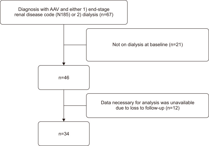
A total of 34 patients with AAV who were on dialysis at baseline were included in the present study (Figure 1). Of these patients, 13 discontinued dialysis after treatment for AAV, and the remaining 21 continued to be dialysis-dependent. The baseline characteristics of total patients and a comparison of the two groups according to dialysis dependency are shown in Table 1. The median age was 64.5 years (IQR, 60.0~72.3 years), and the proportion of female patients was 61.8% (21/34). The mean BVAS score was 20.3±5.6 at baseline, which was not significantly different between the two groups. The duration from symptom onset to treatment was 33.0 days (IQR, 18.5~51.5 days). There was no significant difference between the two groups with respect to the clinical features, including AAV subtypes, co-morbidities, and organ involvement, except ear-nose-throat manifestation. In terms of renal parameters, the baseline eGFR value of the dialysis-discontinued group was 10.0 mL/min/1.73 m² (IQR 9.0~13.0 mL/min/1.73 m²), which was significantly higher than that of the dialysis-dependent group (7.0 mL/min/1.73 m² [IQR 5.5–8.5 mL/min/1.73 m²]; p<0.001).
Fig. 1.
Patient selection flowchart. AVV: anti-neutrophil cytoplasmic antibody-associated vasculitis.
Table 1.
Baseline characteristics of patients with or without the discontinuation of dialysis
| All (n=34) | Discontinuation of dialysis (n=13) | On dialysis (n=21) |
p-value | |
|---|---|---|---|---|
| Age (yr) | 64.5 (60.0~72.3) | 64.0 (60.0~69.5) | 67.0 (60.0~75.5) | 0.467 |
| Female | 21 (61.8) | 7 (53.8) | 14 (66.7) | 0.455 |
| Body mass index (kg/m²) | 23.2±3.1 | 22.8±3.3 | 23.4±3.0 | 0.567 |
| BVAS | 20.3±5.6 | 21.3±5.0 | 19.7±6.0 | 0.415 |
| Duration of follow up (mo) | 35.5 (16.8~93.0) | 81.0 (21.5~105.5) | 28.0 (12.5~71.0) | 0.132 |
| Time from symptom onset to treatment (day) | 33.0 (18.5~51.5) | 29.0 (16.0~35.0) | 34.0 (22.5~56.5) | 0.279 |
| Achievement of remission | 24 (70.6) | 10 (76.9) | 14 (66.7) | 0.704 |
| Duration from symptom onset to treatment ≤1 mo | 19 (55.9) | 5 (38.5) | 14 (66.7) | 0.107 |
| ANCA-associated vasculitis type | 0.313 | |||
| MPA | 25 (73.5) | 9 (69.2) | 16 (76.2) | |
| GPA | 8 (23.5) | 3 (23.1) | 5 (23.8) | |
| ANCA type by ELISA | 0.434 | |||
| Anti-PR3 | 7 (20.6) | 2 (15.4) | 5 (23.8) | |
| Anti-MPO | 25 (73.5) | 9 (69.2) | 16 (76.2) | |
| Both anti-PR3 and anti-MPO | 1 (2.9) | 1 (7.7) | 0 (0.0) | |
| Manifestations at enrollment | ||||
| Pulmonary involvement | 24 (70.6) | 9 (69.2) | 15 (71.4) | 0.891 |
| Alveolar hemorrhage | 6 (17.6) | 1 (7.7) | 5 (23.8) | 0.370 |
| Ear, nose, and throat | 3 (8.8) | 3 (23.1) | 0 (0.0) | 0.048* |
| Neurologic involvement | 5 (14.7) | 3 (23.1) | 2 (9.5) | 0.348 |
| Comorbidity | ||||
| Hypertension | 14 (41.2) | 8 (61.5) | 6 (28.6) | 0.058 |
| Diabetes mellitus | 4 (11.8) | 3 (23.1) | 1 (4.8) | 0.274 |
| Chronic kidney disease | 4 (11.8) | 2 (15.4) | 2 (9.5) | 0.627 |
| Coronary artery disease | 1 (2.9) | 0 (0.0) | 1 (4.8) | >0.999 |
| Renal profiles at baseline | ||||
| eGFR (mL/min/1.73 m²) | 8.0 (6.8~10.3) | 10.0 (9.0~13.0) | 7.0 (5.5~8.5) | <0.001* |
| Hematuria (>10/HPF)† | 23 (74.2) | 9 (75.0) | 14 (73.7) | 0.437 |
| Proteinuria (dipstick, > +1) | 19 (55.9) | 5 (38.5) | 14 (66.7) | 0.067 |
| Laboratory data | ||||
| ESR, (mm/h) | 61.39±39.83 | 70.0±41.0 | 54.8±39.2 | 0.376 |
| CRP (mg/dL) | 7.6 (2.5~3.5) | 9.5 (4.0~12.9) | 5.4 (1.2~13.8) | 0.507 |
Values are median (interquartile range), number (%), or mean±standard deviation. BVAS: Birmingham Vasculitis Activity Score, ANCA: antineutrophil cytoplasmic antibody, MPA: microscopic polyangiitis, GPA: granulomatosis polyangiitis, Anti-PR3: anti-proteinase 3, Anti- MPO: anti-myeloperoxidase, eGFR: estimated glomerular filtration rate, HPF: high-power field, ESR: erythrocyte sedimentation rate, CRP: C-reactive protein. *Statistically significant (p<0.05). †The value of microscopic hematuria had three missing values.
Treatment of total patients with or without the discontinuation of dialysis
Thirty-two patients were treated with intravenous or oral cyclophosphamide as an induction therapy for AAV (Table 2). Rituximab was administered as first-line induction therapy in two patients. During induction therapy, 50% of the patients were administered pulse (≥500 mg/day) corticosteroids, and high- and medium-dose corticosteroids were used in 41.2% and 8.8% of the patients, respectively. Plasmapheresis was performed in 11 patients (32.4%). As maintenance therapy, azathioprine and mycophenolate mofetil were used in 52.9% and 5.9% of the patients, respectively. There was no significant difference between the two groups in terms of immunomodulatory therapy, including plasmapheresis, for AAV.
Table 2.
Treatment of patients with or without the discontinuation of dialysis
| All (n=34) | Discontinuation of dialysis (n=13) | On dialysis (n=21) | p-value | |
|---|---|---|---|---|
| Induction therapy | ||||
| IV cyclophosphamide | 7 (20.6) | 3 (23.1) | 4 (19.0) | 0.781 |
| PO cyclophosphamide | 26 (76.5) | 10 (76.9) | 16 (76.2) | 0.962 |
| Rituximab | 2 (5.9) | 1 (7.7) | 1 (4.8) | 0.728 |
| Plasmapheresis | 11 (32.4) | 3 (23.1) | 8 (38.1) | 0.370 |
| Steroid doses during induction | 0.984 | |||
| Medium (≥0.5 mg/kg/day) | 3 (8.8) | 2 (15.4) | 1 (4.8) | |
| High (≥1 mg/kg/day) | 14 (41.2) | 4 (30.8) | 10 (47.6) | |
| Pulse (≥ 500 mg/day) | 17 (50.0) | 7 (53.8) | 10 (47.6) | |
| Maintenance therapy | ||||
| Azathioprine | 18 (52.9) | 10 (76.9) | 8 (38.1) | 0.055 |
| Mycophenolate mofetil | 2 (5.9) | 2 (15.4) | 0 (0.0) | 0.068 |
Values are presented as number (%). IV: intravenous, PO: per oral.
Histopathologic findings of renal biopsy of patients with AAV
Of the 34 patients, a kidney biopsy was performed in 24 patients, and the histopathologic results were compared between patients who continued dialysis and those who discontinued dialysis (Table 3). The median number of total glomeruli present in the biopsy specimens was 16.0 (IQR, 11.0~20.5), and the number of normal glomeruli was 2.0 (IQR 0.3~5.0). Interestingly, patients in the dialysis discontinuation group had significantly higher numbers (5.0 [2.5~11.5] vs. 1.0 [0.0~3.0]; p=0.002) and percentages (46.7% [18.0%~69.2%] vs. 8.2% [0.0%~12.8%], p<0.001) of normal glomeruli than those in the dialysis-dependent group. While eight patients had interstitial fibrosis of 2+ or higher in the group who continued dialysis, interstitial fibrosis was found in only one patient in the group who discontinued dialysis. There was, however, no significant difference in certain types of histopathologic findings according to the four different classifications (sclerotic, focal, crescentic, and mixed) [23].
Table 3.
Histopathologic findings of patients with or without the discontinuation of dialysis
| All (n=24) | Discontinuation of dialysis (n=9) | On dialysis (n=15) |
p-value | |
|---|---|---|---|---|
| Total glomeruli (n) | 16.0 (11.0~20.5) | 13.0 (10.5~22.0) | 18.0 (12.0~21.0) | 0.482 |
| Normal glomeruli (n) | 2.0 (0.3~5.0) | 5.0 (2.5~11.5) | 1.0 (0.0~3.0) | 0.002* |
| Normal glomeruli (%) | 12.2 (4.7~36.4) | 46.7 (18.0~69.2) | 8.2 (0.0~12.8) | <0.001* |
| Fibrinoid necrosis (n) | 2.0 (0.0~3.0) | 0.0 (0.0~2.5) | 2.0 (0.0~3.0) | 0.410 |
| Fibrinoid necrosis (%) | 4.1 (0.0~20.7) | 0.0 (0.0~21.6) | 7.3 (0.0~21.1) | 0.741 |
| Cellular crescents | 7.3 (0.0~37.8) | 9.1 (0.0~46.5) | 5.6 (0.0~35.7) | 0.976 |
| Fibrous crescents | 0.0 (0.0~0.0) | 0.0 (0.0~0.0) | 0.0 (0.0~0.0) | 0.795 |
| Fibrocellular crescents | 14.3 (0.0~36.0) | 0.0 (0.0~29.4) | 20.0 (11.1~49.0) | 0.101 |
| Glomerulosclerosis | 3.0 (3.0~3.0) | 3.0 (3.0~3.0) | 3.0 (3.0~3.0) | 0.646 |
| Interstitial infiltration (–/+/++/+++) | 7/9/4/4 | 4/4/1/0 | 3/5/3/4 | 0.061 |
| Interstitial fibrosis (–/+/++/+++) | 8/9/4/5 | 5/4/1/0 | 3/5/3/5 | 0.024* |
| Tubular atrophy (–/+/++/+++) | 6/10/4/4 | 3/5/0/1 | 3/5/4/3 | 0.168 |
| Arteriosclerosis (–/+) | 11/13 | 4/5 | 7/8 | 0.918 |
| Type | 0.525 | |||
| Sclerotic | 3 (12.5) | 0 (0.0) | 3 (20.0) | |
| Focal | 4 (16.7) | 4 (44.4) | 0 (0) | |
| Crescentic | 14 (58.3) | 4 (44.4) | 10 (66.7) | |
| Mixed | 3 (12.5) | 1 (11.1) | 2 (13.3) |
Values are median (interquartile range) or number (%). *Statistically significant (p<0.05).
Predictors associated with discontinuation of dialysis
To determine the clinicopathologic factors related to the discontinuation of dialysis in patients who were under dialysis at the time of AAV diagnosis, we performed a logistic regression analysis (Table 4). Clinical features, including age, sex, and BVAS, did not show a significant association with dialysis discontinuation. In addition, treatment modality and baseline eGFR were not associated with recovery from dialysis. In the pathologic findings, the percentage of normal glomeruli tended to be associated with dialysis discontinuation at follow-up (odds ratio=1.29, 95% confidence interval 0.99~1.68, p=0.063), although this association did not reach statistical significance.
Table 4.
Uni- and multi-variate analysis of predictors associated with the discontinuation of dialysis
| Predictor | Univariate | Multi-variate | |||
|---|---|---|---|---|---|
| OR (95% CI) | p-value | OR (95% CI) | p-value | ||
| Age | 0.97 (0.91~1.03) | 0.370 | |||
| Sex, male | 0.58 (0.14~2.41) | 0.583 | |||
| ANCA type | 0.71 (0.11~4.44) | 0.715 | |||
| Duration between symptom and treatment | 1.00 (0.98~1.01) | 0.714 | |||
| BVAS | 1.06 (0.93~1.20) | 0.406 | |||
| Percentage of normal glomeruli | 1.23 (0.99~1.54) | 0.059 | 1.29 (0.99~1.68) | 0.063 | |
| Presence of interstitial fibrosis | 0.31 (0.05~1.94) | 0.212 | |||
| Presence of tubular atrophy | 0.50 (0.08~3.27) | 0.469 | |||
| Steroid, high dose compared with pulse | 0.57 (0.13~2.58) | 0.467 | |||
| Maintenance therapy | 4.44 (0.94~21.00) | 0.060 | 9.41 (0.25~356.54) | 0.227 | |
| Plasma exchange | 0.49 (0.10~2.33) | 0.367 | |||
| Achievement of remission | 1.67 (0.34~8.07) | 0.526 | |||
| Duration from symptom onset to treatment ≤ 1 mo | 0.31 (0.07~1.32) | 0.113 | |||
| eGFR (mL/min/1.73 m²) | 1.12 (0.95~1.32) | 0.192 | 0.97 (0.81~1.16) | 0.736 | |
OR: odds ratio, CI: confidence interval, ANCA: antineutrophil cytoplasmic antibody, BVAS: Birmingham Vasculitis Activity Score, eGFR: estimated glomerular filtration rate.
Long-term follow-up of patients who discontinued dialysis
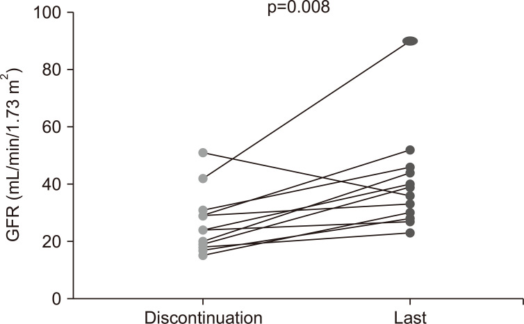
Thirteen patients with AAV who had discontinued the dialysis were further followed up for a median of 81 months (21.5~105.5 months). The total duration of dialysis was a median of 36.0 days (IQR, 33.5~126.5 days), and all patients stopped dialysis within 278 days after dialysis initiation (Table 5). At the time of dialysis discontinuation, the median creatinine level of patients was 2.60 mg/dL (2.05~3.16 mg/dL), and the median eGFR value was 24.0 mL/min/1.73 m² (18.5~30.0 mL/min/1.73 m²). During follow-up, only one patient restarted dialysis due to acute kidney injury with septic shock at 2,337 days after discontinuation. The remaining 12 patients were independent of dialysis, with a median eGFR value of 37.5 mL/min/1.73 m² (28.5~45.5 mL/min/1.73 m²) at the last follow-up. Notably, eGFR values were significantly higher at the last follow-up than at the time of dialysis discontinuation (Figure 2). At the last follow-up, 11 patients were still in remission based on the BVAS (0 or persistent 1).
Table 5.
Long-term follow-up of patients who discontinued the dialysis
| All (n=13) | |
|---|---|
| Age (yr) | 64.0 (60.0~69.5) |
| Female | 7 (53.8) |
| Duration of follow up (mo) | 81.0 (21.5~105.5) |
| Duration of dialysis (day) | 36.0 (33.5~126.5) |
| Remission | 10 (76.9) |
| Renal profile at the time of dialysis discontinuation | |
| Creatinine (mg/dL) | 2.60 (2.05~3.16) |
| eGFR (mL/min/1.73 m²) | 24.0 (18.5~30.0) |
| Induction therapy | |
| IV cyclophosphamide | 3 (23.1) |
| PO cyclophosphamide | 10 (76.9) |
| Rituximab | 1 (4.8) |
| Maintenance therapy | |
| Azathioprine | 10 (76.9) |
| Mycophenolate mofetil | 2 (15.4) |
| Duration of therapy (day) | 594.0 (119.5~2,160.5) |
| Steroid doses | |
| Duration of therapy (day) | 1,067.0 (427.5~1,825.5) |
| Cumulative dose (g) | 10.23 (8.74~12.70) |
| Renal profile at last follow-up | |
| Creatinine (mg/dL) | 1.85 (1.34~2.02) |
| eGFR (mL/min/1.73 m²) | 36.0 (27.5~45.0) |
| On re-dialysis | 1 (7.7) |
| BVAS at last follow-up | |
| 0 | 9 (69.2) |
| 1 (persistent) | 2 (15.4) |
| 4 | 1 (7.7) |
| 6 | 1 (7.7) |
Values are presented as median (interquartile range) or number (%). eGFR: estimated glomerular filtration rate, IV: intravenous, PO: per oral, BVAS: Birmingham Vasculitis Activity Score.
Fig. 2.
The comparison of values of estimated glomerular filtration rate (eGFR) of patients (n=12) with dialysis discontinuation between the time of dialysis discontinuation and time after the follow-up period.
DISCUSSION
In the present study, the data of 34 patients on dialysis at the time of AAV diagnosis were analyzed. We found that 38.2% of patients (n=13) were able to discontinue dialysis after treatment for AAV. The patients who discontinued dialysis had significantly higher numbers and proportions of normal glomeruli than those who depended on dialysis, and there was a trend, although not statistically significant, between the proportion of normal glomeruli and dialysis discontinuation. Upon analysis of follow-up data of patients with dialysis discontinuation, we found that most patients remained dialysis-free with an improvement in renal function.
The present study demonstrated that in the long-term follow-up of patients who discontinued dialysis, most patients remained off dialysis. Previous studies mainly focused on short-term outcomes of whether dialysis could be discontinued in AAV patients after induction treatment [14,15]. A previous study with data of patients on dialysis from the Plasma Exchange for Renal Vasculitis (MEPEX) trial aimed to identify risk factors associated with the chances of dialysis independence over death after 12 months of treatment [14]. This study showed that recovery from dialysis dependence was related to histological features, including the proportion of normal glomeruli and the extent of tubular atrophy. In a study of a similar design from a single Chinese center, histological features (such as, the proportion of normal glomeruli, extent of tubular atrophy, and extent of interstitial fibrosis) but not clinical factors (such as, age and sex) were independently associated with dialysis discontinuation 6 months after diagnosis [15]. Similar to these previous studies, we found that the proportion of normal glomeruli in the kidney tissue was related to recovery from dialysis in the present study. Considering that kidney biopsies are not routinely performed when AAV patients are already on dialysis but were performed in 70.6% (24/34) of the patients in our study, indicates that kidney biopsies can be more actively utilized to predict renal prognosis even in patients with severe renal dysfunction.
In terms of treatment, data on patients with AAV on dialysis was limited. There have been conflicting results on the efficacy of plasmapheresis as adjuvant therapy in severe AAV. While early data in the MEPEX trial showed a benefit of plasmapheresis in reducing the requirement of dialysis at 12 months, recent studies, including the PEXIVAS (plasma exchange and glucocorticoid dosing in the treatment of antineutrophil cytoplasm antibody associated vasculitis) trial conducted in patients with severe AAV including dialysis, demonstrated that plasmapheresis did not reduce the incidence of death or ESRD in patients with AAV [16-18]. In contrast, in a recent study involving 66 patients under dialysis at baseline, the use of plasmapheresis in patients receiving cyclophosphamide was associated with a higher dialysis-free rate at 12 months [13]. In the present study, there were no significant differences in therapeutic modalities, including induction therapy, plasmapheresis, and corticosteroid dosage between the dialysis-discontinuation and dialysis-dependent groups. Further studies are needed to identify the subgroups of patients with AAV on dialysis who would benefit from particular types of therapy.
Our study is notable in that it showed that dialysis was no longer required in the group that stopped dialysis for a follow-up of a median of 81 months. Additionally, except for one patient who restarted dialysis due to acute kidney injury with septic shock, patients who discontinued dialysis showed stable or even improving kidney function during follow-up. In the dialysis-discontinuation group, dialysis was required for a median of 36 days (IQR 33.5~126.5 days) and a maximum of 278 days. Thus, these findings suggest that long-term follow-up is necessary to determine the possibility of discontinuing dialysis. Furthermore, this study provides robust evidence regarding the long-term outcomes of renal AAV patients in South Korea.
The present study has several limitations. First, this study was conducted in a single center and included a small number of patients. Thus, it is difficult to generalize our results to the overall population. However, to the best of our knowledge, this is the first study covering the long-term data of patients after dialysis discontinuation. Second, although histologic findings in a kidney biopsy can be important, it was not performed in all patients.
In conclusion, 13 of 34 (38.2%) patients who required dialysis at the time of AAV diagnosis were able to discontinue dialysis. The higher proportion of normal glomeruli in kidney pathology and cessation of dialysis were positively correlated. Importantly, analysis of follow-up data of patients who discontinued dialysis showed that most patients had improved renal function without re-dialysis. Our findings suggest that patients with AAV on dialysis should be given the possibility of dialysis discontinuation and renal recovery, especially those with normal glomeruli in kidney pathology.
ACKNOWLEDGMENTS
None.
Footnotes
FUNDING
This study was supported by a grant (2022IP0062-1) from the Asan Institute for Life Sciences, Asan Medical Center, Seoul, Korea.
CONFLICT OF INTEREST
No potential conflict of interest relevant to this article was reported.
AUTHOR CONTRIBUTIONS
Conceptualization: S.H., B.Y. Data curation or/and Analysis: Y.J.L. and J.S.O. Writing - original draft: Y.J.L., S.M.A. Writing - Review & Editing: Y.J.L., Y.G.K., C.K.L., and S.H. All authors read and approved the final manuscript.
REFERENCES
- 1.Almaani S, Fussner LA, Brodsky S, Meara AS, Jayne D. ANCA-associated vasculitis: an update. J Clin Med. 2021;10:1446. doi: 10.3390/jcm10071446. [DOI] [PMC free article] [PubMed] [Google Scholar]
- 2.Sinico RA, Di Toma L, Radice A. Renal involvement in anti-neutrophil cytoplasmic autoantibody associated vasculitis. Autoimmun Rev. 2013;12:477–82. doi: 10.1016/j.autrev.2012.08.006. [DOI] [PubMed] [Google Scholar]
- 3.Day CJ, Howie AJ, Nightingale P, Shabir S, Adu D, Savage CO, et al. Prediction of ESRD in pauci-immune necrotizing glomerulonephritis: quantitative histomorphometric assessment and serum creatinine. Am J Kidney Dis. 2010;55:250–8. doi: 10.1053/j.ajkd.2009.10.047. [DOI] [PMC free article] [PubMed] [Google Scholar]
- 4.Rhee RL, Hogan SL, Poulton CJ, McGregor JA, Landis JR, Falk RJ, et al. Trends in long-term outcomes among patients with antineutrophil cytoplasmic antibody-associated vasculitis with renal disease. Arthritis Rheumatol. 2016;68:1711–20. doi: 10.1002/art.39614. [DOI] [PMC free article] [PubMed] [Google Scholar]
- 5.Mohammad AJ, Segelmark M. A population-based study showing better renal prognosis for proteinase 3 antineutrophil cytoplasmic antibody (ANCA)-associated nephritis versus myeloperoxidase ANCA-associated nephritis. J Rheumatol. 2014;41:1366–73. doi: 10.3899/jrheum.131038. [DOI] [PubMed] [Google Scholar]
- 6.Booth AD, Almond MK, Burns A, Ellis P, Gaskin G, Neild GH, et al. Outcome of ANCA-associated renal vasculitis: a 5-year retrospective study. Am J Kidney Dis. 2003;41:776–84. doi: 10.1016/S0272-6386(03)00025-8. [DOI] [PubMed] [Google Scholar]
- 7.Mukhtyar C, Flossmann O, Hellmich B, Bacon P, Cid M, Cohen-Tervaert JW, et al. Outcomes from studies of antineutrophil cytoplasm antibody associated vasculitis: a systematic review by the European League Against Rheumatism systemic vasculitis task force. Ann Rheum Dis. 2008;67:1004–10. doi: 10.1136/ard.2007.071936. [DOI] [PubMed] [Google Scholar]
- 8.Sánchez Álamo B, Moi L, Bajema I, Faurschou M, Flossmann O, Hauser T, et al. Long-term outcomes and prognostic factors for survival of patients with ANCA-associated vasculitis. Nephrol Dial Transplant. 2023;38:1655–65. doi: 10.1093/ndt/gfac320. [DOI] [PubMed] [Google Scholar]
- 9.Bourgarit A, Toumelin PL, Pagnoux C, Cohen P, Mahr A, Guern VL, et al. Deaths occurring during the first year after treatment onset for polyarteritis nodosa, microscopic polyangiitis, and Churg-Strauss syndrome: a retrospective analysis of causes and factors predictive of mortality based on 595 patients. Medicine (Baltimore) 2005;84:323–30. doi: 10.1097/01.md.0000180793.80212.17. [DOI] [PubMed] [Google Scholar]
- 10.Flossmann O, Berden A, de Groot K, Hagen C, Harper L, Heijl C, et al. Long-term patient survival in ANCA-associated vasculitis. Ann Rheum Dis. 2011;70:488–94. doi: 10.1136/ard.2010.137778. [DOI] [PubMed] [Google Scholar]
- 11.Lionaki S, Hogan SL, Jennette CE, Hu Y, Hamra JB, Jennette JC, et al. The clinical course of ANCA small-vessel vasculitis on chronic dialysis. Kidney Int. 2009;76:644–51. doi: 10.1038/ki.2009.218. [DOI] [PMC free article] [PubMed] [Google Scholar]
- 12.Weidner S, Geuss S, Hafezi-Rachti S, Wonka A, Rupprecht HD. ANCA-associated vasculitis with renal involvement: an outcome analysis. Nephrol Dial Transplant. 2004;19:1403–11. doi: 10.1093/ndt/gfh161. [DOI] [PubMed] [Google Scholar]
- 13.Morel P, Karras A, Porcher R, Belenfant X, Audard V, Rafat C, et al. Management of severe renal disease in anti-neutrophil-cytoplasmic-antibody-associated vasculitis: the place of rituximab and plasma exchange? Rheumatology (Oxford) 2022;61:4056–64. doi: 10.1093/rheumatology/keac046. [DOI] [PubMed] [Google Scholar]
- 14.de Lind van Wijngaarden RA, Hauer HA, Wolterbeek R, Jayne DR, Gaskin G, Rasmussen N, et al. Chances of renal recovery for dialysis-dependent ANCA-associated glomerulonephritis. J Am Soc Nephrol. 2007;18:2189–97. doi: 10.1681/ASN.2007010066. Erratum in: J Am Soc Nephrol 2007;18:3206. [DOI] [PubMed] [Google Scholar]
- 15.Li ZY, Gou SJ, Chen M, Zhao MH. Predictors for outcomes in patients with severe ANCA-associated glomerulonephritis who were dialysis-dependent at presentation: a study of 89 cases in a single Chinese center. Semin Arthritis Rheum. 2013;42:515–21. doi: 10.1016/j.semarthrit.2012.09.005. [DOI] [PubMed] [Google Scholar]
- 16.Jayne DR, Gaskin G, Rasmussen N, Abramowicz D, Ferrario F, Guillevin L, et al. Randomized trial of plasma exchange or high-dosage methylprednisolone as adjunctive therapy for severe renal vasculitis. J Am Soc Nephrol. 2007;18:2180–8. doi: 10.1681/ASN.2007010090. [DOI] [PubMed] [Google Scholar]
- 17.Casal Moura M, Irazabal MV, Eirin A, Zand L, Sethi S, Borah BJ, et al. Efficacy of rituximab and plasma exchange in antineutrophil cytoplasmic antibody-associated vasculitis with severe kidney disease. J Am Soc Nephrol. 2020;31:2688–704. doi: 10.1681/ASN.2019111197. [DOI] [PMC free article] [PubMed] [Google Scholar]
- 18.Walsh M, Merkel PA, Peh CA, Szpirt WM, Puéchal X, Fujimoto S, et al. Plasma exchange and glucocorticoids in severe ANCA-associated vasculitis. N Engl J Med. 2020;382:622–31. doi: 10.1056/NEJMoa1803537. [DOI] [PMC free article] [PubMed] [Google Scholar]
- 19.Jennette JC, Falk RJ, Bacon PA, Basu N, Cid MC, Ferrario F, et al. 2012 revised International Chapel Hill Consensus Conference Nomenclature of Vasculitides. Arthritis Rheum. 2013;65:1–11. doi: 10.1002/art.37715. [DOI] [PubMed] [Google Scholar]
- 20.Mukhtyar C, Lee R, Brown D, Carruthers D, Dasgupta B, Dubey S, et al. Modification and validation of the Birmingham Vasculitis Activity Score (version 3) Ann Rheum Dis. 2009;68:1827–32. doi: 10.1136/ard.2008.101279. [DOI] [PubMed] [Google Scholar]
- 21.Hellmich B, Flossmann O, Gross WL, Bacon P, Cohen-Tervaert JW, Guillevin L, et al. EULAR recommendations for conducting clinical studies and/or clinical trials in systemic vasculitis: focus on anti-neutrophil cytoplasm antibody-associated vasculitis. Ann Rheum Dis. 2007;66:605–17. doi: 10.1136/ard.2006.062711. [DOI] [PMC free article] [PubMed] [Google Scholar]
- 22.Kemna MJ, Damoiseaux J, Austen J, Winkens B, Peters J, van Paassen P, et al. ANCA as a predictor of relapse: useful in patients with renal involvement but not in patients with nonrenal disease. J Am Soc Nephrol. 2015;26:537–42. doi: 10.1681/ASN.2013111233. [DOI] [PMC free article] [PubMed] [Google Scholar]
- 23.Berden AE, Ferrario F, Hagen EC, Jayne DR, Jennette JC, Joh K, et al. Histopathologic classification of ANCA-associated glomerulonephritis. J Am Soc Nephrol. 2010;21:1628–36. doi: 10.1681/ASN.2010050477. [DOI] [PubMed] [Google Scholar]


