Abstract
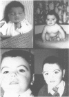
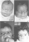
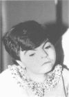
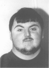
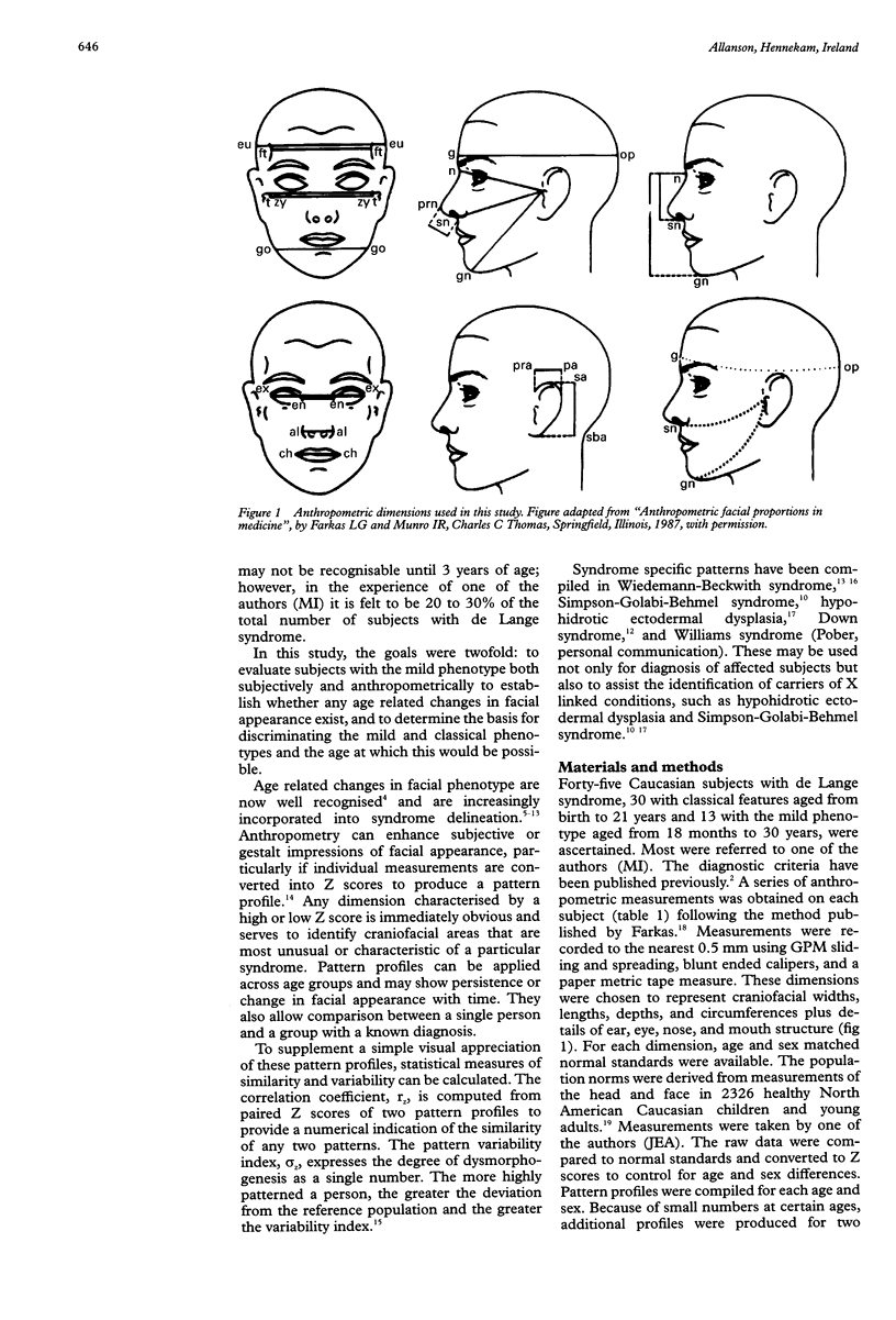
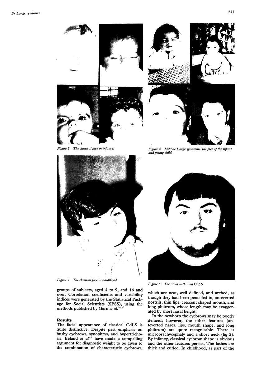
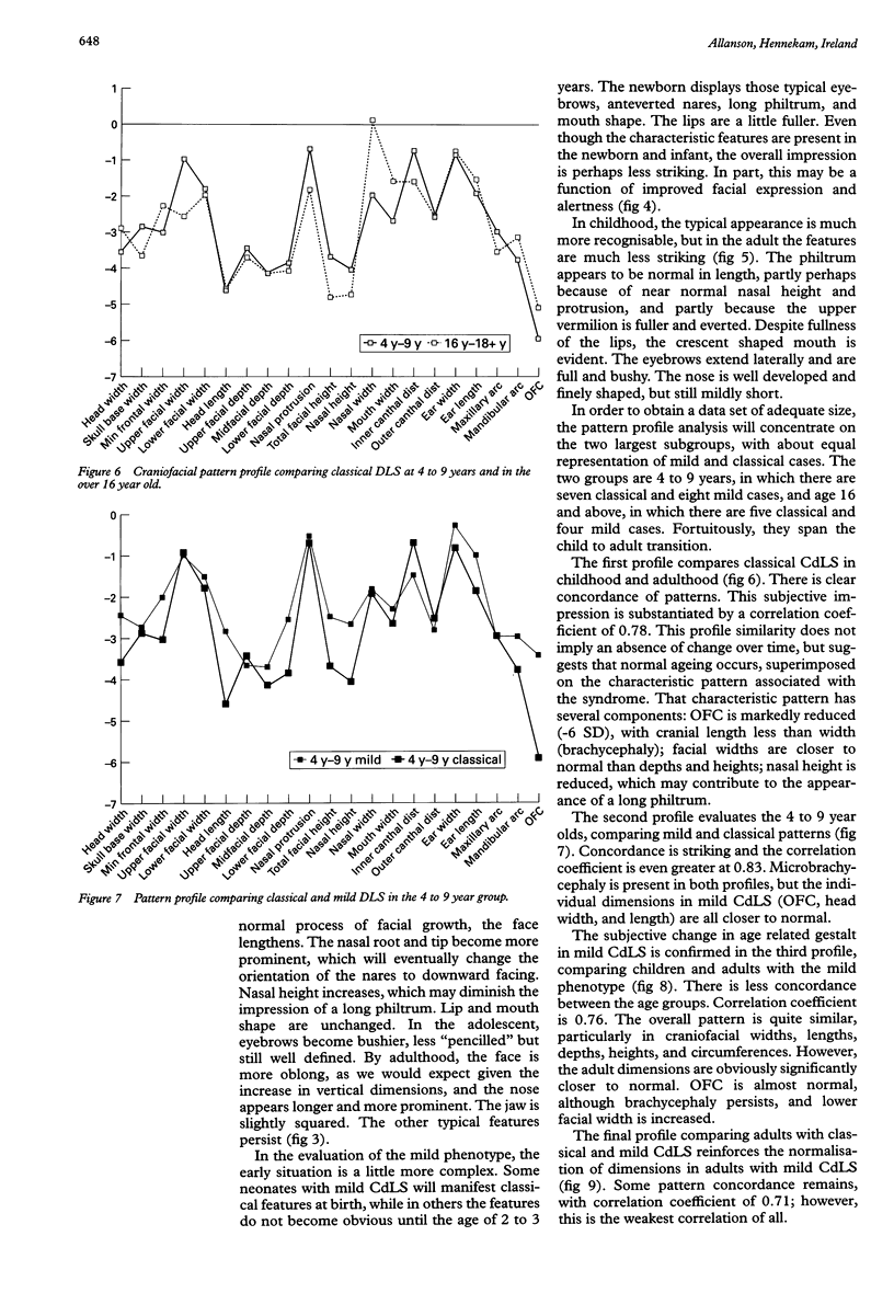
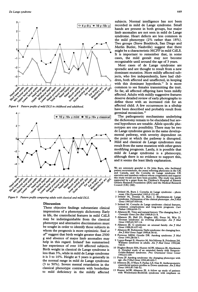
Classical de Lange syndrome presents with a striking face, pronounced growth and mental retardation, and variable limb deficiencies. Over the past five years, a mild variant has been defined, with less significant psychomotor retardation, less marked pre- and postnatal growth deficiency, and an uncommon association with major malformations, although mild limb anomalies may be present. We have evaluated 43 subjects with de Lange syndrome, 30 with classical features, aged from birth to 21 years, and 13 with the mild phenotype, aged from 18 months to 30 years. In addition to assessment of gestalt and facial change with time, detailed craniofacial measurements have been obtained on each subject and composite pattern profiles compiled. The characteristic face of classical de Lange syndrome is present at birth and changes little throughout life, although there is some lengthening of the face with age and the jaw becomes squared. In mild de Lange syndrome, the characteristic classical appearance may be present at birth, but in some subjects it may be two or three years before the typical face is obvious. In general, the overall impression is less striking, perhaps because of increased facial expression and greater alertness. With age, the face loses the characteristic appearance, the nasal height increases, the philtrum does not seem as long, and the upper vermilion is full and everted, although the crescent shaped mouth with downturned corners remains. Eyebrows may be full and bushy. Objective comparison of the face in mild and classical de Lange syndrome, through the use of craniofacial pattern profiles, shows marked similarity of patterns at 4 to 9 years; both groups have microbrachycephaly, but the individual dimensions of the mild group are slightly closer to normal than their classical counterparts. The correlation coefficient is high (0.83). In the adult groups, similarity of patterns remains but is less marked. The normalisation of scores in the mild group is more dramatic. The correlation coefficient is lower (0.71). These objective findings substantiate clinical impressions of a phenotypic dichotomy. Early in life, the craniofacial features in mild de Lange syndrome may be indistinguishable from the classical phenotype and alternative discriminators must be sought in order to identify those subjects in whom the prognosis is more optimistic. Birth weight of more than 2500 g and absence of major limb anomalies may help in this regard.
Full text
PDF

Images in this article
Selected References
These references are in PubMed. This may not be the complete list of references from this article.
- Allanson J. E. G syndrome: an unusual family. Am J Med Genet. 1988 Nov;31(3):637–642. doi: 10.1002/ajmg.1320310319. [DOI] [PubMed] [Google Scholar]
- Allanson J. E., Hall J. G., Hughes H. E., Preus M., Witt R. D. Noonan syndrome: the changing phenotype. Am J Med Genet. 1985 Jul;21(3):507–514. doi: 10.1002/ajmg.1320210313. [DOI] [PubMed] [Google Scholar]
- Allanson J. E., O'Hara P., Farkas L. G., Nair R. C. Anthropometric craniofacial pattern profiles in Down syndrome. Am J Med Genet. 1993 Oct 1;47(5):748–752. doi: 10.1002/ajmg.1320470530. [DOI] [PubMed] [Google Scholar]
- Allanson J. E. Rubinstein-Taybi syndrome: the changing face. Am J Med Genet Suppl. 1990;6:38–41. doi: 10.1002/ajmg.1320370606. [DOI] [PubMed] [Google Scholar]
- Allanson J. E. Time and natural history: the changing face. J Craniofac Genet Dev Biol. 1989;9(1):21–28. [PubMed] [Google Scholar]
- Chodirker B. N., Chudley A. E. Male-to-male transmission of mild Brachmann-de Lange syndrome. Am J Med Genet. 1994 Sep 1;52(3):331–333. doi: 10.1002/ajmg.1320520315. [DOI] [PubMed] [Google Scholar]
- Fryns J. P. Aarskog syndrome: the changing phenotype with age. 1992 Apr 15-May 1Am J Med Genet. 43(1-2):420–427. doi: 10.1002/ajmg.1320430164. [DOI] [PubMed] [Google Scholar]
- Fryns J. P., Dereymaeker A. M., Hoefnagels M., D'Hondt F., Mertens G., van den Berghe H. The Brachmann-de Lange syndrome in two siblings of normal parents. Clin Genet. 1987 Jun;31(6):413–415. doi: 10.1111/j.1399-0004.1987.tb02835.x. [DOI] [PubMed] [Google Scholar]
- Garn S. M., Lavelle M., Smith B. H. Quantification of dysmorphogenesis: pattern variability index, sigma z. AJR Am J Roentgenol. 1985 Feb;144(2):365–369. doi: 10.2214/ajr.144.2.365. [DOI] [PubMed] [Google Scholar]
- Garn S. M., Smith B. H., LaVelle M. Applications of pattern profile analysis to malformations of the head and face. Radiology. 1984 Mar;150(3):683–690. doi: 10.1148/radiology.150.3.6695067. [DOI] [PubMed] [Google Scholar]
- Hunter A. G., Allanson J. E. Follow-up study of patients with Wiedemann-Beckwith syndrome with emphasis on the change in facial appearance over time. Am J Med Genet. 1994 Jun 1;51(2):102–107. doi: 10.1002/ajmg.1320510205. [DOI] [PubMed] [Google Scholar]
- Ireland M., Burn J. Cornelia de Lange syndrome--photo essay. Clin Dysmorphol. 1993 Apr;2(2):151–160. [PubMed] [Google Scholar]
- Ireland M., Donnai D., Burn J. Brachmann-de Lange syndrome. Delineation of the clinical phenotype. Am J Med Genet. 1993 Nov 15;47(7):959–964. doi: 10.1002/ajmg.1320470705. [DOI] [PubMed] [Google Scholar]
- Lopez-Rangel E., Maurice M., McGillivray B., Friedman J. M. Williams syndrome in adults. Am J Med Genet. 1992 Dec 1;44(6):720–729. doi: 10.1002/ajmg.1320440605. [DOI] [PubMed] [Google Scholar]
- Porteous M. E., Goudie D. R. Aarskog syndrome. J Med Genet. 1991 Jan;28(1):44–47. doi: 10.1136/jmg.28.1.44. [DOI] [PMC free article] [PubMed] [Google Scholar]
- Saal H. M., Samango-Sprouse C. A., Rodnan L. A., Rosenbaum K. N., Custer D. A. Brachmann-de Lange syndrome with normal IQ. Am J Med Genet. 1993 Nov 15;47(7):995–998. doi: 10.1002/ajmg.1320470711. [DOI] [PubMed] [Google Scholar]
- Ward R. E., Bixler D. Anthropometric analysis of the face in hypohidrotic ectodermal dysplasia: a family study. Am J Phys Anthropol. 1987 Dec;74(4):453–458. doi: 10.1002/ajpa.1330740404. [DOI] [PubMed] [Google Scholar]


