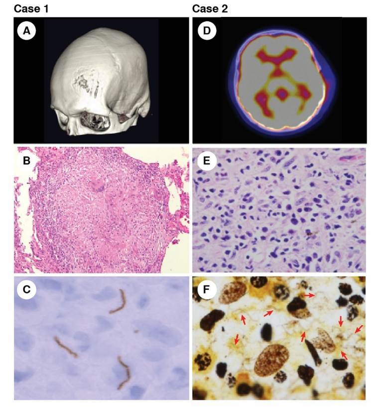
Figure 1. Left column: Case 1 - Tertiary syphilis. (A) Three-dimensional reconstruction of the skull computed tomography showing the bone lesion in the frontal bone. (B) Syphilitic gumma with chronic granulomatous infiltrate, hematoxylin eosin, 10X. (C) Immunochemistry of the lytic bone lesion showing spirochetes in the tissue.

Right column: Case 2 - Secondary syphilis. (D) PET scan revealing lytic lesions with increased metabolism on the frontal and temporal bones. (E) Lymphoplasmocytic infiltrate in the bone biopsy, hematoxylin eosin, 100X. (F) Warthin-Starry stain revealing the presence of numerous spirochetes, 100X
