PLOS ONE logoLink to PLOS ONE
. 2023 Sep 26;18(9):e0281561. doi: 10.1371/journal.pone.0281561

Early death after a diagnosis of metastatic solid cancer–raising awareness and identifying risk factors from the SEER database

Opher Globus 1,2,#, Shira Sagie 1,3,4,#, Noy Lavine 5, Daniel Itshak Barchana 5, Damien Urban 1,2,*
Editor: Mirosława Püsküllüoğlu6
PMCID: PMC10522015  PMID: 37751439

Abstract

Background

Cancer death rates are declining, in part due to smoking cessation, better detection and new treatments; nevertheless, a large fraction of metastatic cancer patients die soon after diagnosis. Few studies and interventions focus on these patients. Our study aims to characterize early mortality in a wide range of metastatic solid tumors.

Methods

We retrieved data on adult patients diagnosed with pathologically confirmed de- novo metastatic solid tumors between the years 2004–2016 from the Surveillance, Epidemiology, and End Results database (SEER). Our primary outcome was cancer specific early death rate (defined as death within two months of diagnosis). Additional data extracted included socio-demographical data, tumor primary, sites of metastases, and cause of death.

Results

109,207 (20.8%) patients died of de-novo metastatic cancer within two months of diagnosis. The highest rates of early death were found in hepatic (36%), pancreato-biliary (31%) and lung (25%) primaries. Factors associated with early death included primary site, liver, and brain metastases, increasing age, and lower income. Cancer was the cause of death in 92.1% of all early deaths. Two-month mortality rates have moderately improved during the study period (from 22.4% in 2004 to 18.8% in 2016).

Conclusion

A fifth of de-novo metastatic cancer patients die soon after diagnosis, with little improvement over the last decade. Further research is required to better classify and identify patients at risk for early mortality, which patients might benefit from faster diagnostic tracks, and which might avoid invasive and futile diagnostic procedures.

Introduction

Despite improvements in early detection and treatment, cancer remains one of the leading causes of death in the United States and around the world [1, 2]. Major advances in the personalization of systemic treatment of cancer have demonstrated rapid responses and long-term survival in some patients, even in the metastatic setting [3]. However, clinicians, often encounter patients with suspected de-novo metastatic disease who are extremely ill at the time of presentation. In these clinical situations, physicians, patients, and families are faced with fundamental decisions. On the one hand, particularly due to advances in personalized oncology, some select patients can rapidly improve with targeted treatments. On the other hand, diagnostic procedures are not without financial costs and potential complications [4], often resulting in hospital admissions and poor end-of-life care. As many of the pivotal improvements require predictive biomarkers, which can take weeks to return, the benefit of these treatment advances may elude many patients.

The aim of our current study is to describe early mortality in solid tumors and to characterize which patients with de-novo metastatic disease die within two months of diagnosis. As early mortality from cancer is due to a combination of complex tumor-related, patient-related, and healthcare-related mechanisms, our objective is to present and disseminate the scope of this phenomenon to healthcare professionals involved in the diagnosis and treatment of cancer and to identify characteristics associated with early mortality, thereby helping guide physicians, patients, and families in appropriate clinical decisions.

Methods

Study population and design

Patients with histologically confirmed de-novo metastatic cancer were identified from the Surveillance, Epidemiology, and End Results (SEER) database from 2004 to 2016.

The SEER database, maintained by the NCI in the U.S., is a comprehensive and publicly accessible cancer database. It offers detailed information on cancer incidence, prevalence, survival, and treatment patterns. Since 1973, SEER facilitates research, policymaking, and clinical decision-making by providing valuable insights into cancer patterns and outcomes. The 18 population based SEER cancer registries cover 28% of the US population [5]. Given the nature of this study, there was no requirement for institutional review board submission. Access to the SEER data was in accordance with the SEER data agreement.

Patients included were at least 18 years of age and presented with a first diagnosis of pathologically confirmed metastatic cancer. Patients with recurrent metastatic disease were excluded. As the focus of our study was metastatic solid tumors, we excluded patients with primary neurological tumors, hematological malignancies, and rare solid tumors (peripheral nervous system tumors, tumors stemming from the retroperitoneum, non-lung cancer chest tumors, tumors identified as not otherwise specified from the male genital, female genital, and the urinary tract).

Study variables

Our primary outcome was cancer-specific two-month mortality from the date of diagnosis.

Cause-specific survival in the SEER data is a measure that focuses on the survival of individuals attributed to a specific cause of death, excluding other causes. It involves specifying the cause of death and considering individuals who die from causes other than the specified cause as censored. The cancer registries utilize algorithms to process information from death certificates and identify a single, disease-specific underlying cause of death. However, there can be challenges in accurately attributing a single cause of death, leading to potential misattribution. To address this, SEER utilizes cause-specific death classification variables that consider tumor sequence, the site of the original cancer diagnosis, and comorbidities to capture deaths related to the specific cancer even if not explicitly coded as such. This approach leads to a relatively small bias [6].

Additional data extracted included age, sex, race, residence, income, tumor primary site, metastasis location (available only after 2010 for liver, bone, brain, and lung), year of diagnosis (grouped in four time periods 2004–2006, 2007–2009, 2010–2012, 2013–2016), cause of death, and survival by months.

We separately analyzed twelve groups of tumor locations: bladder, breast, colorectal, gastro-esophageal, hepatocellular, head and neck, kidney, lung, melanoma, ovary, pancreato-biliary, and prostate, all defined by primary tumor location except for melanoma which was defined by both histology and location.

Statistical analysis

The Mann-Whitney U test was used for variables that did not follow a normal distribution and the chi square test and Fisher Exact test were used as appropriate for categorical variables. We fitted multivariable logistic models. Data for metastasis sites was available since 2010, thus we fitted separate multivariable models for the whole population and for patients diagnosed after 2010. Adjusted OR and 95% confidence intervals (CI) were obtained for the independent variables from these models. P<0.05 was considered statistically significant. The statistical analyses were performed using R version 4.0.3.

Results

Baseline characteristics

We identified 525,780 patients diagnosed with de-novo metastatic solid malignancies between 2004 and 2016 of which 109,207 (20.77%) died of cancer within two months of diagnosis. Patient characteristics are presented in Table 1. Most patients were male (52.1%), white (78.6%), and 50–70 years old (51.7%). The most common primary site was lung (40%) followed by pancreato-biliary (11.2%) and colorectal cancer (10.8%). Patients that died within two months compared to those who did not, were older (median 70 vs 65), more frequently male (54.3% vs 51.5%), had lower income (56.8% vs 54.9%) and more frequently had brain (15% vs 12.9%), lung (49.2% vs 33.5%) and liver metastasis (33.5% vs 28%).

Table 1. Characteristics of the study group classified by early mortality.

    Early mortality  
  Overall No Yes p
N 525780 416573 109207  
Sex, Male (%) 273976 (52.1) 214661 (51.5) 59315 (54.3) <0.001
Race (%)       <0.001
White 413207 (78.6) 325598 (78.2) 87609 (80.2)  
Black 69371 (13.2) 55525 (13.3) 13846 (12.7)  
American Indian/Alaska Native 3576 (0.7) 2814 (0.7) 762 (0.7)  
Asian or Pacific Islander 38627 (7.3) 31769 (7.6) 6858 (6.3)  
Unknown 999 (0.2) 867 (0.2) 132 (0.1)  
Age (Median [IQR])) 66 [57, 75] 65 [56, 74] 70 [61, 78] <0.001
Residence Category (%)       <0.001
Metropolitan 313730 (59.8) 248942 (59.8) 64788 (59.4)  
Rural 27729 (5.3) 21788 (5.2) 5941 (5.4)  
Urban 183506 (35.0) 145222 (34.9) 38284 (35.1)  
Income below $65K/year (%) 284945 (55.3) 224293 (54.9) 60652 (56.8) <0.001
Grouped Years (%)       <0.001
2004–2006 113549 (21.6) 88288 (21.2) 25261 (23.1)  
2007–2009 118303 (22.5) 93352 (22.4) 24951 (22.8)  
2010–2012 119990 (22.8) 95143 (22.8) 24847 (22.8)  
2013–2016 173938 (33.1) 139790 (33.6) 34148 (31.3)  
Bone Metastasis, Yes (%) 93642 (33.0) 75821 (33.3) 17821 (31.7) <0.001
Brain Metastasis, Yes (%) 37565 (13.3) 29214 (12.9) 8351 (15.0) <0.001
Lung Metastasis, Yes (%) 104138 (36.6) 76277 (33.5) 27861 (49.2) <0.001
Liver Metastasis, Yes (%) 81644 (29.1) 63075 (28.0) 18569 (33.5) <0.001
Primary Cancer Location (%)      
Bladder 4819 (0.9) 3791 (0.9) 1028 (0.9)  
Breast 34387 (6.5) 31066 (7.5) 3321 (3.0)  
CRC 56763 (10.8) 48506 (11.6) 8257 (7.6)  
Gastro-esophageal 35563 (6.8) 27269 (6.5) 8294 (7.6)  
HCC 7964 (1.5) 5119 (1.2) 2845 (2.6)  
Head and Neck 7647 (1.5) 6968 (1.7) 679 (0.6)  
Kidney 17399 (3.3) 14803 (3.6) 2596 (2.4)  
Lung 210027 (39.9) 156898 (37.7) 53129 (48.6)  
Melanoma 6807 (1.3) 5755 (1.4) 1052 (1.0)  
Ovary 17854 (3.4) 15027 (3.6) 2827 (2.6)  
Pancreato-biliary 59071 (11.2) 40683 (9.8) 18388 (16.8)  
Prostate 28219 (5.4) 27283 (6.5) 936 (0.9)  

*Income- omitted 10,615 NA’s, Residence-omitted 811 NA’s, Bone metastasis -omitted 242413 NA’s, Brain metastasis—Omitted 244295 NA’s, Lung metastasis—Omitted 245322 NA’s, Liver metastasis—Omitted 241850 NA’s. HCC: Hepatocellular carcinoma. CRC: Colorectal cancer. NA = Not available

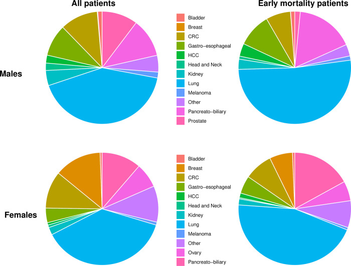
Breast and prostate cancer primaries were less frequent in the early mortality group (3% and 0.9%, respectively); in contrast, lung and pancreato-biliary primaries were more frequent (48.6% and 16.8%, respectively). Primary tumor distribution in the whole cohort and in the early mortality group by gender appears in Fig 1.

Fig 1. Disease site distribution by gender in the whole cohort and in the early mortality group.

Fig 1

Left Panel-525,780 Patients with de-novo metastatic cancer were analyzed by disease site origin, separated by gender. Right Panel -109,207 patients which died in the first two months after disease diagnosis analyzed by disease site origin, separated by gender. Primary site color code differs between genders and appears in the middle.

Causes of death

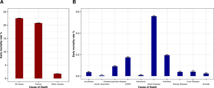
Within our cohort, 109,207 (20.8%) patients diagnosed with de-novo metastatic solid cancer died of cancer within two months of diagnosis. Cancer was the cause of death in 92.1% of early deaths (Fig 2A). The most common non-cancer cause of early death was heart disease n = 2,896 (0.56% of all patients) followed by infection n = 1,010 (0.19% of all patients). The ten most common non-cancer causes of early death appear in Fig 2B.

Fig 2. Major causes of early death in de novo diagnosed metastatic cancer patients.

Fig 2

All cause and cancer specific early death rates (A). Ten major causes of non-cancer early death (B). Y axes present percentages in both plots.

Primary tumor specific early mortality rates

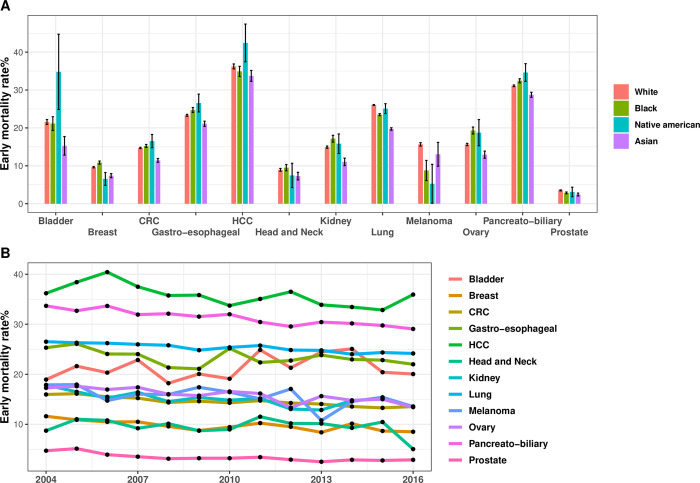
The two-month cancer specific mortality rate was highest in patients with hepatocellular (36%), pancreato-biliary (31%), lung (25%), gastro-esophageal (23%) and bladder (21%) primaries and lowest in patients with prostate (3%), head and neck (9%) and breast (10%) primaries (Fig 3). Black patients had a significantly higher early mortality rate in breast, gastroesophageal, pancreato-biliary, ovarian and kidney cancer, whereas white patients had a significantly higher mortality rate in lung, prostate cancer, and melanoma. These differences remain statistically significant in multivariate analysis. Fig 3 displays early mortality rates by primary tumor, race, and year of diagnosis.

Fig 3. Early mortality rates by primary tumor, race, and year of diagnosis.

Fig 3

Early mortality rates were calculated separately for each primary tumor group in each race group (A). Early mortality trends by primary tumor site and year of diagnosis (B). Y axes present percentages.

In most cancer types a statistically significant reduction in early mortality was observed between 2004 and 2016, with the overall two-month cancer specific mortality rate improving during the period of our study from 22.4% in 2004 to 18.8% in 2016. The strongest relative reductions in early mortality were documented in prostate, kidney, and breast cancers. Cancers without a statistically significant reduction in early mortality include bladder cancer, head and neck cancer and melanoma (Fig 3B).

Multivariate analysis of all cancers

On multivariate analysis, factors significantly associated with increased two-month mortality (Table 2) included site of metastases, particularly liver metastases (data starting from 2010, OR 1.91 CI 1.87–1.96, p<0.001), male sex (OR 1.17 CI 1.16–1.19, p<0.001), income below $65K a year (OR 1.12 CI 1.11.13, p<0.001), race (black and native American, OR 1.07 CI 1.04–1.09, p<0.001 and OR 1.13 CI 1.02–1.24, p<0.001 respectively), and increasing age (OR 1.03 per year p<0.001).

Table 2. Multivariable logistic regression analysis of the correlates of early mortality in all patients.

  Multivariable 2010+ Multivariable all data
  OR, CI p-value OR, CI p-value
Sex:        
Women   Ref.   Ref.
Men 1.19 [1.16,1.21] <0.001 1.17 [1.16,1.19] <0.001
Grouped Years:    
2004–2006 NA NA Ref. Ref.
2007–2009 NA NA 0.92 [0.91,0.94] <0.001
2010–2012 Ref. Ref. 0.91 [0.89,0.93] <0.001
2013–2016 0.94 [0.92,0.96] <0.001 0.85 [0.84,0.87] <0.001
Race:    
White Ref. Ref. Ref. Ref.
Black 1.05 [1.02,1.08] 0 1.07 [1.04,1.09] <0.001
American Indian/Alaska Native 1.01 [0.87,1.16] 0.46 1.13 [1.02,1.24] 0.02
Asian or Pacific Islander 0.8 [0.77,0.84] <0.001 0.8 [0.78,0.82] <0.001
Unknown 0.78 [0.6,1] <0.001 0.76 [0.62,0.91] <0.001
Age 1.03 [1.03,1.03] <0.001 1.03 [1.03,1.03] <0.001 
Income Category:    
Above 65K Ref. Ref. Ref. Ref.
Below 65K 1.16 [1.14,1.19] <0.001 1.12 [1.1,1.13] <0.001
Residence Category    
Metropolitan Ref. Ref. Ref. Ref.
Rural 0.94 [0.9,0.99] 0.8 0.94 [0.91,0.98] <0.001
Urban 0.97 [0.95,0.99] 0.19 0.98 [0.96,0.99] <0.001
Bone Metastasis:   NA NA
No Ref. Ref. NA NA
Yes 1.17 [1.14,1.2] <0.001 NA NA
Brain Metastasis:   NA NA
No Ref. Ref. NA NA
Yes 1.17 [1.14,1.21] <0.001 NA NA
Lung Metastasis:   NA NA
No Ref. Ref. NA NA
Yes 1.29 [1.26,1.32] <0.001 NA NA
Liver Metastasis:   NA NA
No Ref. Ref. NA NA
Yes 1.91 [1.87,1.96] <0.001 NA NA

*We generated two logistic models, the first including data after 2010 that contains information about metastatic sites, the second model includes all data since 2004.

Discussion

Our study reports that approximately 20% of patients with biopsy proven de-novo metastatic solid cancer die within two months of diagnosis. Previous studies of individual cancers have also reported high early mortality rates. An analysis of the national cancer database (NCDB) showed that between 2006 and 2014, 13% of patients with metastatic non-small cell lung cancer (NSCLC) died within 30 days of diagnosis [7]. A SEER analysis of patients with metastatic breast cancer showed that in 2013, 13.4% of patients died within one month, while a SEER analysis of patients with pancreatic adenocarcinoma showed that approximately half of patients died within two months of diagnosis [8, 9]. These studies demonstrated that early mortality is a prevalent phenomenon, associated with increased age, insurance status, geographic location, and comorbidity burden. Nevertheless, the scope of early mortality across many solid tumors and the total magnitude of this phenomenon is underrecognized.

Death so quickly after a de-novo metastatic solid cancer diagnosis may be due to causes related to the cancer itself or because of non-cancer related comorbidities. In our study over 90% of early mortalities (Fig 2) were due to the cancer itself, a figure significantly higher than previously reported [10]; likely because our cohort included only patients who had a histologically confirmed diagnosis, indicating they were well enough to undergo invasive testing during the diagnostic process. We chose to examine two-month mortality because this is the period often required to see an improvement from many cancer therapies, while death within this period suggests that patients are unlikely to benefit from many systemic treatments. In our study patients with pancreato-biliary, lung, and liver primaries had the highest proportion of early mortality. Patients with liver metastases were almost twice as likely to suffer early mortality independent of other factors.

Our findings also shed light on the role of income in cancer outcomes. Lower income was identified as a significant risk factor for early mortality in de-novo metastatic cancer patients. From a policy perspective, this presents a crucial area for intervention. Lower income patients might face barriers to access timely diagnosis, treatment, and comprehensive care, contributing to worse outcomes. Addressing this issue requires broader policy interventions aimed at minimizing healthcare disparities and improving access and affordability of care, particularly for those most vulnerable.

The cancer diagnostic process itself is costly and time consuming, taking a median of 35 days for a diagnosis and an additional 23 days for molecular testing results in certain tumor types, such as non-small cell lung cancer [11, 12]. Unfortunately for many patients, early cancer death in inevitable, even with immediate treatment. Early identification of high-risk patients can help avoid costly evaluations and prompt consideration of end of life care, even without tissue diagnosis. Conversely, rapid treatment can yield significant responses in certain patient subsets. For instance, lung cancer accounts for nearly 50% of patients dying within 2 months of a de-novo metastatic diagnosis. The remarkable advances in non-small cell lung cancer targeted therapy can sometimes prompt rapid responses in up to 70% of patients. Given that 20–30% of patients have a targetable driver mutation, there lies a potential to significantly extend survival if such mutations are detected promptly. Using minimally invasive diagnostic tools, such as liquid biopsies identifying genomic alterations [13, 14], it is possible to identify patients that have a high chance of targetable mutations and a subsequently significant response to treatments, while potentially avoiding more invasive and time-consuming diagnostic interventions such as tissue biopsies. The nuanced distinction between patients who are nearing death, for whom both diagnosis and treatment are likely to be futile versus those who could potentially benefit from treatment, carries significant clinical implications. Identifying this fine line between treatable and non-treatable conditions is of utmost importance.

Despite the substantial early mortality rate in our study, we also observed a small yet encouraging trend of improvement over time amongst most solid tumors. While the observed trend in our analysis is likely to persist, delayed diagnosis and healthcare seeking behaviors during the COVID-19 pandemic may have a dampening effect on this positive trend [15, 16]. Looking into the future, the advent of blood based multicancer early detection assays stands to improve early mortality from de-novo metastatic cancer by potentially lowering costs, enhancing early detection and streamlining the diagnostic process in patients with suspected metastatic disease. Our study further emphasizes the profound implications this technology may have on cancer care [1719].

Our study, like others utilizing the SEER database, has several limitations [20]. We do not have information on important confounders including performance status, comorbidities, socioeconomic environment, and healthcare facilities treated. In addition, we do not have data on physician referral or treatment patterns. Our cohort includes only patients with de-novo metastatic cancer and is not applicable to patients with metastatic recurrences.

In conclusion, our study highlights the magnitude of early mortality among de-novo metastatic cancer patients and identifies risk factors associated with early mortality including cancer primary, liver and brain metastases, advanced age, and lower income. We report that the principal cause of death for these patients was their metastatic disease, rather than any co-existing medical conditions. Further studies are required to better identify patients at high risk for early mortality, facilitating educated discussion with patients and caregivers. If properly identified, a unique and more efficient diagnostic pathway for this patient population may help improve the outcomes of these patients.

Supporting information

S1 Data

(CSV)

Acknowledgments

Disclosures: Dr Urban reports receiving consulting fees from Merck, Sharpe & Dohme, Roche Israel, Takeda, Nucleai, Rhenium Oncotest, and lecture fees from Astrazeneca, Merck, Sharpe & Dohme, Roche, Takeda, Bristol Myers Squibb, and Merck Seronoe. Dr Globus reports receiving lecture fees from Pfizer Lilly, Roche, Astra Zeneca, Novartis, Gilead, and Merck, Sharpe & Dohme, consulting fees from Lilly, Gilead, Pfizer, and Novartis, and expenses for conferences from Pfizer, Medison, Rhenium Onctotest, and Gilead. Dr Sagie, Lavine, and Barchana report no potential conflicting interests.

Data Availability

All relevant data are within the paper and its Supporting Information files.

Funding Statement

The authors received no specific funding for this work.

References

  • 1.Sung H, Ferlay J, Siegel RL, Laversanne M, Soerjomataram I, Jemal A, et al. Global Cancer Statistics 2020: GLOBOCAN Estimates of Incidence and Mortality Worldwide for 36 Cancers in 185 Countries. CA Cancer J Clin. 2021. May;71(3):209–49. doi: 10.3322/caac.21660 [DOI] [PubMed] [Google Scholar]
  • 2.Siegel RL, Miller KD, Fuchs HE, Jemal A. Cancer statistics, 2022. CA: A Cancer Journal for Clinicians. 2022;72(1):7–33. doi: 10.3322/caac.21708 [DOI] [PubMed] [Google Scholar]
  • 3.Shah M, Nunes MR, Stearns V. CDK4/6 Inhibitors: Game Changers in the Management of Hormone Receptor–Positive Advanced Breast Cancer? Oncology (Williston Park). 2018. May 15;32(5):216–22. [PMC free article] [PubMed] [Google Scholar]
  • 4.Lokhandwala T, Dann R, Johnson M, D’Souza AO. Costs of the Diagnostic Workup for Lung Cancer: A Medicare Claims Analysis: Diagnosis/Staging. International Journal of Radiation Oncology, Biology, Physics. 2014. Nov 15;90(5):S9–10. [Google Scholar]
  • 5.Surveillance, Epidemiology, and End Results (SEER) Program (www.seer.cancer.gov) SEER*Stat Database: Incidence - SEER Research Data, 18 Registries, Nov 2019 Sub (2000–2017)—Linked To County Attributes—Time Dependent (1990–2017) Income/Rurality, 1969–2018 Counties, National Cancer Institute, DCCPS, Surveillance Research Program, released April 2020, based on the November 2019 submission
  • 6.Hu CY, Xing Y, Cormier JN, Chang GJ. Assessing the utility of cancer-registry-processed cause of death in calculating cancer-specific survival. Cancer. 2013. May 15;119(10):1900–7. doi: 10.1002/cncr.27968 [DOI] [PMC free article] [PubMed] [Google Scholar]
  • 7.Amini A, Verma V, Glaser SM, Shinde A, Sampath S, Stokes WA, et al. Early mortality of stage IV non-small cell lung cancer in the United States. Acta Oncologica. 2019. Aug 3;58(8):1095–101. doi: 10.1080/0284186X.2019.1599138 [DOI] [PubMed] [Google Scholar]
  • 8.Vaz-Luis I, Lin NU, Keating NL, Barry WT, Winer EP, Freedman RA. Factors Associated with Early Mortality Among Patients with De Novo Metastatic Breast Cancer: A Population-Based Study. Oncologist. 2017. Apr;22(4):386–93. doi: 10.1634/theoncologist.2016-0369 [DOI] [PMC free article] [PubMed] [Google Scholar]
  • 9.Golan T, Sella T, Margalit O, Amit U, Halpern N, Aderka D, et al. Short- and Long-Term Survival in Metastatic Pancreatic Adenocarcinoma, 1993–2013. J Natl Compr Canc Netw. 2017. Aug;15(8):1022–7. doi: 10.6004/jnccn.2017.0138 [DOI] [PubMed] [Google Scholar]
  • 10.Brewster DH, Clark DI, Stockton DL, Munro AJ, Steele RJC. Characteristics of patients dying within 30 days of diagnosis of breast or colorectal cancer in Scotland, 2003–2007. Br J Cancer. 2011. Jan;104(1):60–7. doi: 10.1038/sj.bjc.6606036 [DOI] [PMC free article] [PubMed] [Google Scholar]
  • 11.Advanced‐stage cancer and time to diagnosis: An International Cancer Benchmarking Partnership (ICBP) cross‐sectional study—Tørring—2019—European Journal of Cancer Care—Wiley Online Library [Internet]. [cited 2022 Mar 9]. Available from: https://onlinelibrary.wiley.com/doi/abs/10.1111/ecc.13100?casa_token=dVEkBBAtDUYAAAAA:Z8tX8eKvrjma_Ie2gzoOgPab3DRIXCZaSpENm4BVuoHEfFjPXa_Ivf0YCA_iQM7obiTRq5J9etpqodg [DOI] [PubMed]
  • 12.Gutierrez ME, Choi K, Lanman RB, Licitra EJ, Skrzypczak SM, Benito RP, et al. Genomic Profiling of Advanced Non–Small Cell Lung Cancer in Community Settings: Gaps and Opportunities. Clinical Lung Cancer. 2017. Nov 1;18(6):651–9. doi: 10.1016/j.cllc.2017.04.004 [DOI] [PubMed] [Google Scholar]
  • 13.Schouten RD, Vessies DCL, Bosch LJW, Barlo NP, van Lindert ASR, Cillessen SAGM, et al. Clinical Utility of Plasma-Based Comprehensive Molecular Profiling in Advanced Non–Small-Cell Lung Cancer. JCO Precision Oncology. 2021. Jul 1;(5):1112–21. [DOI] [PMC free article] [PubMed] [Google Scholar]
  • 14.Kilgour E, Rothwell DG, Brady G, Dive C. Liquid Biopsy-Based Biomarkers of Treatment Response and Resistance. Cancer Cell. 2020. Apr 13;37(4):485–95. doi: 10.1016/j.ccell.2020.03.012 [DOI] [PubMed] [Google Scholar]
  • 15.Maringe C, Spicer J, Morris M, Purushotham A, Nolte E, Sullivan R, et al. The impact of the COVID-19 pandemic on cancer deaths due to delays in diagnosis in England, UK: a national, population-based, modelling study. The Lancet Oncology. 2020. Aug 1;21(8):1023–34. doi: 10.1016/S1470-2045(20)30388-0 [DOI] [PMC free article] [PubMed] [Google Scholar]
  • 16.Reyes R, López-Castro R, Auclin E, García T, Chourio MJ, Rodriguez A, et al. MA03.08 Impact of COVID-19 Pandemic in the Diagnosis and Prognosis of Lung Cancer. J Thorac Oncol. 2021. Mar;16(3):S141. [Google Scholar]
  • 17.Hackshaw A, Cohen SS, Reichert H, Kansal AR, Chung KC, Ofman JJ. Estimating the population health impact of a multi-cancer early detection genomic blood test to complement existing screening in the US and UK. Br J Cancer. 2021. Nov;125(10):1432–42. doi: 10.1038/s41416-021-01498-4 [DOI] [PMC free article] [PubMed] [Google Scholar]
  • 18.Klein EA, Richards D, Cohn A, Tummala M, Lapham R, Cosgrove D, et al. Clinical validation of a targeted methylation-based multi-cancer early detection test using an independent validation set. Ann Oncol. 2021. Sep;32(9):1167–77. doi: 10.1016/j.annonc.2021.05.806 [DOI] [PubMed] [Google Scholar]
  • 19.Nicholson BD, Oke J, Virdee PS, Harris DA, O’Doherty C, Park JE, et al. Multi-cancer early detection test in symptomatic patients referred for cancer investigation in England and Wales (SYMPLIFY): a large-scale, observational cohort study. Lancet Oncol. 2023. Jul;24(7):733–43. doi: 10.1016/S1470-2045(23)00277-2 [DOI] [PubMed] [Google Scholar]
  • 20.Yu JB, Gross CP, Wilson LD, Smith BD. NCI SEER public-use data: applications and limitations in oncology research. Oncology (Williston Park). 2009. Mar;23(3):288–95. [PubMed] [Google Scholar]

Decision Letter 0

Rita Desanctis

26 May 2023

PONE-D-23-02061Early death after a diagnosis of metastatic solid cancer – raising awareness and identifying risk factors from the SEER database.PLOS ONE

Dear Dr. Urban,

Thank you for submitting your manuscript to PLOS ONE. After careful consideration, we feel that it has merit but does not fully meet PLOS ONE’s publication criteria as it currently stands. Therefore, we invite you to submit a revised version of the manuscript that addresses the points raised during the review process.

We have completed the review process for your manuscript titled "Early death after a diagnosis of metastatic solid cancer – raising awareness and identifying risk factors from the SEER database." (Manuscript ID: PONE-D-23-02061), and I would like to thank you for submitting your work to PLOS ONE.

Your manuscript was reviewed by two independent experts in the field. I have carefully considered the reviewers' feedback, which provided valuable insights into the strengths and weaknesses of your work. The reviewers' comments have highlighted both the potential for major revisions and concerns leading to a recommendation for rejection. I will summarize the reviewers' evaluations and provide guidance regarding the next steps for your manuscript.

Reviewer 1:

Reviewer 1 has provided a thorough review and identified several significant issues that need to be addressed. The reviewer has pointed out areas where clarification, further analysis, or additional experiments are necessary to strengthen the paper's claims. The reviewer's suggestions and constructive criticism offer a clear path towards improving the manuscript. I highly encourage you to carefully consider these comments and implement the proposed revisions in order to enhance the scientific rigor and overall quality of your work.

Reviewer 2:

Reviewer 2, on the other hand, expressed reservations about the manuscript and recommended rejection. While their feedback may appear discouraging, it is important to note that scientific opinions can vary, and reviewer evaluations do not always align. However, their concerns cannot be overlooked, and it is essential to carefully address them in any revision attempt. Please take their comments into consideration and attempt to address their main concerns to the best of your ability.

Considering the time in which the paper was under review, and the varying evaluations from the reviewers, I propose that you make a broad and substantial modification of the paper, in accordance with the comments of both reviewers. By addressing their concerns, you will have the opportunity to strengthen your work and increase its chances of acceptance.

It is important to emphasize that despite your revisions, we cannot guarantee that the paper will be finally accepted. However, by incorporating the suggested changes and responding adequately to the reviewers' comments, you will greatly improve the manuscript's chances of a favorable outcome.

I kindly request that you submit a revised version of your manuscript, which should address all the concerns raised by the reviewers. In your resubmission, please include a detailed response letter that outlines how you have addressed each comment, providing clear references to the corresponding revisions in the manuscript. This will assist the reviewers and the editorial team in evaluating the improvements made.

Should you decide to submit a revised version, please adhere to the journal's guidelines and ensure that all required elements, such as figures, tables, and supplementary materials, are appropriately included.

I understand that major revisions can be challenging, but I encourage you to view this process as an opportunity for growth and enhancement of your work. If you require any assistance or have any questions, please do not hesitate to reach out to us.

Once again, I appreciate your dedication and the significant effort you have put into your research. I look forward to receiving your revised manuscript.

Please submit your revised manuscript by Jul 10 2023 11:59PM. If you will need more time than this to complete your revisions, please reply to this message or contact the journal office at plosone@plos.org. When you're ready to submit your revision, log on to https://www.editorialmanager.com/pone/ and select the 'Submissions Needing Revision' folder to locate your manuscript file.

Please include the following items when submitting your revised manuscript:

  • A rebuttal letter that responds to each point raised by the academic editor and reviewer(s). You should upload this letter as a separate file labeled 'Response to Reviewers'.

  • A marked-up copy of your manuscript that highlights changes made to the original version. You should upload this as a separate file labeled 'Revised Manuscript with Track Changes'.

  • An unmarked version of your revised paper without tracked changes. You should upload this as a separate file labeled 'Manuscript'.

If you would like to make changes to your financial disclosure, please include your updated statement in your cover letter. Guidelines for resubmitting your figure files are available below the reviewer comments at the end of this letter.

If applicable, we recommend that you deposit your laboratory protocols in protocols.io to enhance the reproducibility of your results. Protocols.io assigns your protocol its own identifier (DOI) so that it can be cited independently in the future. For instructions see: https://journals.plos.org/plosone/s/submission-guidelines#loc-laboratory-protocols. Additionally, PLOS ONE offers an option for publishing peer-reviewed Lab Protocol articles, which describe protocols hosted on protocols.io. Read more information on sharing protocols at https://plos.org/protocols?utm_medium=editorial-email&utm_source=authorletters&utm_campaign=protocols.

We look forward to receiving your revised manuscript.

Kind regards,

Rita De Sanctis

Academic Editor

PLOS ONE

Journal Requirements:

When submitting your revision, we need you to address these additional requirements.

1. Please ensure that your manuscript meets PLOS ONE's style requirements, including those for file naming. The PLOS ONE style templates can be found at 

https://journals.plos.org/plosone/s/file?id=wjVg/PLOSOne_formatting_sample_main_body.pdf and 

https://journals.plos.org/plosone/s/file?id=ba62/PLOSOne_formatting_sample_title_authors_affiliations.pdf

2. Thank you for stating the following in the Competing Interests section: 

Dr Urban reports receiving consulting fees from Merck, Sharpe & Dohme, Roche Israel, Takeda, Nucleai, Rhenium Oncotest, and lecture fees from Astrazeneca, Merck, Sharpe & Dohme, Roche, Takeda, Bristol Myers Squibb, and Merck Seronoe. Dr Globus reports receiving lecture fees from Pfizer Lilly, Roche, Astra Zeneca, Novartis, Gilead, and Merck, Sharpe & Dohme, consulting fees from Lilly, Gilead and Novartis, and expenses for conferences from Pfizer, Medison, Rhenium Onctotest, and Gilead. Dr Sagie, Levina, and Brachana report no potential conflicting interests.   

We note that you received funding from a commercial source: [Name of Company]

Please provide an amended Competing Interests Statement that explicitly states this commercial funder, along with any other relevant declarations relating to employment, consultancy, patents, products in development, marketed products, etc. 

Within this Competing Interests Statement, please confirm that this does not alter your adherence to all PLOS ONE policies on sharing data and materials by including the following statement: "This does not alter our adherence to PLOS ONE policies on sharing data and materials.” (as detailed online in our guide for authors http://journals.plos.org/plosone/s/competing-interests).  If there are restrictions on sharing of data and/or materials, please state these. Please note that we cannot proceed with consideration of your article until this information has been declared. 

Please include your amended Competing Interests Statement within your cover letter. We will change the online submission form on your behalf.

[Note: HTML markup is below. Please do not edit.]

Reviewers' comments:

Reviewer's Responses to Questions

Comments to the Author

1. Is the manuscript technically sound, and do the data support the conclusions?

The manuscript must describe a technically sound piece of scientific research with data that supports the conclusions. Experiments must have been conducted rigorously, with appropriate controls, replication, and sample sizes. The conclusions must be drawn appropriately based on the data presented.

Reviewer #1: Yes

Reviewer #2: No

**********

2. Has the statistical analysis been performed appropriately and rigorously?

Reviewer #1: Yes

Reviewer #2: No

**********

3. Have the authors made all data underlying the findings in their manuscript fully available?

The PLOS Data policy requires authors to make all data underlying the findings described in their manuscript fully available without restriction, with rare exception (please refer to the Data Availability Statement in the manuscript PDF file). The data should be provided as part of the manuscript or its supporting information, or deposited to a public repository. For example, in addition to summary statistics, the data points behind means, medians and variance measures should be available. If there are restrictions on publicly sharing data—e.g. participant privacy or use of data from a third party—those must be specified.

Reviewer #1: Yes

Reviewer #2: Yes

**********

4. Is the manuscript presented in an intelligible fashion and written in standard English?

PLOS ONE does not copyedit accepted manuscripts, so the language in submitted articles must be clear, correct, and unambiguous. Any typographical or grammatical errors should be corrected at revision, so please note any specific errors here.

Reviewer #1: Yes

Reviewer #2: No

**********

5. Review Comments to the Author

Please use the space provided to explain your answers to the questions above. You may also include additional comments for the author, including concerns about dual publication, research ethics, or publication ethics. (Please upload your review as an attachment if it exceeds 20,000 characters)

Reviewer #1: The Authors present the results of a very interesting analysis of the SEER database. The methods are sound and the results - the main of which is, one fifth of de novo diagnosed cancer patients die withih the first two months since diagnosis - deserve dissemination.

I can offer the following comments:

- the analysis spans from 2004 to 2016. Twelve years in which the early death rate decreased by 4%. Which are Authors' expectations for the last 6 years, 2017-2023, taking into account also the pandemic period?

- Table 1, the results do not match with the text (e.g., 54.3% of those with early dying appear to be women, but the same figures was reported for men in the text). In the same Table, what does the p-value for race refer to? there seem to be major differences in terms of ethnicities in the overall population, but no in the early vs non early mortalty groups (e.g., 80.2% of patients with early morality was white, compared with 78.2% in those without early mortality - a difference unlikely to be significant). Can the Authors carefully reconsider all data?

- the Authors should always clarify that their findings apply to patients with de novo diagnosed metastatic solid cancer (see for instance the first paragraph of the Discussion, this is not clear).

Minor:

- Why does the Introduction start with a word on cancer death rate in the US? (the Authors are from Israel and Cyprus). Can this be generalized?

- Can the Authors expand on the SEER database?

- in the sentence (Results section) 'Patients that died early were older (70 vs 65), more frequently male (54.3% vs 51.5%), had lower income (56.8% vs 54.9% had an income below $65K a year) and more frequently had brain (15% vs 12.9%), lung (49.2% vs 33.5%) and liver metastasis (33.5% vs 28%).', do the Authors mean 'than those who died after two months since diagnosis'?

- Quality of figures should be improved.

Reviewer #2: This a retrospective study based on available SEER data, investigating factors of early mortality (2-months) in solid tumors. As a general comment, I believe that the clinical question regarding the drivers of early mortality is crucial, but it is extremely difficult to assess in a cohort of patients with different solid tumors. Indeed, the biology, the response to treatments, the diagnostic process as well as symptoms and possible complication cancer and treatment related significantly differ across cancer subtypes. Therefore, it is difficult to draw generalized conclusions for all solid tumors. In addition, treatment information is missing, and as well as adverse events that may have a role in early mortality. Another missing element that can affect mortality is comorbidities status: patients with a compromised general status or organ impairment due to other diseases are expected to be more likely to rapid decline. An example is COPD in patients with lung cancer: I expect that a patient with an obstructive lung disease and lung cancer is more likely to develop respiratory failure than a patient with normal respiratory function at the time of the cancer diagnosis. In addition, the choice of cut-off of 2 months have created an intrinsic selection bias for aggressive cancer more likely to develop rapid progression. Indeed, authors in the discussion states that 2 months is the usual cut-off to see response in patients in solid tumors. If the baseline is date of biopsy as it seems, this means that patients had the time to probably received 1 cycle of therapy (in average, 1-2 weeks for pathology report, 3-4 weeks to have genomic profile/biological characterization of the tumor). With 2 months as cut-off, many patients with potentially early death are missed. Ideally, for accuracy, the cut-off for early death should be defined according to the cancer subtype.

Another issue difficult to overcome in this type of analyses is the following: how is cancer-related death defined? This information should be included in the methods since it is the main objective of the study. In cancer patients, death can be associated with different scenarios including organ failure due to cancer invasion, or secondary consequences of the cancer dissemination (i.e. hypercoagulative state, compression issues etc..). This element is difficult to retrieve from SEER published data.

Authors specified that they included only metastatic de novo, but they do not explain why. This choice introduces another selection bias: there is a potential underestimation of early death in those cancer subtype who are less likely to present as de novo (for example, head and neck cancer, that are actually expected to progress rapidly and be lethal for loco-regional complications).

The results are consistent with what expected: higher prevalence of pancreatic cancer and lung cancer, presence of visceral metastases, advanced age, and lower income were associated with early death (the latter element, very relevant form policy standpoint and has not been properly commented in the discussion). In the discussion section, it is not clear what is the main finding (new finding) and comments on clinical implications are questionable. In my opinion, palliative support needs to be activated in all patients with symptoms, in parallel to the active treatment regardless of if patient it is at risk of early death. I provide a similar argument for invasive diagnostic procedure: for the “primum non nocere” principle, less invasive procedures should be considered for all patients. Liquid biopsy can be diagnostic only for some cancer subtype (i.e. colon cancer, lung cancer) and it is not validated for all type of solid tumors, and in those case in which is validated, it has been performed as standard of care and routinely at the diagnosis for all patients. On the other hand, if a patient is considered ineligible for any active treatment for performance status/organ impairments, nor invasive nor non-invasive (but high cost) procedures should be recommended. This decision needs to be established case by case and cannot be generalized. There are some situations in which the effectiveness of the cancer treatment is impressive and the toxicity burden relatively low (for example immunotherapy in melanoma), where the treatment can significantly improve survival of patients also in case of extensive organs involvement. Therefore, I do not believe we can extrapolate from this analysis any factor that can drive or help the difficult decision of omission of active treatment, that should be taken with the patient, after extensive discussion, and influenced by many different variables.

I suggest running a median survival for all de novo patients, similarly as it has been done in this paper recently published on JNCI, https://academic.oup.com/jnci/advance-article/doi/10.1093/jnci/djad020/7086066. Another way to improve the quality of the work and the message delivered could be to limit the analysis to a few cancers subtype with a similar prognosis.

**********

6. PLOS authors have the option to publish the peer review history of their article (what does this mean?). If published, this will include your full peer review and any attached files.

If you choose “no”, your identity will remain anonymous but your review may still be made public.

Do you want your identity to be public for this peer review? For information about this choice, including consent withdrawal, please see our Privacy Policy.

Reviewer #1: Yes: Luca Giacomelli

Reviewer #2: No

**********

[NOTE: If reviewer comments were submitted as an attachment file, they will be attached to this email and accessible via the submission site. Please log into your account, locate the manuscript record, and check for the action link "View Attachments". If this link does not appear, there are no attachment files.]

While revising your submission, please upload your figure files to the Preflight Analysis and Conversion Engine (PACE) digital diagnostic tool, https://pacev2.apexcovantage.com/. PACE helps ensure that figures meet PLOS requirements. To use PACE, you must first register as a user. Registration is free. Then, login and navigate to the UPLOAD tab, where you will find detailed instructions on how to use the tool. If you encounter any issues or have any questions when using PACE, please email PLOS at figures@plos.org. Please note that Supporting Information files do not need this step.

PLoS One. 2023 Sep 26;18(9):e0281561. doi: 10.1371/journal.pone.0281561.r002

Author response to Decision Letter 0


20 Jul 2023

Respond to reviewers and to the editor

We thank the reviewers and the editor for the in-depth review of our manuscript and the constructive remarks. Below, please find our response to the specific critiques.

Reviewer #1

The Authors present the results of a very interesting analysis of the SEER database. The methods are sound and the results - the main of which is, one fifth of de novo diagnosed cancer patients die within the first two months since diagnosis - deserve dissemination.

Reply: We thank the reviewer for the positive comment.

I can offer the following comments:

- the analysis spans from 2004 to 2016. Twelve years in which the early death rate decreased by 4%. Which are Authors' expectations for the last 6 years, 2017-2023, taking into account also the pandemic period?

Reply: We thank the reviewer for raising this point. The observed trend in our analysis is likely to have persisted, given the advancements in targeted therapies and immunotherapy during the study period. The COVID-19 pandemic has led to delayed diagnosis and delayed healthcare seeking behaviors. This factor likely contributes to a dampening effect on the positive trend we would expect.

We think these analyses are of great interest, and should be performed in future research, however the SEER data takes a long time to be fully updated with all variables and is not currently available for the pandemic years.

We have updated our discussion addressing these valuable comments page 11 lines 237-245.

- Table 1, the results do not match with the text (e.g., 54.3% of those with early dying appear to be women, but the same figures was reported for men in the text).

Reply: We are sorry for this mistake, and we thank the reviewer for noticing it. The label in the second line of Table 1 was wrong due to a typing error. We verified the table content again and changed the label to male, accordingly, as mentioned also in the text.

In the same Table, what does the p-value for race refer to?

there seem to be major differences in terms of ethnicities in the overall population, but no in the early vs non early mortality groups (e.g., 80.2% of patients with early morality was white, compared with 78.2% in those without early mortality - a difference unlikely to be significant). Can the Authors carefully reconsider all data?

Reply: The p values refer to the results of a Chi square test comparing the distribution of race groups between the early mortality patient and the non-early mortality. We agree the difference is not of major clinical significance, however, due to the large sample size the difference is statistically significant.

The Authors should always clarify that their findings apply to patients with de novo diagnosed metastatic solid cancer (see for instance the first paragraph of the Discussion, this is not clear).

Reply: We have made the appropriate clarifications throughout the text. Changed in the following places: Abstract, Page 6 line 129, Page 8 line 174, page 8 line 187.

Minor:

- Why does the Introduction start with a word on cancer death rate in the US? (the Authors are from Israel and Cyprus). Can this be generalized?

Reply: We agree with the reviewer’s remark and we changed the introduction accordingly (page 3 lines 43-44).

- Can the Authors expand on the SEER database?

Reply: we added this information in the methods section page 3 lines 66-71.

- in the sentence (Results section) 'Patients that died early were older (70 vs 65), more frequently male (54.3% vs 51.5%), had lower income (56.8% vs 54.9% had an income below $65K a year) and more frequently had brain (15% vs 12.9%), lung (49.2% vs 33.5%) and liver metastasis (33.5% vs 28%).', do the Authors mean 'than those who died after two months since diagnosis'?

Reply: Yes. We updated this sentence that it would read better (page 5 line 116), thanks for the comment.

- Quality of figures should be improved.

Reply: Higher resolution figures were attached.

Reviewer #2: This a retrospective study based on available SEER data, investigating factors of early mortality (2-months) in solid tumors. As a general comment, I believe that the clinical question regarding the drivers of early mortality is crucial, but it is extremely difficult to assess in a cohort of patients with different solid tumors. Indeed, the biology, the response to treatments, the diagnostic process as well as symptoms and possible complication cancer and treatment related significantly differ across cancer subtypes. Therefore, it is difficult to draw generalized conclusions for all solid tumors. In addition, treatment information is missing, and as well as adverse events that may have a role in early mortality. Another missing element that can affect mortality is comorbidities status: patients with a compromised general status or organ impairment due to other diseases are expected to be more likely to rapid decline. An example is COPD in patients with lung cancer: I expect that a patient with an obstructive lung disease and lung cancer is more likely to develop respiratory failure than a patient with normal respiratory function at the time of the cancer diagnosis.

Reply:

Thank you for your valuable comments. We agree that analyzing a diverse cohort of solid tumors presents its unique challenges and requires cautious interpretation of our findings. Nevertheless, the primary objective of our study was to illustrate the prevalence of early mortality across an array of tumors. This comprehensive approach, though not focused on specific tumor types, provides critical insights not only for oncologists but also for general practitioners who frequently encounter patients with suspected metastatic cancer during the diagnostic process. This understanding can guide them in making judicious decisions regarding the utility of certain diagnostic tests, thereby potentially preventing unnecessary procedures. Furthermore, a grasp of early mortality trends across various tumors can improve communication between clinicians, patients, and their families regarding prognosis, which is a crucial aspect of patient care.

With regards to treatment details and adverse events, we fully acknowledge their importance in understanding their impact on early mortality. However, our study's reliance on the Surveillance, Epidemiology, and End Results (SEER) database presented certain limitations, notably the lack of treatment data and adverse events documentation. This constraint prevents us from exploring the potential influences of specific treatments and treatment-induced complications on early mortality. We identify this as a significant gap in our current research and strongly advocate for future investigations that incorporate extensive treatment and adverse event data. This would yield a more comprehensive understanding of early mortality in de-novo metastatic solid cancer patients. Your constructive criticism enhances the discourse surrounding this important topic, and we are appreciative of your inputs. We believe the issue of early mortality is underrecognized and underreported and we hope disseminating our data may provoke further research on the subject.

In addition, the choice of cut-off of 2 months have created an intrinsic selection bias for aggressive cancer more likely to develop rapid progression. Indeed, authors in the discussion states that 2 months is the usual cut-off to see response in patients in solid tumors. If the baseline is date of biopsy as it seems, this means that patients had the time to probably received 1 cycle of therapy (in average, 1-2 weeks for pathology report, 3-4 weeks to have genomic profile/biological characterization of the tumor). With 2 months as cut-off, many patients with potentially early death are missed. Ideally, for accuracy, the cut-off for early death should be defined according to the cancer subtype.

Reply: We appreciate the critical feedback provided by the reviewer. The primary objective of our study was to shed light on the prevalence of early mortality among patients with metastatic solid tumors. While we acknowledge the potential variation in defining early death within different cancer types, our intention was to investigate the number of individuals across various diagnoses who present for treatment in a critically advanced stage, where the time between diagnosis and death is minimal. By focusing on this aspect, we aimed to highlight the urgency and severity of the situation for these patients. This information is important for all medical practitioners who are involved in the diagnostic process of patients with various cancer types.

Another issue difficult to overcome in this type of analyses is the following: how is cancer-related death defined? This information should be included in the methods since it is the main objective of the study. In cancer patients, death can be associated with different scenarios including organ failure due to cancer invasion, or secondary consequences of the cancer dissemination (i.e. hypercoagulative state, compression issues etc..). This element is difficult to retrieve from SEER published data.

Reply: Cancer related death is defined as death caused by the specific cancer diagnosis. The cancer registries utilize algorithms to process information from death certificates and identify a single, disease-specific underlying cause of death.

However, there can be challenges in accurately attributing a single cause of death, leading to potential misattribution. To address this, SEER utilizes cause-specific death classification variables that consider tumor sequence, the site of the original cancer diagnosis, and comorbidities to capture deaths related to the specific cancer even if not explicitly coded as such. This approach leads to a relatively small bias.

As seen in our data, most patients (>90%) were considered to have died from their cancer but the specific details are not available. Further studies looking at the specific cause of cancer death would be interesting and could highlight specific mechanisms of early mortality in cancer.

We added this information to the methods section page 4 lines 86-92.

Authors specified that they included only metastatic de novo, but they do not explain why. This choice introduces another selection bias: there is a potential underestimation of early death in those cancer subtype who are less likely to present as de novo (for example, head and neck cancer, that are actually expected to progress rapidly and be lethal for loco-regional complications).

Reply: We specifically chose to concentrate on de novo metastatic cancers to emphasize the significance of the diagnostic process, which is fundamentally different in recurrent cases. The former is usually first seen by non-oncology medical staff whereas the latter are often under oncology follow-up which could introduce bias to the diagnostic process, particularly in cancers that have a higher likelihood of recurrence. We believe our report of early mortality in de novo metastatic cancer patients is of particular importance to primary care physicians and internists who often manage these patients during the diagnostic process.

The results are consistent with what was expected: higher prevalence of pancreatic cancer and lung cancer, presence of visceral metastases, advanced age, and lower income were associated with early death (the latter element, very relevant form policy standpoint and has not been properly commented in the discussion).

Reply: The significance of our study lies in its ability to shed light on the widespread occurrence of early mortality, the consistency of our results with the expected, strengthens the validity of our analysis. We acknowledge the reviewer's suggestion to place more focus on the lower-income aspect, and in response, we have incorporated this aspect into the discussion section on page 8 lines 185-191.

In the discussion section, it is not clear what is the main finding (new finding) and comments on clinical implications are questionable. In my opinion, palliative support needs to be activated in all patients with symptoms, in parallel to the active treatment regardless of if patient it is at risk of early death. I provide a similar argument for invasive diagnostic procedure: for the “primum non nocere” principle, less invasive procedures should be considered for all patients. Liquid biopsy can be diagnostic only for some cancer subtype (i.e. colon cancer, lung cancer) and it is not validated for all type of solid tumors, and in those case in which is validated, it has been performed as standard of care and routinely at the diagnosis for all patients. On the other hand, if a patient is considered ineligible for any active treatment for performance status/organ impairments, nor invasive nor non-invasive (but high cost) procedures should be recommended. This decision needs to be established case by case and cannot be generalized.

Reply:

Our study brings to light the alarming prevalence of early mortality among patients diagnosed with de-novo metastatic solid cancer. In our experience, this phenomenon is commonly encountered, particularly in the inpatient hospital setting, but has not been well described. The associated risk factors found were the type of cancer, presence of liver or brain metastases, advanced age, and lower income. These findings emphasize the urgent need for speedier and less invasive diagnostic methods to identify patients who are likely to significantly benefit from specific treatments. We agree that while palliative care activation should indeed be considered for all patients with symptoms, the emphasis in this study is on early mortality and shedding light on this this common clinical scenario increasing discussion on the best approach to these patients. This issue has significant ramifications not only for the patients themselves, but also for the healthcare professionals treating them and the broader healthcare system.

We aim to identify those who might benefit most from an immediate shift in focus towards end-of-life care rather than pursuing potentially burdensome and less beneficial diagnostic and treatment options.

In our discussion we mention the option of liquid biopsy as a potential alternative in some cases where sequencing is routinely conducted on the tumor tissue after a histological diagnosis (i.e. lung cancer, colon cancer). In our experience, the use of liquid biopsies in histologically undiagnosed cancer patients is currently not routinely done. While this suggestion is not the focus of our study, we mentioned it to stimulate further research on decreasing early mortality.

There are some situations in which the effectiveness of the cancer treatment is impressive and the toxicity burden relatively low (for example immunotherapy in melanoma), where the treatment can significantly improve survival of patients also in case of extensive organs involvement. Therefore, I do not believe we can extrapolate from this analysis any factor that can drive or help the difficult decision of omission of active treatment, that should be taken with the patient, after extensive discussion, and influenced by many different variables.

I suggest running a median survival for all de novo patients, similarly as it has been done in this paper recently published on JNCI, https://academic.oup.com/jnci/advance-article/doi/10.1093/jnci/djad020/7086066.

Reply:

While we have included the median survival statistics above, we are concerned that integrating them into our results may shift the focus of our research away from the high "early mortality" in de novo metastatic cancer.

Another way to improve the quality of the work and the message delivered could be to limit the analysis to a few cancers subtype with a similar prognosis.

Reply: Thank you for your suggestion. Narrowing down the analysis to cancer subtypes with similar prognosis could help to control for the significant variability in disease trajectories and patient outcomes across different types of cancers. This approach might enable a more detailed examination of the factors associated with early mortality within these specific patient populations.

However, as previously noted the intention of our current study was to provide a broad overview of early mortality across a wide range of de-novo metastatic solid cancers. By doing this, we hoped to highlight the overall scale of the issue and identify some general risk factors. In future research we certainly see the value in conducting more focused analyses as you suggest.

Attachment

Submitted filename: Respond to reviewers and to the editor Plos_One_Globus .docx

Decision Letter 1

Mirosława Püsküllüoğlu

10 Sep 2023

Early death after a diagnosis of metastatic solid cancer – raising awareness and identifying risk factors from the SEER database.

PONE-D-23-02061R1

Dear Dr. Urban,

We’re pleased to inform you that your manuscript has been judged scientifically suitable for publication and will be formally accepted for publication once it meets all outstanding technical requirements.

Within one week, you’ll receive an e-mail detailing the required amendments. When these have been addressed, you’ll receive a formal acceptance letter and your manuscript will be scheduled for publication.

An invoice for payment will follow shortly after the formal acceptance. To ensure an efficient process, please log into Editorial Manager at http://www.editorialmanager.com/pone/, click the 'Update My Information' link at the top of the page, and double check that your user information is up-to-date. If you have any billing related questions, please contact our Author Billing department directly at authorbilling@plos.org.

If your institution or institutions have a press office, please notify them about your upcoming paper to help maximize its impact. If they’ll be preparing press materials, please inform our press team as soon as possible -- no later than 48 hours after receiving the formal acceptance. Your manuscript will remain under strict press embargo until 2 pm Eastern Time on the date of publication. For more information, please contact onepress@plos.org.

Kind regards,

Mirosława Püsküllüoğlu, MD, PhD

Academic Editor

PLOS ONE

Additional Editor Comments (optional):

Reviewers' comments:

Reviewer's Responses to Questions

Comments to the Author

1. If the authors have adequately addressed your comments raised in a previous round of review and you feel that this manuscript is now acceptable for publication, you may indicate that here to bypass the “Comments to the Author” section, enter your conflict of interest statement in the “Confidential to Editor” section, and submit your "Accept" recommendation.

Reviewer #1: All comments have been addressed

Reviewer #2: All comments have been addressed

**********

2. Is the manuscript technically sound, and do the data support the conclusions?

The manuscript must describe a technically sound piece of scientific research with data that supports the conclusions. Experiments must have been conducted rigorously, with appropriate controls, replication, and sample sizes. The conclusions must be drawn appropriately based on the data presented.

Reviewer #1: Yes

Reviewer #2: Yes

**********

3. Has the statistical analysis been performed appropriately and rigorously?

Reviewer #1: Yes

Reviewer #2: Yes

**********

4. Have the authors made all data underlying the findings in their manuscript fully available?

The PLOS Data policy requires authors to make all data underlying the findings described in their manuscript fully available without restriction, with rare exception (please refer to the Data Availability Statement in the manuscript PDF file). The data should be provided as part of the manuscript or its supporting information, or deposited to a public repository. For example, in addition to summary statistics, the data points behind means, medians and variance measures should be available. If there are restrictions on publicly sharing data—e.g. participant privacy or use of data from a third party—those must be specified.

Reviewer #1: Yes

Reviewer #2: Yes

**********

5. Is the manuscript presented in an intelligible fashion and written in standard English?

PLOS ONE does not copyedit accepted manuscripts, so the language in submitted articles must be clear, correct, and unambiguous. Any typographical or grammatical errors should be corrected at revision, so please note any specific errors here.

Reviewer #1: Yes

Reviewer #2: Yes

**********

6. Review Comments to the Author

Please use the space provided to explain your answers to the questions above. You may also include additional comments for the author, including concerns about dual publication, research ethics, or publication ethics. (Please upload your review as an attachment if it exceeds 20,000 characters)

Reviewer #1: The Authors did a great job in addressing my comments and those of the other reviewer. I think that this paper will be a major contribution to the field.

Reviewer #2: Thanks so much for the detailed response.

I do not have additional comments.

All point have been properly addressed.

The early mortality in solid tumor is an important data that is missing in literature.

**********

7. PLOS authors have the option to publish the peer review history of their article (what does this mean?). If published, this will include your full peer review and any attached files.

If you choose “no”, your identity will remain anonymous but your review may still be made public.

Do you want your identity to be public for this peer review? For information about this choice, including consent withdrawal, please see our Privacy Policy.

Reviewer #1: Yes: Luca Giacomelli

Reviewer #2: No

**********

Acceptance letter

Mirosława Püsküllüoğlu

14 Sep 2023

PONE-D-23-02061R1

Early death after a diagnosis of metastatic solid cancer – raising awareness and identifying risk factors from the SEER database.

Dear Dr. Urban:

I'm pleased to inform you that your manuscript has been deemed suitable for publication in PLOS ONE. Congratulations! Your manuscript is now with our production department.

If your institution or institutions have a press office, please let them know about your upcoming paper now to help maximize its impact. If they'll be preparing press materials, please inform our press team within the next 48 hours. Your manuscript will remain under strict press embargo until 2 pm Eastern Time on the date of publication. For more information please contact onepress@plos.org.

If we can help with anything else, please email us at plosone@plos.org.

Thank you for submitting your work to PLOS ONE and supporting open access.

Kind regards,

PLOS ONE Editorial Office Staff

on behalf of

Dr. Mirosława Püsküllüoğlu

Academic Editor

PLOS ONE

Associated Data

    This section collects any data citations, data availability statements, or supplementary materials included in this article.

    Supplementary Materials

    S1 Data

    (CSV)

    Attachment

    Submitted filename: Respond to reviewers and to the editor Plos_One_Globus .docx

    Data Availability Statement

    All relevant data are within the paper and its Supporting Information files.


    Articles from PLOS ONE are provided here courtesy of PLOS

    RESOURCES