Abstract
Simple Summary
Our objective was to investigate the impact of pN0(i+) or pN1mi in HER2-positive breast cancer patients undergoing up-front surgery on their outcomes. Survival was not adversely affected by pN0(i+) and pN1mi in 1771 HER2-positive patients. However, in the case of pT1a-b HER2-positive breast cancers, a negative impact on recurrence-free survival was observed specifically for patients with pN0(i+) and pN1mi diseases, particularly among those with pT1b tumors without adjuvant chemotherapy. Our findings highlight the importance of considering the pN0(i+) and pN1mi status in the decision-making process when discussing trastuzumab-based adjuvant chemotherapy for these patients.
Abstract
(1) Background: The independent negative prognostic value of isolated tumor cells or micro-metastases in axillary lymph nodes has been established in triple-negative breast cancers (BC). However, the prognostic significance of pN0(i+) or pN1mi in HER2-positive BCs treated by primary surgery remains unexplored. Therefore, our objective was to investigate the impact of pN0(i+) or pN1mi in HER2-positive BC patients undergoing up-front surgery on their outcomes. (2) Methods: We retrospectively analyzed 23,650 patients treated in 13 French cancer centers from 1991 to 2013. pN status was categorized as pN0, pN0(i+), pN1mi, and pNmacro. The effect of pN0(i+) or pN1mi on outcomes was investigated both in the entire cohort of patients and in pT1a-b tumors. (3) Results: Of 1771 HER2-positive BC patients included, pN status distributed as follows: 1047 pN0 (59.1%), 60 pN0(i+) (3.4%), 118 pN1mi (6.7%), and 546 pN1 macro-metastases (30.8%). pN status was significantly associated with sentinel lymph node biopsy, axillary lymph node dissection, age, ER status, tumor grade, and size, lymphovascular invasion, adjuvant systemic therapy (ACt), and radiation therapy. With 61 months median follow-up (mean 63.2; CI 95% 61.5–64.9), only pN1 with macro-metastases was independently associated with a negative impact on overall, disease-free, recurrence-free, and metastasis-free survivals in multivariate analysis. In the pT1a-b subgroup including 474 patients, RFS was significantly decreased in multivariate analysis for pT1b BC without ACt (HR 2.365, 1.04–5.36, p = 0.039) and for pN0(i+)/pN1mi patients (HR 2.518, 1.03–6.14, p = 0.042). (4) Conclusions: Survival outcomes were not adversely affected by pN0(i+) and pN1mi in patients with HER2-positive BC. However, in the case of pT1a-b HER2-positive BC, a negative impact on RFS was observed specifically for patients with pN0(i+) and pN1mi diseases, particularly among those with pT1b tumors without ACt. Our findings highlight the importance of considering the pN0(i+) and pN1mi status in the decision-making process when discussing trastuzumab-based ACt for these patients.
Keywords: breast cancer, HER2-positive, sentinel node, micro-metastases, survival
1. Introduction
The prognostic value of axillary lymph node invasion by isolated tumor cells (ITC) or micro-metastases has been the subject of numerous studies with divergent results but without any distinction between breast cancer (BC) molecular subtypes [1]. Since the introduction of sentinel lymph node biopsy (SLNB), ITC and micro-metastases have been detected more often in patients with BC. This limited metastatic lymph node involvement is observed in 8–10% of patients with early BC and sentinel lymph node biopsy (SLNB), representing 10–28% of patients with involved sentinel node [1,2,3,4,5,6,7,8,9,10,11,12,13,14,15,16,17,18,19,20,21,22,23,24,25,26,27,28,29]. Immuno-histo chemistry (IHC) analysis increased the SN involvement rate from 9% to 47% when compared with HES only [30]. However, different rates of LN involvement according to tumor subtypes were reported with lower rates in triple-negative BC and higher rates in HER2-positive BC [31,32,33]. The presence of ITC or micro-metastases in the axillary lymph nodes of triple-negative cancers has an independent negative prognostic value, particularly in association with the presence of LVI in patients treated by up-front surgery [34,35]. Conversely, no independent negative prognostic value has been shown for the presence of ITC or micro-metastases in the axillary lymph nodes of endocrine receptor (ER)-positive HER2-negative cancers [36]. The prognostic value of ITC or micro-metastatic axillary lymph node involvement in HER2-positive BCs treated by primary surgery has not been specifically studied. HER2-positive BCs larger than 2 cm or with clinical and/or ultrasound involvement of the axillary lymph nodes are currently treated with neoadjuvant chemotherapy [37], except where contraindicated by age, comorbidities, and, in particular, physiological age. However, as the rate of complete pathological response does not correlate with the initial clinical size of the tumor, neoadjuvant chemotherapy is increasingly proposed for tumors with no clinical axillary lymph node involvement of more than 15 mm, or even more than 10 mm [38]. This study aimed to determine, from a multicenter cohort, the prognostic value of axillary node invasion by ITC or micro-metastases in HER2-positive BCs treated by primary surgery for all patients, as well as for patients with pT1a-b cancer.
2. Methods
2.1. Study Design and Data Source
The medical records of 23,650 patients that were treated from January 1991 to December 2013 were retrieved from the clinical databases of 13 cancer centers in France for retrospective analysis. Of this initial cohort, all patients treated with primary surgery for HER2-positive BC, with or without adjuvant chemotherapy and trastuzumab, who had undergone breast conservative surgery (BCS) or mastectomy, were included. Data were collected on patient and tumor characteristics, treatments received, and clinical outcomes.
2.2. Pathological Assessment
The determination of ER and HER2 status followed national guidelines, where estrogen and/or progesterone receptor positivity was assessed using IHC with a 10% threshold for ER positivity. HER2 positivity was identified by either a 3+ IHC score or HER2 amplification detected through in situ hybridization. To determine lymphovascular invasion (LVI), trained pathologists examined HES slides and identified the presence of lymphovascular emboli, characterized as tumor cells within an endothelium-lined space in the peritumoral area [39]. All sentinel lymph node biopsies were analyzed by serial sections with standard HES after fixation. No intraoperatively analysis were performed. If all the serial sections were negative, an additional IHC analysis was carried out.
2.3. Statistical Analysis
Analyses were performed separately for all patients and by ER status, on factors associated with pN status (categorized in four groups as pN0, pN0(i+), pN1mi, and pNmacro—defined as any pN+ greater than 2 mm) according to patient, disease, and clinical characteristics such as age, tumor size, Scarff–Bloom–Richardson (SBR) grade, IHC surrogate of molecular subtypes luminal B-like/HER2-positive and Her-positive/ER-negative, breast and axillary surgery, endocrine therapy (ET), adjuvant chemotherapy (ACt), and radiotherapy. Overall survival (OS) was defined as the time interval from the date of surgery to death or last follow-up; disease-free survival (DFS) was defined as the time interval from the date of surgery to any event (recurrence, metastasis, or death) or last follow-up; recurrence-free survival (RFS) was defined as the time interval from the date of surgery to local, regional, or distant recurrence whichever comes first or last follow-up; metastasis-free survival (MFS) was defined as the time interval from the date of surgery to distant recurrence or death as a first event or last follow-up. Patients lost to follow-up were considered as alive as of the date of last contact. The associations between categorical values were evaluated via χ2 tests. Factors significantly associated with pN status were determined by binary logistic regression adjusted for all significant variables determined by univariate analysis. Kaplan–Meier method and log-rank tests were employed to analyze survival functions, and multivariate survival analyses were performed using the Cox proportional-hazard regression model adjusted for significant variables. Subsequent analyses focused on patients with pT1a-b pN0-pN0(i+) or pN1mi. A significance level of p ≤ 0.05 was set, and SPSS 16.0 (SPSS Inc., Chicago, IL, USA) was used for all analyses. All procedures performed in this study involving human participants were carried out by French ethical standards and with the 2008 Helsinki Declaration. As this was a retrospective non-interventional study, no formal personal consent was required. Authorization to use the database was obtained from the strategic orientation committee of the Paoli-Calmettes Institute (ClinicalTrials.gov NCT02869607).
3. Results
3.1. Association of pN Status with Other Clinical and Pathological Features
HER2-positivity for patients treated by up-front surgery was reported in 1771 cases: 1047 pN0 (59.1%), 60 pN0(i+) (3.4%), 118 pN1mi (6.7%), and 546 pN1 with macro-metastases (30.8%). Pathologic tumor size was less than 20 mm for 1082 patients (61.1%). Median age was 55 years (mean 55.64; CI 95% 55.1–56.2). Median tumor size was 16.0 mm (mean 20.1; CI 95% 19.3–21.0): 13.0 (mean 15.1), 15.0 (mean 17.1), 16.0 (mean 18.9), and 23.5 (mean 29.9) for pN0, pN0(i+), pN1mi, and pN1 with macro-metastases, respectively. pN status was significantly associated with clinical and pathological characteristics (Table 1): sentinel lymph node biopsy (SLNB), axillary lymph node dissection (ALND), age groups, endocrine receptors status, SBR grade, tumor size < or ≥20 mm, lymphovascular invasion (LVI), type of surgery, adjuvant chemotherapy with trastuzumab, radiotherapy or not, regional node irradiation (RNI) (Table 1).
Table 1.
Characteristics of patients according to pathologic nodal status.
| All Patients Her2-Positive | pN0 | pN0(i+) | pN1mi | pN1 Macro | Chi 2 | |||||
|---|---|---|---|---|---|---|---|---|---|---|
| Nb | % | Nb | % | Nb | % | Nb | % | p | ||
| Total | 1047 | 59.1 | 60 | 3.4 | 118 | 6.7 | 546 | 30.8 | ||
| Age | median | 56 | 54 | 54.3 | 54 | |||||
| mean | 56.2 | 53.4 | 55.2 | 55 | ||||||
| Age groups | ≤40 | 113 | 10.8 | 12 | 20.0 | 17 | 14.4 | 77 | 14.1 | 0.007 |
| 40.1–50 | 234 | 22.3 | 12 | 20.0 | 35 | 29.7 | 135 | 24.7 | ||
| 50.1–74.9 | 628 | 60.0 | 32 | 53.3 | 53 | 44.9 | 281 | 51.5 | ||
| ≥75 | 72 | 6.9 | 4 | 6.7 | 13 | 11.0 | 53 | 9.7 | ||
| Tumor size | median | 13 | 15 | 16 | 23.5 | |||||
| mean | 15.1 | 17.1 | 18.9 | 29.9 | ||||||
| pT size groups | <20 mm | 781 | 74.6 | 35 | 58.3 | 72 | 61.0 | 194 | 35.5 | <0.0001 |
| ≥20 mm | 240 | 22.9 | 25 | 41.7 | 43 | 36.4 | 344 | 63.0 | ||
| Unknown | 26 | 2.5 | 0 | 0 | 3 | 2.5 | 8 | 1.5 | ||
| SLNB | No | 113 | 10.8 | 0 | 0 | 4 | 3.4 | 183 | 33.5 | <0.0001 |
| Yes | 934 | 89.2 | 60 | 100 | 114 | 96.6 | 363 | 66.5 | ||
| ALND | No | 829 | 79.2 | 24 | 40.0 | 27 | 22.9 | 47 | 8.6 | <0.0001 |
| Yes | 218 | 20.8 | 36 | 60.0 | 91 | 77.1 | 499 | 91.4 | ||
| Grade SBR | 1 | 103 | 9.8 | 5 | 8.3 | 11 | 9.3 | 23 | 4.2 | <0.0001 |
| 2 | 490 | 46.8 | 33 | 55.0 | 50 | 42.4 | 220 | 40.3 | ||
| 3 | 425 | 40.6 | 21 | 35.0 | 56 | 47.5 | 298 | 54.6 | ||
| Unknown | 29 | 2.8 | 1 | 1.7 | 1 | 0.8 | 5 | 0.9 | ||
| LVI | No | 745 | 71.2 | 32 | 53.3 | 61 | 51.7 | 226 | 41.4 | <0.0001 |
| Yes | 170 | 16.2 | 25 | 41.7 | 52 | 44.1 | 283 | 51.8 | ||
| Unknown | 132 | 12.6 | 3 | 5.0 | 5 | 4.2 | 37 | 6.8 | ||
| Subtype | ER-negative | 331 | 31.6 | 21 | 35.0 | 33 | 28.0 | 225 | 41.2 | 0.001 |
| ER-positive | 715 | 68.4 | 39 | 65.0 | 85 | 72.0 | 321 | 58.8 | ||
| Chemotherapy | No | 345 | 33.0 | 11 | 18.3 | 8 | 6.8 | 34 | 6.2 | <0.0001 |
| Yes | 702 | 67.0 | 49 | 81.7 | 110 | 93.2 | 512 | 93.8 | ||
| Trastuzumab | No | 487 | 46.5 | 20 | 33.3 | 27 | 22.9 | 178 | 32.6 | <0.0001 |
| Yes | 560 | 53.5 | 40 | 66.7 | 91 | 77.1 | 368 | 67.4 | ||
| Endocrine | ||||||||||
| Therapy | No | 385 | 36.8 | 29 | 48.3 | 39 | 33.1 | 246 | 45.1 | 0.002 |
| Yes | 662 | 63.2 | 31 | 51.7 | 79 | 66.9 | 300 | 54.9 | ||
| Surgery | BCS | 758 | 72.4 | 35 | 58.3 | 79 | 66.9 | 260 | 47.6 | <0.0001 |
| Mastectomy | 268 | 25.6 | 25 | 41.7 | 39 | 33.1 | 275 | 50.4 | ||
| Unknown | 21 | 2.0 | 0 | 0 | 0 | 0 | 11 | 2.0 | ||
| Radiotherapy | No | 195 | 18.6 | 11 | 18.3 | 12 | 10.2 | 23 | 4.2 | <0.0001 |
| Yes (1449) | 780 | 74.5 | 49 | 81.7 | 106 | 89.8 | 514 | 94.1 | ||
| Unknown | 72 | 6.9 | 0 | 0 | 0 | 0 | 9 | 1.6 | ||
| RNI * | No | 477 | 72.5 | 19 | 50.0 | 26 | 28.9 | 39 | 8.5 | <0.0001 |
| Yes | 181 | 27.5 | 19 | 50.0 | 64 | 71.1 | 422 | 91.5 | ||
| Follow-up | median | 58.79 | 62.70 | 67.00 | 63.95 | |||||
| Recurrence | No | 970 | 92.6 | 54 | 90.0 | 104 | 88.1 | 444 | 81.3 | <0.0001 |
| Yes | 77 | 7.4 | 6 | 10.0 | 14 | 11.9 | 102 | 18.7 | ||
| Metastases | No | 1001 | 95.9 | 57 | 95.0 | 106 | 90.6 | 458 | 84.5 | <0.0001 |
| Yes | 43 | 4.1 | 3 | 5.0 | 11 | 9.4 | 84 | 15.5 | ||
| Death | No | 994 | 94.9 | 58 | 96.7 | 115 | 97.5 | 470 | 86.1 | <0.0001 |
| Yes | 53 | 5.1 | 2 | 3.3 | 3 | 2.5 | 76 | 13.9 | ||
Legend: RNI: regional nodal irradiation, * RNI 1247 known for 1449 with radiotherapy, SLNB: sentinel lymph node biopsy, ALND: axillary lymph node dissection, LVI: lympho vascular invasion, ER: endocrine receptors, BCS: breast conservative surgery.
In logistic regression analysis, pN status was significantly associated with pT size ≥ 20 mm (p < 0.0001), ER status (p = 0.005), and LVI (p < 0.0001). Age groups and SBR grade were not statistically associated with pN status.
For patients with known LVI status, LVI was present in 33.2% (530/1594) of cases: 18.6% (170/915) of pN0, 43.8% (25/57) of pN0(i+), 46.0% (52/113) of pN1mi, and 55.6% (283/509) of pN1 macro-metastases. Similar results were observed by ER status: 17.1% (48/280), 47.4% (9/19), 46.9% (15/32), 56.8% (117/206) (p < 0.0001), and 19.2% (122/634), 42.1% (16/38), 45.7% (37/81), 54.8% (166/303) (p < 0.0001), in ER-negative and ER-positive BC respectively.
ACt was administered in 67.0% of pN0 patients (702/1047), 81.7% of pN0(i+) (49/60), 93.2% of pN1mi (110/118), and 93.8% of pN1 with macro-metastases (512/546) (p < 0.0001). In binary logistic regression analysis, ACt was significantly associated with pT ≥ 20 mm (odds ratio, OR 2.476, CI 95% 1.74–3.52, p < 0.0001), grade 2 (OR 3.288, 2.15–5.03, p < 0.0001), grade 3 (OR 10.040, 6.26–16.10, p < 0.0001), LVI (OR 2.066, 1.39–3.06, p < 0.0001), ER-positivity (OR 0.739, 0.55–1.00, p = 0.050), age >40–50 (OR 0.498, 0.28–0.89, p = 0.019), age >50–74.9 (OR 0.327, 0.19–0.56, p < 0.0001), and age ≥ 75 (OR 0.044, 0.02–0.09, p < 0.0001) compared to an age ≤40, as well as pN status: pN1mi (OR 6.727, 3.03–14.94, p < 0.0001) and pN1 with macro-metastases (OR 4.657, 3.04–7.13, p < 0.0001) compared to pN0 status; pN0(i+) was not statistically associated with ACt (OR 1.582, 0.76–3.28, p = 0.219) (Supplementary Table S1).
3.2. Prognostic Impact of pN Status on OS, DFS, RFS, MFS in the Entire Population: Univariate Analysis
Median follow-up was 61 months (mean 63.2; CI 95% 61.5–64.9): 58.8 (mean 59.9), 62.7 (mean 61.7), 67 (mean 68.9), and 64.0 (mean 68.3) for pN0, pN0(i+), pN1mi, and pN1 with macro-metastases, respectively.
OS was significantly associated with pN status (p < 0.0001): the 2, 5, 7, and 10-year OS were 98.9%, 95.5%, 93.0%, and 88.4% for pN0, 96.6% at 2-5-7 years for pN0(i+), 100%, 98.0%, 95.7%, and 95.7% for pN1mi, and 97.3%, 88.2%, 82.9%, and 76.8% for pN1 with macro-metastases. Several criteria were associated with lower OS: ER-negativity (p = 0.008), mastectomy (p < 0.0001), age (p < 0.0001), pT ≥ 20 mm (p < 0.0001), and LVI (p < 0.0001). Grade and ACt were not statistically associated with OS (p = 0.636 and 0.371, respectively).
3.3. Prognostic Impact of pN Status on OS, DFS, RFS, MFS in the Entire Population: Multivariate Analyses
Only pN1 with macro-metastases was significantly associated with a negative impact on OS (HR 1.583, 1.01–2.49, p = 0.048), DFS (HR 1.737, 1.25–2.41, p = 0.001), RFS (HR 2.044, 1.42–2.95, p < 0.0001), and MFS (HR 1.824, 1.26–2.64, p = 0.001). Other significant factors associated with worse OS and DFS were LVI, pT ≥ 20 mm, ER-negativity, absence of ACt, and age ≥ 75 years (Table 2).
Table 2.
Survival results in multivariate analyses.
| All Patients | Cox OS | Cox DFS | Cox RFS | Cox MFS | |||||||||
|---|---|---|---|---|---|---|---|---|---|---|---|---|---|
| HR | CI 95% | p | HR | CI 95% | p | HR | CI 95% | p | HR | CI 95% | p | ||
| Age groups | ≤40 | 1 | 1 | 1 | 1 | ||||||||
| 40.1–50 | 0.842 | 0.41–1.74 | 0.643 | 0.767 | 0.49–1.21 | 0.256 | 0.721 | 0.45–1.15 | 0.172 | 0.852 | 0.49–1.47 | 0.566 | |
| 50.1–74.9 | 1.768 | 0.97–3.21 | 0.061 | 1.056 | 0.72–1.56 | 0.783 | 0.840 | 0.56–1.26 | 0.402 | 1.377 | 0.86–2.19 | 0.178 | |
| ≥75 ≥ | 4.918 | 2.38–10.17 | <0.0001 | 2.157 | 1.30–3.58 | 0.003 | 1.265 | 0.71–2.26 | 0.427 | 2.796 | 1.57–4.99 | 0.001 | |
| pT size groups | <20 mm | 1 | 1 | 1 | 1 | ||||||||
| ≥20 mm | 1.687 | 1.12–2.55 | 0.013 | 1.605 | 1.20–2.15 | 0.002 | 1.684 | 1.22–2.33 | 0.002 | 1.868 | 1.34–2.61 | <0.0001 | |
| LVI | No | 1 | 1 | 1 | 1 | ||||||||
| Yes | 2.122 | 1.42–3.17 | <0.0001 | 1.520 | 1.14–2.03 | 0.004 | 1.401 | 1.02–1.93 | 0.038 | 1.813 | 1.32–2.50 | <0.0001 | |
| Subtype | ER-negative | 1 | 1 | 1 | 1 | ||||||||
| ER-positive | 0.683 | 0.48–0.97 | 0.032 | 0.682 | 0.53–0.88 | 0.004 | 0.649 | 0.49–0.87 | 0.003 | 0.798 | 0.60–1.07 | 0.130 | |
| pN status | pN0 | 1 | 1 | 1 | 1 | ||||||||
| pN0(i+) | 0.440 | 0.11–1.84 | 0.261 | 0.780 | 0.34–1.80 | 0.560 | 1.120 | 0.48–2.61 | 0.793 | 0.494 | 0.15–1.59 | 0.236 | |
| pN1mi | 0.414 | 0.13–1.36 | 0.147 | 1.138 | 0.64–2.03 | 0.663 | 1.556 | 0.86–2.82 | 0.146 | 1.114 | 0.58–2.16 | 0.748 | |
| pN1macro | 1.583 | 1.00–2.50 | 0.048 | 1.737 | 1.25–2.41 | 0.001 | 2.044 | 1.42–2.95 | <0.0001 | 1.824 | 1.26–2.64 | 0.001 | |
| Surgery | BCS | 1 | 1 | 1 | 1 | ||||||||
| Mastectomy | 1.369 | 0.94–2.00 | 0.104 | 1.081 | 0.82–1.43 | 0.583 | 1.016 | 0.75–1.38 | 0.922 | 1.345 | 0.99–1.83 | 0.060 | |
| Unknown | 1.806 | 0.64–5.06 | 0.261 | 1.126 | 0.49–2.58 | 0.780 | 0.980 | 0.36–2.69 | 0.970 | 1.633 | 0.70–3.78 | 0.252 | |
| Chemotherapy | No | 1 | 1 | 1 | 1 | ||||||||
| Yes | 0.536 | 0.33–0.87 | 0.012 | 0.500 | 0.36–0.70 | <0.0001 | 0.556 | 0.37–0.83 | 0.004 | 0.533 | 0.36–0.79 | 0.002 | |
Legend: LVI: lympho vascular invasion, ER: endocrine receptors, BCS: breast conservative surgery, OS: overall survival, DFS: disease free survival, RFS: recurrence free survival, MFS: metastases free survival.
3.4. Subgroup Analysis for pT1a-b Tumors with pN0 or (pN0(i+) and pN1mi)
This subgroup analysis included 474 patients, 178 pT1a (37.6%), and 296 pT1b (62.4%) with 426 pN0 (89.9%), 20 pN0(i+) (4.2%), and 28 pN1mi (5.9%). Characteristics of patients according to pN0 and pN0(i+) or pN1mi are reported in Table 3. A statistically significant difference was observed for ACt, type of surgery, age groups, and LVI. SBR grade and ER status were not statistically different.
Table 3.
Characteristics of patients according to pN0 and pN0(i+) or pN1mi status.
| pT1a-b | pN0 | pN0(i+) & pN1mi | Chi 2 | |||
|---|---|---|---|---|---|---|
| Nb | % | Nb | % | p | ||
| 426 | 89.9 | 48 | 10.1 | |||
| Grade | 1 | 57 | 13.4 | 5 | 10.4 | 0.887 |
| 2 | 222 | 52.1 | 27 | 56.2 | ||
| 3 | 129 | 30.3 | 15 | 31.2 | ||
| unknown | 18 | 4.3 | 1 | 2.1 | ||
| Age | ≤40 | 35 | 8.2 | 9 | 18.8 | 0.035 |
| 40.1–50 | 90 | 21.1 | 14 | 29.2 | ||
| 50.1–74.9 | 279 | 65.5 | 23 | 47.9 | ||
| ≥75 | 22 | 5.2 | 2 | 4.2 | ||
| Subtype | ER- | 169 | 39.7 | 22 | 45.8 | 0.250 |
| ER+ | 257 | 60.3 | 26 | 54.2 | ||
| Chemotherapy | No | 218 | 51.2 | 11 | 22.9 | <0.0001 |
| Yes | 208 | 48.8 | 37 | 77.1 | ||
| Surgery | BCS | 320 | 75.1 | 30 | 62.5 | 0.031 |
| Mastectomy | 93 | 21.8 | 18 | 37.5 | ||
| unknown | 13 | 3.1 | 0 | 0 | ||
| LVI | No | 308 | 72.3 | 31 | 64.6 | <0.0001 |
| Yes | 36 | 8.5 | 14 | 29.2 | ||
| unknown | 82 | 19.2 | 3 | 6.2 | ||
Legend: LVI: lympho vascular invasion, ER: endocrine receptors, BCS: breast conservative surgery.
ACt was statistically significantly associated with SBR grade, ER status, age groups, LVI, and pN status (Supplementary Table S2). The type of surgery did not impact ACt use. In a binary logistic regression analysis, ACt was statistically significantly associated with SBR grade, ER status, age groups, LVI, and pT1. Statistical difference for pN status was not reached but a trend was observed (Table 4).
Table 4.
Regression analyses for adjuvant chemotherapy in pT1a-b, pN0 or (pN0(i+) and pN1mi) breast cancer.
| Chemotherapy: Regression | OR | CI 95% | p | |
|---|---|---|---|---|
| Grade | 1 | 1 | ||
| 2 | 3.834 | 1.83–8.04 | <0.0001 | |
| 3 | 12.822 | 5.64–29.16 | <0.0001 | |
| Subtype | Type_Mol(1) | 2.118 | 1.31–3.42 | 0.002 |
| Age | ≤40 | 1 | ||
| 40.1–50 | 0.411 | 0.16–1.05 | 0.063 | |
| 50.1–74.9 | 0.273 | 0.12–0.65 | 0.003 | |
| ≥75 | 0.101 | 0.03–0.38 | 0.001 | |
| LVI | No | 1 | ||
| Yes | 6.721 | 2.27–19.90 | 0.001 | |
| unknown | 0.334 | 0.18–0.60 | <0.0001 | |
| pT size | pT1b | 3.189 | 1.98–5.13 | <0.0001 |
| pN | pN0(i+) & pN1mi | 2.137 | 0.94–4.88 | 0.071 |
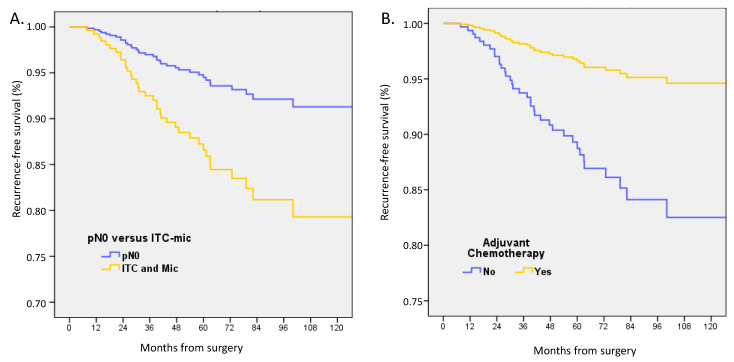
At a median follow-up of 59.8 months (mean 62.7), the following events had occurred: 22 deaths, 39 recurrences, 52 death or recurrences, and 16 metastases. In univariate analysis, pN status, pT size, type of surgery, ER status, and grade were non-statistically significantly associated with OS. LVI, age, and no ACt were significantly associated with decreased OS. In multivariate analysis adjusted on ACt, ER-status, age groups, LVI, pT size, and pN status (patients with pN0(i+) or pN1mi) were associated with decreased RFS (HR 2.548, CI 95% 1.06–6.13, p = 0.037) and patients with ACt had better RFS (HR 0.288, 0.13–0.65, p = 0.003) (Figure 1, Table 5). pN status was not associated with a difference in OS and DFS.
Figure 1.
Recurrence-free survival adjusted on ER status, age, LVI, and tumor size between (A) patients with pT1a-b pN0 or pN0(i+)/pN1mi tumors, and (B) with or without adjuvant chemotherapy.
Table 5.
Multivariate survival analyses for pT1a-b pN0 or pN0(i+) and pN1mi breast cancers.
| pT1a-b pN0 or pN0(i+) & pN1mi | Cox OS | Cox DFS | Cox RFS | |||||||
|---|---|---|---|---|---|---|---|---|---|---|
| HR | CI 95% | p | HR | CI 95% | p | HR | CI 95% | p | ||
| Age groups | ≤40 | 1 | 1 | 1 | ||||||
| 40.1–50 | 0.00–1306 | 0.969 | 0.387 | 0.15–1.03 | 0.057 | 0.418 | 0.15–1.15 | 0.090 | ||
| 50.1–74.9 | 4.134 | 0.42–40.7 | 0.224 | 0.445 | 0.20–0.98 | 0.043 | 0.336 | 0.14–0.80 | 0.014 | |
| ≥75 | 19.643 | 1.67–230.5 | 0.018 | 1.020 | 0.33–3.17 | 0.973 | 0.237 | 0.03–1.95 | 0.181 | |
| pT size groups | pT1a | 1 | 1 | 1 | ||||||
| pT1b | 1.940 | 0.72–5.20 | 0.187 | 1.718 | 0.94–3.13 | 0.077 | 1.747 | 0.87–3.53 | 0.120 | |
| LVI | No | 1 | 1 | 1 | ||||||
| Yes | 2.168 | 0.40–11.74 | 0.369 | 0.983 | 0.33–2.96 | 0.976 | 0.566 | 0.13–2.52 | 0.455 | |
| Subtype | ER-positive | 1 | 1 | 1 | ||||||
| ER-negative | 1.246 | 0.50–3.09 | 0.635 | 1.677 | 0.95–2.97 | 0.077 | 1.667 | 0.84–3.31 | 0.144 | |
| pN status | pN0 | 1 | 1 | 1 | ||||||
| pN0(i+)/pN1mi | 0.764 | 0.09–6.50 | 0.806 | 1.843 | 0.79–4.31 | 0.158 | 2.548 | 1.06–6.13 | 0.037 | |
| Chemotherapy | No | 1 | 1 | 1 | ||||||
| Yes | 0.259 | 0.07–0.97 | 0.046 | 0.272 | 0.13–0.56 | <0.0001 | 0.288 | 0.13–0.65 | 0.003 | |
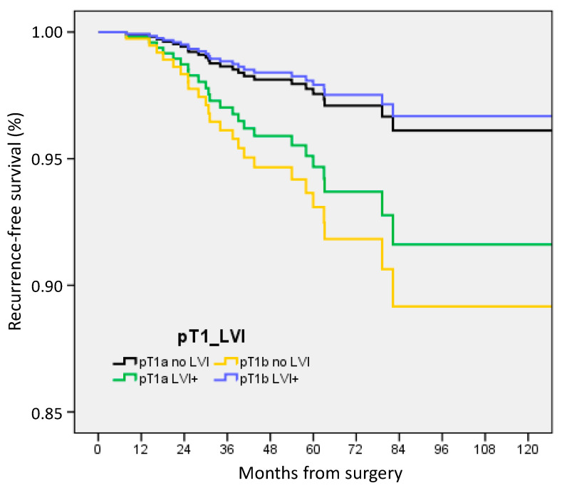
ACt was associated with pT and LVI status: 36% for pT1a/no LVI (45/125), 55.6% for pT1a/LVI+ (5/9), 61.2% for pT1b/no LVI (131/214), and 92.7% for pT1b/LVI+ (38/41) (p < 0.0001). In multivariate analysis, RFS was decreased in pT1b tumors without LVI (HR 2.893, CI 95% 1.09–7.67, p = 0.033), in patients without ACt (HR 3.758, 1.41–9.99, p = 0.008), in patients with ER-negative BC (HR 2.702, 1.10–6.63, p = 0.030), and patients with pN0(i+) and pN1mi (HR 2.780, 1.10–7.04, p = 0.031) (Figure 2). A better RFS was observed for patients over 50 and under 75 years (HR 0.189, 0.06–0.55, p = 0.002).
Figure 2.
Recurrence-free survival according to tumor size and nodal status in pT1a-b patients.
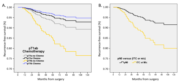
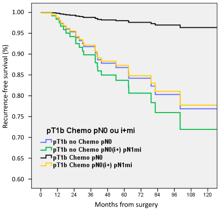
In multivariate analysis adjusted on LVI, ER status, age groups, pN status, and pT size with or without ACt, RFS was significantly decreased for pT1b BC without ACt (HR 2.365, 1.04–5.36, p = 0.039) and for pN0(i+) or pN1mi (HR 2.518, 1.03–6.14, p = 0.042) (Figure 3).
Figure 3.
Recurrence-free survival adjusted on LVI, ER status, age groups, pN status, and pT size in (A) patients with pT1a-b pN0 or pN0(i+)/pN1mi tumors according to adjuvant chemotherapy, and (B) in patients with pT1a-b tumors according to nodal status.
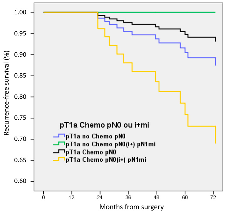
For patients with pT1a BC, no significant difference was observed between patients stratified according to ACt and pN status. Patients with pN0(i+) or pN1mi had a lower RFS but without a statistical difference (only nine patients in this later group) (Figure 4).
Figure 4.
Recurrence-free survival according to adjuvant chemotherapy and nodal status in pT1a patients.
For patients with pT1b BC, only patients with pN0 and ACt exhibit better RFS (Figure 5): HR 0.140, 0.04–0.48, p = 0.002 in comparison with patients with pN0 without ACt.
Figure 5.
Recurrence-free survival according to adjuvant chemotherapy and nodal status in pT1b patients.
4. Discussion
The main result of this study is the absence of prognostic value of pN0(i+) or pN1mi in HER2-positive BC in the whole cohort, but a negative impact on RFS in pT1a-b patients, particularly for those with pT1b tumors. Our results support the consideration of pN0(i+) and pN1mi status in the decision-making process when discussing trastuzumab-based ACt for these patients.
4.1. Incidence of pN0(i+) and pN1mi and Non-Sentinel Node (NSN) Involvement Rates
In a study reported by Reyal et al. [33], positive SN rates were 34.7% (439/1264) for ER-positive/HER2-negative tumors, 31.6% (25/79) for ER-positive/HER2-positive tumors, 41.5% (22/53) for ER-negative/HER2-positive tumors, and 20.4% (30/147) for triple-negative tumors, but without distinction between pN0(i+), pN1mi, and pN1-macro-metastases (Supplementary Table S3). In the French cohort of 12,572 patients with SLNB [40], pN1mi rate was 8% (n = 970) and pN0(i+) was 3% (n = 355) with 66% of pN0 (n = 8253) and 24% of pN-macro-metastases (n = 2994). By tumor subtypes, pN0(i+) and pN1mi rates were 11.08% (996/8988) and 34.72% (996/2869) among all patients and among patients with involved lymph nodes and Luminal-A-like tumors, 12.64% (149/1178) and 24.96% (149/597) among patients with Luminal-B-HER2-negative-like tumors, 8.88% (68/766) and 23.45% (68/290) among patients with ER-positive/HER2-positive tumors, 8.89% (40/450) and 19.42% (40/206) among patients with ER-negative/HER2-positive tumors, and 6.42% (70/1091) and 20.71% (70/338) among patients with triple-negative tumors. In the SERC trial [41] that includes patients with SN involvement, SN ITC was present in 5.91% of patients, micro-metastases in 28.12%, and macro-metastases in 65.97%. According to tumor phenotypes, ITC, micro-metastases, and macro-metastases rates were respectively 5.5% (75/1354), 27.5% (373/1354), and 66.9% (906/1354) among patients with ER-positive/HER2-negative tumors, 6.67% (9/135), 27.4% (37/135), and 65.9% (89/135) among patients with ER-positive/HER2-positive tumors, 7.3% (3/41), 24.4% (10/41), and 68.3% (28/41) among patients with ER-negative/HER2-positive tumors, and 8.2% (8/97), 29.9% (29/97), and 61.8% (60/97) among patients with triple-negative tumors. Crude rates of positive NSN according to SN status were 6.1% for patients with ITC (2/33), 10.3% for SN micro-metastases (22/214), and 25.7% for SN macro-metastases (134/522). Positive-NSN rates in multivariate analysis for patients with completion ALND were statistically significantly associated with ACt when performed after ALND (OR 2.99, p < 0.0001) in comparison to the absence of ACt and non-statistically significant when ACt was performed before ALND (OR 1.51, p = 0.232), in comparison with the absence of ACt use. No differences between ITC, micro-metastases, and macro-metastases SN status were reported.
In summary, there was no strong difference in ITC and micro-metastases rates and no strong difference in NSN rates, according to tumor subtypes. However, a strong downstaging on NSN involvement rate was reported when completion ALND was performed after ACt, whatever tumor subtype.
4.2. Prognostic Value of Micro-Metastases in Triple-Negative and ER-Positive/HER2-Negative Patients
Axillary lymph node involvement is a key prognostic feature in early TNBC when ITC and micro-metastases are identified. The independent prognostic features identified for DFS were tumor size ≥ 20 mm (HR = 1.86; 95% CI: 1.11–3.10, p = 0.018), LVI (HR = 1.69; 95% CI: 1.21–2.34, p = 0.002), and axillary lymph node involvement both in case of macro-metastases (HR = 1.97; 95% CI: 1.38–2.81, p < 0.0001) and occult metastases (HR = 1.72; 95% CI: 1.1–2.71, p = 0.019) [34]. In a recent study, [36] including 13,773 ER-positive/HER2-negative patients, with 546 pN0(i+) and 1446 pN1mi cases, LN micro-metastases had no detectable prognostic impact leading to the conclusion that they should not be considered as a determining factor in indicating ACt. This observation was also suggested in other recent studies [21,23,24].
Indication of ACt and Trastuzumab in HER2-Positive BC
Axillary micro-metastases do not impact the indication of adjuvant treatment with chemotherapy ± trastuzumab in HER2-positive and triple-negative tumors with pathologic size >2 cm, and even > 10 mm [42,43]. Neo-adjuvant chemotherapy is usually administered for patients with pT2 or pT1a-b-c N1 BC [37]. ACt after up-front surgery is indicated for patients with ≥ pT1c or pN1 macro-metastases but should also be discussed for patients with pT1a-b pN0 or with ITC or micro-metastases.
4.3. Available Literature on HER2-Positive, pT1ab BC
Adjuvant trastuzumab-based chemotherapy clearly improved survival in randomized clinical trials [44,45,46,47], but there were few or no patients with pT1abN0 stages and this specific situation was only addressed in relatively rare retrospective cohort studies [48,49,50,51,52,53]. In our study [42], ACt ± trastuzumab was associated with a significantly reduced risk of recurrence, including distant recurrence, in infra-centimetric node-negative HER2-positive BC and most of the benefit may have been driven by pT1b tumors.
No data were identified specifically for pT1a-b, HER2-positive, pN0(i+), or pN1mi in our literature search.
4.4. Negative Impact of LVI
A strong interaction between the presence of LVI and axillary lymph node involvement was reported whatever tumor subtypes [40] and both for ER-positive and ER-negative tumors [39]. In the present study, for patients with HER2-positive tumors and known LVI status, LVI was present in 33.2% (530/1594): 18.6% (170/915) of pN0, 43.8% (25/57) of pN0(i+), 46.0% (52/113) of pN1mi, and 55.6% (283/509) of pN1 macro-metastases.
In our recent study [39], LVI was present in 24% (4205/17,322) of patients and was most prevalent in patients with luminal B-like tumors, specifically high-grade HER2-negative tumors (44%)—22% (3279/14,655) of ER-positive and 35% (926/2667) of ER-negative tumors—(Supplementary Table S4). In the present study, LVI was present in 33.2% (530/1594) of HER2-positive BC—35.2% (189/537) of ER-positive and 32.3% (341/1056) of ER-negative tumors In the subgroup of pT1a-b BC, LVI was present in 10.5% (36/344) of pN0 BC and 31.1% (14/45) of pN0(i+) and pN1mi BC. The presence of LVI was statistically significantly associated with worse OS, DFS, and MFS in all patients, both with and without ACt and both for ER-positive and ER-negative tumors [39]. These findings are consistent with those of other recent studies that investigated the prognostic significance of LVI [46,47,48,49,50,51,52,53,54,55,56,57]. Moreover, Gujam et al., in a systematic review concluded that LVI was a powerful prognostic factor of poorer survival, whose impact was mainly seen in patients with node-negative BC [58]. The significance of LVI as an independent prognostic factor for patients with triple-negative tumors was also reported [34,35]. In the present study, LVI was also significantly associated with a negative impact on OS, DFS, RFS, and MFS in multivariate analysis for all HER2-positive patients.
4.4.1. Value of IHC Testing for ITC and Micro-Metastases in Triple-Negative and HER2-Positive BC
Detecting ITCs and micro-metastases is challenging due to their small size and limited presence in tissue samples. Conventional hematoxylin and eosin (HE) staining may lack the necessary sensitivity to accurately identify these subpopulations. Considering the prognosis significance of ITC and micro-metastases in triple-negative BC, it becomes crucial to employ more sensitive techniques, such as serial sections and IHC examination of axillary lymph nodes, to improve their detection and characterization. This is especially important for small-sized tumors without other criteria warranting ACt administration. Considering the negative impact of ITCs and micro-metastases on prognosis in our cohort, our findings suggest that the need for serial sections and IHC examination of axillary lymph nodes may also apply to HER2-positive pT1a-b breast cancer to assist in the decision-making process regarding ACt.
4.4.2. Limitations
There are several limitations to our study, including its retrospective design, lack of differentiation between the types of chemotherapy used, the need for a larger cohort to analyze the impact of ER-status and LVI on pT1a-b BC, and the inability to evaluate RFS specifically for pN0(i+) and pN1mi due to the limited number of patients in this subgroup. Moreover, we were not able to report the presence of ductal carcinoma in situ (DCIS). The clinical and biological significance of HER2 overexpression in patients with DCIS remains poorly defined, and current practice guidelines do not recommend HER2 testing in DCIS patients. Nevertheless, evidence suggests that HER2-positive DCIS cases may be associated with adverse clinicopathological parameters and increased recurrence rates [59].
5. Conclusions
Survival outcomes were not adversely affected by pN0(i+) and pN1mi in patients with HER2-positive breast cancer. Adjuvant systemic therapy was commonly used for patients with BC ≥ pT1c. However, in the case of small HER2-positive breast cancer (pT1a-b), a negative impact on RFS was observed specifically for patients with pN0(i+) and pN1mi axillary nodes, particularly among those with pT1b tumors. These findings support the need to consider pN0(i+) and pN1mi status in the decision-making process when discussing trastuzumab-based ACt for patients with pT1b HER2-positive breast cancer.
Supplementary Materials
The following supporting information can be downloaded at: https://www.mdpi.com/article/10.3390/cancers15184567/s1, Table S1: Regression analyses for adjuvant chemotherapy in all patients; Table S2: Characteristics of patients with pT1a-b tumors according to adjuvant chemotherapy: univariate analyses; Table S3: Positive SN rates in the studies mentioned in the discussion; Table S4: lymphovascular invasion rates according to endocrine receptor (ER) status in the studies mentioned in the discussion.
Author Contributions
Conceptualization, G.H., M.C. and A.d.N.; methodology, G.H., M.C. and A.d.N.; validation, G.H.; formal analysis, G.H.; resources, G.H., M.C., M.M., F.R., J.-M.C., M.-P.C., P.-E.C., M.H., E.J., P.G., A.-S.A., C.C., A.G. and A.d.N.; data curation, G.H., M.C. and A.d.N.; writing—original draft preparation, G.H., M.C. and A.d.N.; writing—review and editing, All authors; visualization, G.H. and A.d.N.; supervision, G.H. All authors have read and agreed to the published version of the manuscript.
Institutional Review Board Statement
The study was conducted in accordance with the Declaration of Helsinki, and approved by the strategic orientation committee of the Paoli-Calmettes Institute (ClinicalTrials.gov NCT02869607).
Informed Consent Statement
As this was a retrospective non-interventional study, no formal personal consent was required.
Data Availability Statement
The datasets used and/or analyzed during the current study available from the corresponding author on reasonable request.
Conflicts of Interest
The authors declare no conflict of interest.
Funding Statement
This research received no external funding.
Footnotes
Disclaimer/Publisher’s Note: The statements, opinions and data contained in all publications are solely those of the individual author(s) and contributor(s) and not of MDPI and/or the editor(s). MDPI and/or the editor(s) disclaim responsibility for any injury to people or property resulting from any ideas, methods, instructions or products referred to in the content.
References
- 1.Madsen E.V.E., Elias S.G., van Dalen T., van Oort P.M.P., van Gorp J., Gobardhan P.D., Bongers V. Predictive factors of isolated tumor cells and micrometastases in axillary lymph nodes in breast cancer. Breast. 2013;22:748–752. doi: 10.1016/j.breast.2012.12.013. [DOI] [PubMed] [Google Scholar]
- 2.Houvenaeghel G., Classe J.-M., Garbay J.-R., Giard S., Cohen M., Faure C., Hélène C., Belichard C., Uzan S., Hudry D., et al. Prognostic value of isolated tumor cells and micrometastases of lymph nodes in early-stage breast cancer: A French sentinel node multicenter cohort study. Breast. 2014;23:561–566. doi: 10.1016/j.breast.2014.04.004. [DOI] [PubMed] [Google Scholar]
- 3.Andersson Y., Bergkvist L., Frisell J., de Boniface J. Long-term breast cancer survival in relation to the metastatic tumor burden in axillary lymph nodes. Breast Cancer Res. Treat. 2018;171:359–369. doi: 10.1007/s10549-018-4820-0. [DOI] [PubMed] [Google Scholar]
- 4.Van Roozendaal L.M., Schipper R.J., Van de Vijver K.K.B.T., Haekens C.M., Lobbes M.B.I., Tjan-Heijnen V.C.G., de Boer M., Smidt M.L. The impact of the pathological lymph node status on adjuvant systemic treatment recommendations in clinically node negative breast cancer patients. Breast Cancer Res. Treat. 2014;143:469–476. doi: 10.1007/s10549-013-2822-5. [DOI] [PubMed] [Google Scholar]
- 5.Maibenco D.C., Dombi G.W., Kau T.Y., Severson R.K. Significance of micrometastases on the survival of women with T1 breast cancer. Cancer. 2006;107:1234–1239. doi: 10.1002/cncr.22112. [DOI] [PubMed] [Google Scholar]
- 6.Rovera F., Fachinetti A., Rausei S., Chiappa C., Lavazza M., Arlant V., Marelli M., Boni L., Dionigi G., Dionigi R. Prognostic role of micrometastases in sentinel lymph node in patients with invasive breast cancer. Int. J. Surg. 2013;11((Suppl. S1)):S73–S78. doi: 10.1016/S1743-9191(13)60022-9. [DOI] [PubMed] [Google Scholar]
- 7.Meattini I., Desideri I., Saieva C., Francolini G., Scotti V., Bonomo P., Greto D., Mangoni M., Nori J., Orzalesi L., et al. Impact of sentinel node tumor burden on outcome of invasive breast cancer patients. Eur. J. Surg. Oncol. J. Eur. Soc. Surg. Oncol. Br. Assoc. Surg. Oncol. 2014;40:1195–1202. doi: 10.1016/j.ejso.2014.08.471. [DOI] [PubMed] [Google Scholar]
- 8.Giuliano A.E., Hawes D., Ballman K.V., Whitworth P.W., Blumencranz P.W., Reintgen D.S., Morrow M., Leitch A.M., Hunt K.K., McCall L.M., et al. Association of occult metastases in sentinel lymph nodes and bone marrow with survival among women with early-stage invasive breast cancer. JAMA. 2011;306:385–393. doi: 10.1001/jama.2011.1034. [DOI] [PMC free article] [PubMed] [Google Scholar]
- 9.Yang Z.J., Yu Y., Hou X.W., Chi J.R., Ge J., Wang X., Cao X.C. The prognostic value of node status in different breast cancer subtypes. Oncotarget. 2017;8:4563–4571. doi: 10.18632/oncotarget.13943. [DOI] [PMC free article] [PubMed] [Google Scholar]
- 10.Gobardhan P.D., Elias S.G., Madsen E.V.E., van Wely B., van den Wildenberg F., Theunissen E.B.M., Ernst M.F., Kokke M.C., van der Pol C., Borel Rinkes I.H.M., et al. Prognostic value of lymph node micrometastases in breast cancer: A multicenter cohort study. Ann. Surg. Oncol. 2011;18:1657–1664. doi: 10.1245/s10434-010-1451-z. [DOI] [PMC free article] [PubMed] [Google Scholar]
- 11.Weaver D.L., Ashikaga T., Krag D.N., Skelly J.M., Anderson S.J., Harlow S.P., Julian T.B., Mamounas E.P., Wolmark N. Effect of occult metastases on survival in node-negative breast cancer. N. Engl. J. Med. 2011;364:412–421. doi: 10.1056/NEJMoa1008108. [DOI] [PMC free article] [PubMed] [Google Scholar]
- 12.Hansen N.M., Grube B., Ye X., Turner R.R., Brenner R.J., Sim M.-S., Giuliano A.E. Impact of micrometastases in the sentinel node of patients with invasive breast cancer. J. Clin. Oncol. Off. J. Am. Soc. Clin. Oncol. 2009;27:4679–4684. doi: 10.1200/JCO.2008.19.0686. [DOI] [PubMed] [Google Scholar]
- 13.Maaskant-Braat A.J., van de Poll-Franse L.V., Voogd A.C., Coebergh J.W.W., Roumen R.M., Nolthenius-Puylaert M.C.T., Nieuwenhuijzen G.A. Sentinel node micrometastases in breast cancer do not affect prognosis: A population-based study. Breast Cancer Res. Treat. 2011;127:195–203. doi: 10.1007/s10549-010-1086-6. [DOI] [PubMed] [Google Scholar]
- 14.Iqbal J., Ginsburg O., Giannakeas V., Rochon P.A., Semple J.L., Narod S.A. The impact of nodal micrometastasis on mortality among women with early-stage breast cancer. Breast Cancer Res. Treat. 2017;161:103–115. doi: 10.1007/s10549-016-4015-5. [DOI] [PubMed] [Google Scholar]
- 15.Truong P.T., Vinh-Hung V., Cserni G., Woodward W.A., Tai P., Vlastos G., Member of the International Nodal Ratio Working Group The number of positive nodes and the ratio of positive to excised nodes are significant predictors of survival in women with micrometastatic node-positive breast cancer. Eur. J. Cancer. 2008;44:1670–1677. doi: 10.1016/j.ejca.2008.05.011. [DOI] [PubMed] [Google Scholar]
- 16.Tan L.K., Giri D., Hummer A.J., Panageas K.S., Brogi E., Norton L., Hudis C., Borgen P.I., Cody H.S. Occult axillary node metastases in breast cancer are prognostically significant: Results in 368 node-negative patients with 20-year follow-up. J. Clin. Oncol. Off. J. Am. Soc. Clin. Oncol. 2008;26:1803–1809. doi: 10.1200/JCO.2007.12.6425. [DOI] [PubMed] [Google Scholar]
- 17.Kuijt G.P., Voogd A.C., van de Poll-Franse L.V., Scheijmans L.J.E.E., van Beek M.W.P.M., Roumen R.M.H. The prognostic significance of axillary lymph-node micrometastases in breast cancer patients. Eur. J. Surg. Oncol. J. Eur. Soc. Surg. Oncol. Br. Assoc. Surg. Oncol. 2005;31:500–505. doi: 10.1016/j.ejso.2005.01.001. [DOI] [PubMed] [Google Scholar]
- 18.Grabau D., Jensen M.B., Rank F., Blichert-Toft M. Axillary lymph node micrometastases in invasive breast cancer: National figures on incidence and overall survival. APMIS Acta Pathol. Microbiol. Immunol. Scand. 2007;115:828–837. doi: 10.1111/j.1600-0463.2007.apm_442.x. [DOI] [PubMed] [Google Scholar]
- 19.Cox C.E., Kiluk J.V., Riker A.I., Cox J.M., Allred N., Ramos D.C., Dupont E.L., Vrcel V., Diaz N., Boulware D. Significance of sentinel lymph node micrometastases in human breast cancer. J. Am. Coll. Surg. 2008;206:261–268. doi: 10.1016/j.jamcollsurg.2007.08.024. [DOI] [PubMed] [Google Scholar]
- 20.Andersson Y., Frisell J., Sylvan M., de Boniface J., Bergkvist L. Breast cancer survival in relation to the metastatic tumor burden in axillary lymph nodes. J. Clin. Oncol. Off. J. Am. Soc. Clin. Oncol. 2010;28:2868–2873. doi: 10.1200/JCO.2009.24.5001. [DOI] [PubMed] [Google Scholar]
- 21.Tsuda H. Histological examination of sentinel lymph nodes: Significance of macrometastasis, micrometastasis, and isolated tumor cells. Breast Cancer. 2015;22:221–229. doi: 10.1007/s12282-015-0588-9. [DOI] [PubMed] [Google Scholar]
- 22.Wada N., Imoto S. Clinical evidence of breast cancer micrometastasis in the era of sentinel node biopsy. Int. J. Clin. Oncol. 2008;13:24–32. doi: 10.1007/s10147-007-0736-0. [DOI] [PubMed] [Google Scholar]
- 23.De Boer M., van Deurzen C.H.M., van Dijck J.A.A.M., Borm G.F., van Diest P.J., Adang E.M.M., Nortier J.W.R., Rutgers E.J.T., Seynaeve C., Menke-Pluymers M.B.E., et al. Micrometastases or isolated tumor cells and the outcome of breast cancer. N. Engl. J. Med. 2009;361:653–663. doi: 10.1056/NEJMoa0904832. [DOI] [PubMed] [Google Scholar]
- 24.De Boer M., van Dijck J.A.A.M., Bult P., Borm G.F., Tjan-Heijnen V.C.G. Breast cancer prognosis and occult lymph node metastases, isolated tumor cells, and micrometastases. J. Natl. Cancer Inst. 2010;102:410–425. doi: 10.1093/jnci/djq008. [DOI] [PubMed] [Google Scholar]
- 25.Colleoni M., Rotmensz N., Peruzzotti G., Maisonneuve P., Mazzarol G., Pruneri G., Luini A., Intra M., Veronesi P., Galimberti V., et al. Size of breast cancer metastases in axillary lymph nodes: Clinical relevance of minimal lymph node involvement. J. Clin. Oncol. Off. J. Am. Soc. Clin. Oncol. 2005;23:1379–1389. doi: 10.1200/JCO.2005.07.094. [DOI] [PubMed] [Google Scholar]
- 26.Chen S.L., Hoehne F.M., Giuliano A.E. The prognostic significance of micrometastases in breast cancer: A SEER population-based analysis. Ann. Surg. Oncol. 2007;14:3378–3384. doi: 10.1245/s10434-007-9513-6. [DOI] [PubMed] [Google Scholar]
- 27.Salhab M., Patani N., Mokbel K. Sentinel lymph node micrometastasis in human breast cancer: An update. Surg. Oncol. 2011;20:e195–e206. doi: 10.1016/j.suronc.2011.06.006. [DOI] [PubMed] [Google Scholar]
- 28.Jafferbhoy S., McWilliams B. Clinical significance and management of sentinel node micrometastasis in invasive breast cancer. Clin. Breast Cancer. 2012;12:308–312. doi: 10.1016/j.clbc.2012.07.012. [DOI] [PubMed] [Google Scholar]
- 29.Cianfrocca M., Goldstein L.J. Prognostic and predictive factors in early-stage breast cancer. Oncologist. 2004;9:606–616. doi: 10.1634/theoncologist.9-6-606. [DOI] [PubMed] [Google Scholar]
- 30.Dowlatshahi K., Fan M., Anderson J.M., Bloom K.J. Occult metastases in sentinel nodes of 200 patients with operable breast cancer. Ann. Surg. Oncol. 2001;8:675–681. doi: 10.1007/s10434-001-0675-3. [DOI] [PubMed] [Google Scholar]
- 31.Perou C.M., Sørlie T., Eisen M.B., van de Rijn M., Jeffrey S.S., Rees C.A., Pollack J.R., Ross D.T., Johnsen H., Akslen L.A., et al. Molecular portraits of human breast tumours. Nature. 2000;406:747–752. doi: 10.1038/35021093. [DOI] [PubMed] [Google Scholar]
- 32.Cheang M.C.U., Voduc D., Bajdik C., Leung S., McKinney S., Chia S.K., Perou C.M., Nielsen T.O. Basal-like breast cancer defined by five biomarkers has superior prognostic value than triple-negative phenotype. Clin. Cancer Res. Off. J. Am. Assoc. Cancer Res. 2008;14:1368–1376. doi: 10.1158/1078-0432.CCR-07-1658. [DOI] [PubMed] [Google Scholar]
- 33.Reyal F., Rouzier R., Depont-Hazelzet B., Bollet M.A., Pierga J.-Y., Alran S., Salmon R.J., Fourchotte V., Vincent-Salomon A., Sastre-Garau X., et al. The Molecular Subtype Classification Is a Determinant of Sentinel Node Positivity in Early Breast Carcinoma. PLoS ONE. 2011;6:e20297. doi: 10.1371/journal.pone.0020297. [DOI] [PMC free article] [PubMed] [Google Scholar]
- 34.Houvenaeghel G., Sabatier R., Reyal F., Classe J.M., Giard S., Charitansky H., Rouzier R., Faure C., Garbay J.R., Daraï E., et al. Axillary lymph node micrometastases decrease triple-negative early breast cancer survival. Br. J. Cancer. 2016;115:1024–1031. doi: 10.1038/bjc.2016.283. [DOI] [PMC free article] [PubMed] [Google Scholar]
- 35.Sabatier R., Jacquemier J., Bertucci F., Esterni B., Finetti P., Azario F., Birnbaum D., Viens P., Gonçalves A., Extra J.-M. Peritumoural vascular invasion: A major determinant of triple-negative breast cancer outcome. Eur. J. Cancer. 2011;47:1537–1545. doi: 10.1016/j.ejca.2011.02.002. [DOI] [PubMed] [Google Scholar]
- 36.Houvenaeghel G., de Nonneville A., Cohen M., Chopin N., Coutant C., Reyal F., Mazouni C., Gimbergues P., Azuar A.-S., Chauvet M.-P., et al. Lack of prognostic impact of sentinel node micro-metastases in endocrine receptor-positive early breast cancer: Results from a large multicenter cohort. ESMO Open. 2021;6:100151. doi: 10.1016/j.esmoop.2021.100151. [DOI] [PMC free article] [PubMed] [Google Scholar]
- 37.Cardoso F., Kyriakides S., Ohno S., Penault-Llorca F., Poortmans P., Rubio I.T., Zackrisson S., Senkus E. Early breast cancer: ESMO Clinical Practice Guidelines for diagnosis, treatment and follow-up. Ann. Oncol. 2019;30:1194–1220. doi: 10.1093/annonc/mdz173. [DOI] [PubMed] [Google Scholar]
- 38.Houvenaeghel G., de Nonneville A., Cohen M., Viret F., Rua S., Sabiani L., Buttarelli M., Charaffe E., Monneur A., Jalaguier-Coudray A., et al. Neoadjuvant Chemotherapy for Breast Cancer: Evolution of Clinical Practice in a French Cancer Center Over 16 Years and Pathologic Response Rates According to Tumor Subtypes and Clinical Tumor Size: Retrospective Cohort Study. J. Surg. Res. 2022;5:511–525. doi: 10.26502/jsr.10020251. [DOI] [PMC free article] [PubMed] [Google Scholar]
- 39.Houvenaeghel G., Cohen M., Classe J.M., Reyal F., Mazouni C., Chopin N., Martinez A., Daraï E., Coutant C., Colombo P.E., et al. Lymphovascular invasion has a significant prognostic impact in patients with early breast cancer, results from a large, national, multicenter, retrospective cohort study. ESMO Open. 2021;6:100316. doi: 10.1016/j.esmoop.2021.100316. [DOI] [PMC free article] [PubMed] [Google Scholar]
- 40.Houvenaeghel G., Lambaudie E., Classe J.-M., Mazouni C., Giard S., Cohen M., Faure C., Charitansky H., Rouzier R., Daraï E., et al. Lymph node positivity in different early breast carcinoma phenotypes: A predictive model. BMC Cancer. 2019;19:45. doi: 10.1186/s12885-018-5227-3. [DOI] [PMC free article] [PubMed] [Google Scholar]
- 41.Houvenaeghel G., Cohen M., Raro P., De Troyer J., Gimbergues P., Tunon de Lara C., Ceccato V., Vaini-Cowen V., Faure-Virelizier C., Marchal F., et al. Sentinel node involvement with or without completion axillary lymph node dissection: Treatment and pathologic results of randomized SERC trial. NPJ Breast Cancer. 2021;7:133. doi: 10.1038/s41523-021-00336-3. [DOI] [PMC free article] [PubMed] [Google Scholar]
- 42.De Nonneville A., Gonçalves A., Zemmour C., Classe J.M., Cohen M., Lambaudie E., Reyal F., Scherer C., Muracciole X., Colombo P.E., et al. Benefit of adjuvant chemotherapy with or without trastuzumab in pT1ab node-negative human epidermal growth factor receptor 2-positive breast carcinomas: Results of a national multi-institutional study. Breast Cancer Res. Treat. 2017;162:307–316. doi: 10.1007/s10549-017-4136-5. [DOI] [PubMed] [Google Scholar]
- 43.De Nonneville A., Gonçalves A., Zemmour C., Cohen M., Classe J.M., Reyal F., Colombo P.E., Jouve E., Giard S., Barranger E., et al. Adjuvant chemotherapy in pT1ab node-negative triple-negative breast carcinomas: Results of a national multi-institutional retrospective study. Eur. J. Cancer. 2017;84:34–43. doi: 10.1016/j.ejca.2017.06.043. [DOI] [PubMed] [Google Scholar]
- 44.Romond E.H., Perez E.A., Bryant J., Suman V.J., Geyer C.E., Jr., Davidson N.E., Tan-Chiu E., Martino S., Paik S., Kaufman P.A., et al. Trastuzumab plus adjuvant chemotherapy for operable HER2-positive breast cancer. N. Engl. J. Med. 2005;353:1673–1684. doi: 10.1056/NEJMoa052122. [DOI] [PubMed] [Google Scholar]
- 45.Rodrigues M.J., Peron J., Frenel J.-S., Vano Y.-A., Wassermann J., Debled M., Picaud F., Albiges L., Vincent-Salomon A., Cottu P.H. Benefit of adjuvant trastuzumab-based chemotherapy in T1ab node-negative HER2-overexpressing breast carcinomas: A multicenter retrospective series. Ann. Oncol. 2013;24:916–924. doi: 10.1093/annonc/mds536. [DOI] [PubMed] [Google Scholar]
- 46.Liu Y.L., Saraf A., Lee S.M., Zhong X., Hibshoosh H., Kalinsky K., Connolly E.P. Lymphovascular invasion is an independent predictor of survival in breast cancer after neoadjuvant chemotherapy. Breast Cancer Res. Treat. 2016;157:555–564. doi: 10.1007/s10549-016-3837-5. [DOI] [PMC free article] [PubMed] [Google Scholar]
- 47.Kurozumi S., Joseph C., Sonbul S., Alsaeed S., Kariri Y., Aljohani A., Raafat S., Alsaleem M., Ogden A., Johnston S.J., et al. A key genomic subtype associated with lymphovascular invasion in invasive breast cancer. Br. J. Cancer. 2019;120:1129–1136. doi: 10.1038/s41416-019-0486-6. [DOI] [PMC free article] [PubMed] [Google Scholar]
- 48.He K.-W., Sun J.-J., Liu Z.-B., Zhuo P.-Y., Ma Q.-H., Liu Z.-Y., Yu Z.-Y. Prognostic significance of lymphatic vessel invasion diagnosed by D2-40 in Chinese invasive breast cancers. Medicine. 2017;96:e8490. doi: 10.1097/MD.0000000000008490. [DOI] [PMC free article] [PubMed] [Google Scholar]
- 49.Hwang K.-T., Kim Y.A., Kim J., Chu A.J., Chang J.H., Oh S.W., Hwang K.R., Chai Y.J. The influences of peritumoral lymphatic invasion and vascular invasion on the survival and recurrence according to the molecular subtypes of breast cancer. Breast Cancer Res. Treat. 2017;163:71–82. doi: 10.1007/s10549-017-4153-4. [DOI] [PubMed] [Google Scholar]
- 50.Klingen T.A., Chen Y., Stefansson I.M., Knutsvik G., Collett K., Abrahamsen A.L., Aase H., Aas H., Aas T., Wik E., et al. Tumour cell invasion into blood vessels is significantly related to breast cancer subtypes and decreased survival. J. Clin. Pathol. 2017;70:313–319. doi: 10.1136/jclinpath-2016-203861. [DOI] [PubMed] [Google Scholar]
- 51.Chas M., Boivin L., Arbion F., Jourdan M.-L., Body G., Ouldamer L. Clinicopathologic predictors of lymph node metastasis in breast cancer patients according to molecular subtype. J. Gynecol. Obstet. Hum. Reprod. 2018;47:9–15. doi: 10.1016/j.jogoh.2017.10.008. [DOI] [PubMed] [Google Scholar]
- 52.Invernizzi M., Corti C., Lopez G., Michelotti A., Despini L., Gambini D., Lorenzini D., Guerini-Rocco E., Maggi S., Noale M., et al. Lymphovascular invasion and extranodal tumour extension are risk indicators of breast cancer related lymphoedema: An observational retrospective study with long-term follow-up. BMC Cancer. 2018;18:935. doi: 10.1186/s12885-018-4851-2. [DOI] [PMC free article] [PubMed] [Google Scholar]
- 53.Liao G.-S., Hsu H.-M., Chu C.-H., Hong Z.-J., Fu C.-Y., Chou Y.-C., Golshan M., Dai M.-S., Chen T.-W., De-Chian C., et al. Prognostic Role of Lymphovascular Invasion and Lymph Node Status among Breast Cancer Subtypes. J. Med. Sci. 2018;38:54. doi: 10.4103/jmedsci.jmedsci_105_17. [DOI] [Google Scholar]
- 54.Ryu Y.J., Kang S.J., Cho J.S., Yoon J.H., Park M.H. Lymphovascular invasion can be better than pathologic complete response to predict prognosis in breast cancer treated with neoadjuvant chemotherapy. Medicine. 2018;97:e11647. doi: 10.1097/MD.0000000000011647. [DOI] [PMC free article] [PubMed] [Google Scholar]
- 55.Morkavuk Ş.B., Güner M., Çulcu S., Eroğlu A., Bayar S., Ünal A.E. Relationship between lymphovascular invasion and molecular subtypes in invasive breast cancer. Int. J. Clin. Pract. 2021;75:e13897. doi: 10.1111/ijcp.13897. [DOI] [PubMed] [Google Scholar]
- 56.Makower D., Lin J., Xue X., Sparano J.A. Lymphovascular invasion, race, and the 21-gene recurrence score in early estrogen receptor-positive breast cancer. NPJ Breast Cancer. 2021;7:20. doi: 10.1038/s41523-021-00231-x. [DOI] [PMC free article] [PubMed] [Google Scholar]
- 57.Mutai R., Goldvaser H., Shochat T., Peretz I., Sulkes A., Yerushalmi R. Prognostic Value of the Detection of Lymphovascular Invasion in Hormone Receptor-Positive Early Breast Cancer in the Era of Molecular Profiling. Oncology. 2019;96:14–24. doi: 10.1159/000492429. [DOI] [PubMed] [Google Scholar]
- 58.Gujam F.J.A., Going J.J., Edwards J., Mohammed Z.M.A., McMillan D.C. The role of lymphatic and blood vessel invasion in predicting survival and methods of detection in patients with primary operable breast cancer. Crit. Rev. Oncol. Hematol. 2014;89:231–241. doi: 10.1016/j.critrevonc.2013.08.014. [DOI] [PubMed] [Google Scholar]
- 59.Akrida I., Mulita F. The Clinical Significance of HER2 Expression in DCIS. Med. Oncol. 2022;40:16. doi: 10.1007/s12032-022-01876-9. [DOI] [PubMed] [Google Scholar]
Associated Data
This section collects any data citations, data availability statements, or supplementary materials included in this article.
Supplementary Materials
Data Availability Statement
The datasets used and/or analyzed during the current study available from the corresponding author on reasonable request.


