Abstract
The infected host deploys generalized oxidative stress caused by small inorganic reactive molecules as antibacterial weapons. An emerging consensus is that hydrogen sulfide (H2S) and forms of sulfur with sulfur-sulfur bonds termed reactive sulfur species (RSS) provide protection against oxidative stressors and antibiotics, as antioxidants. Here, we review our current understanding of RSS chemistry and its impact on bacterial physiology. We start by describing the basic chemistry of these reactive species and the experimental approaches developed to detect them in cells. We highlight the role of thiol persulfides in H2S-signaling and discuss three structural classes of ubiquitous RSS sensors that tightly regulate cellular H2S/RSS levels in bacteria, with a specific focus on the chemical specificity of these sensors.
Keywords: Reactive sulfur species, persulfide, persulfide sensor, transcriptional regulation, cysteine thiol, transpersulfidation
Introduction
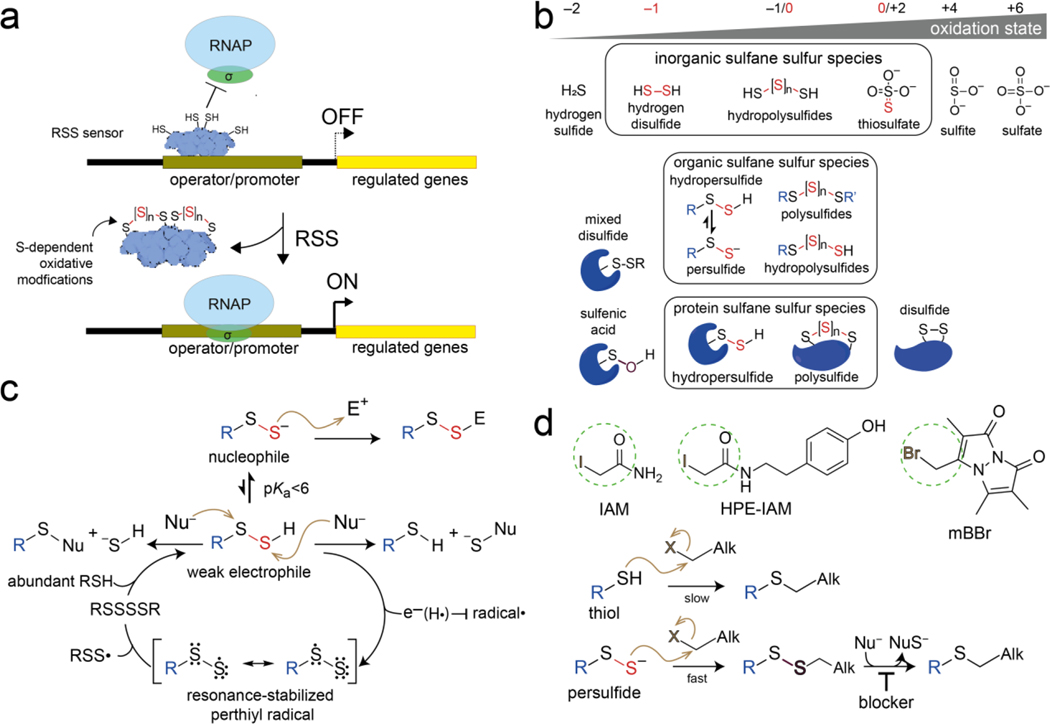
The innate immune system of the vertebrate host has many anti-bacterial weapons in a comprehensive arsenal of strategies designed to restrict or abrogate bacterial growth. These strategies often involve intoxication by highly reactive, deceptively simple inorganic molecules that include the chemically diverse reactive oxygen species (ROS), reactive nitrogen species (RNS) and reactive chlorine species (RCS), each of which possesses its own reactivity profile toward biomolecules. Our understanding of how these toxic species are sensed and detoxified in bacteria continues to grow, but often involves a DNA-binding transcriptional regulator that employs a cysteine thiol-based strategy to sense (react with) one or a small subset of these reactive species. This chemistry, in turn, drives transcriptional de-repression or activation of downstream genes, which encode enzymes tasked with clearing these molecules via cytoplasmic efflux or otherwise metabolize a specific reactive species into a less toxic product (Figure 1a). Reactive sulfur species (RSS) [1–3] are a relatively recent addition to the pantheon of highly reactive, small molecules and metabolites derived from the oxidation of hydrogen sulfide, H2S, and figure prominently in hydrogen sulfide signaling through protein persulfidation [4].
Figure 1.
(a) General regulatory strategy of a bacterial RSS-sensing repressor. (b) Various reactive sulfur species (RSS) with those molecules harboring a sulfur-bonded sulfur (sulfane sulfur) atoms boxed, and grouped into inorganic, organic (where R is a low molecular weight thiol) and proteinaceous species (top to bottom). Three generally reversible oxidative modifications on proteins are also shown. (c) Reactivity of an organic hydropersulfide toward nucleophiles (Nu–) and electrophiles (E+), with the relationship of the perthiyl radical with the persulfide shown. (d) Chemical structures of common alkylating agents (Alk) with the electrophilic moiety circled, used to profile thiols and persulfides in mixtures. A blocker is a functional group on Alk itself that prevents hydrolysis or nucleophilic (Nu–) attack and loss of the persulfide S atom [33,85].
Chemistry of reactive sulfur species
RSS are functionally dominated by species that contain sulfur-bonded or “sulfane” sulfur atoms and can be grouped into organic or inorganic subspecies where sulfur atoms are bonded covalently in chains only to other sulfur atoms [5]. These include the low molecular weight thiol (RSH) hydropersulfides (RSSH), hydropolysulfides (RS-Sn-H, n>1) and polysulfides (RS-Sn-SR’ n≥1), with their inorganic dihydrodisulfide and dihydropolysulfide counterparts, hydrogen disulfide (H2S2) and hydrogen polysulfide (H2Sn, n>2) (Figure 1b). Per- and polysulfides are effectively Janus (two-faced) molecules, where the S-S bond is electrophilic while the terminal proton is acidic, making the terminal sulfur strongly nucleophilic when deprotonated [6,7]. These features distinguish these particular RSS from parent H2S and thiols, dominating their reactivity, since H2S itself can only function as a reductant and cannot oxidize a thiol [8] (Figure 1c).
Indeed, hydropersulfides are excellent H-atom donors, far superior to the corresponding thiol, because of formation of the resonance-stabilized perthiyl radical (Figure 1c) [9]. This property makes hydropersulfides excellent radical scavengers, which may protect mammalian cells against ferroptosis [9,10]. Furthermore, the perthiyl radical self-recombines at diffusion-controlled rates to recreate the tetrasulfide species, which immediately regenerates the hydropersulfide upon reaction with another cell-abundant thiol species (Figure 1c) [9]. This rapid, substoichiometric production of organic thiol persulfides nicely explains why these species, while often present at only ≤0.1–1% of the corresponding thiol [2,11–13], are sensed by specialized RSS-sensing transcriptional regulators [14–17] tuned to respond to small changes in cellular RSS induced by endogenous or exogenous perturbation. These RSS can be quantified in cells using an electrophilic trapping strategy and isotope dilution liquid chromatography-tandem mass spectrometry (LC-MS/MS) in a sulfidomics profiling analysis [2,18]. It is now known that the measured speciation of RSS in cell lysates can be significantly impacted by polysulfide hydrolysis and the nature of the electrophilic trapping agent itself, with hydroxyphenyl-derivatized iodoacetamides now often used for this purpose [19,20] (Figure 1d).
Biogenesis of reactive sulfur species and reversible proteome persulfidation
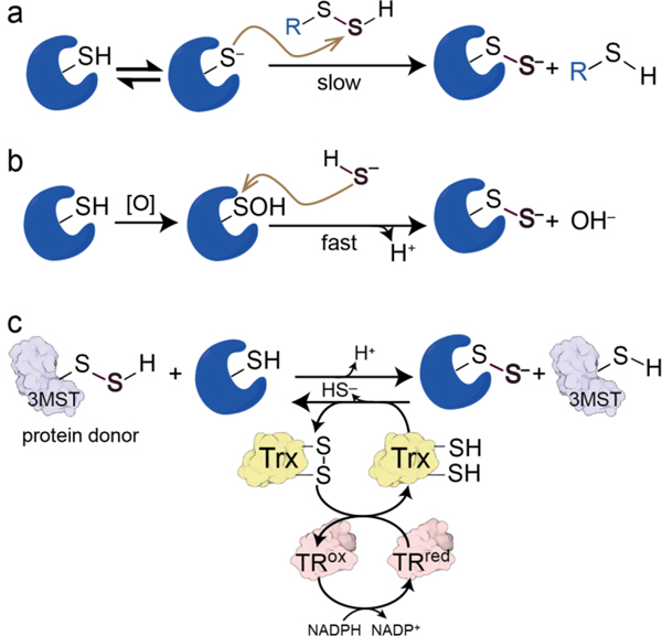
Hydropersulfides and polysulfides are formed endogenously from H2S by a number of mechanisms, including the flavin-dependent sulfide:quinone oxidoreductase (SQR; SQOR) [21] and/or heme-containing proteins [4,22,23]. Thus, when cells are exposed to exogenous H2S or produce endogenous H2S, RSS are formed leading to downstream reactions with both small molecules and proteins. Persulfidation, also known as S-sulfhydration and S-sulfuration, of proteome cysteine residues occurs in all kingdoms of life even under ambient growth conditions not stressed with exogenous Na2S or other sulfur donor [24–28]. The regulatory significance of this modification is a subject of intense debate [29], as is the mechanism by which these sulfur atoms are installed in the proteome. Non-enzymatic transpersulfidation by a low molecular weight (LMW) thiol per- or polysulfide donor (Figure 2a) may be somewhat slow since S-thiolation and the release of H2S may be the preferred reaction [4]. A more nuanced instance of non-enzymatic transpersulfidation (Figure 2a) is the persulfidation of coenzyme A (CoASH)- or acyl-CoA-requiring enzymes though poisoning by bound CoASSH [30], in a mechanism that parallels recently described transnitrosation of α-ketoacid dehydrogenase complex lipoyl arms by S-nitrosated CoA, SNO-CoA [31,32]. Direct attack of HS– on sulfenylated cysteines (Figure 2b) occurs in a monothiolate peroxiredoxin, but the extent to which this impacts global proteome persulfidation is unknown [33,34].
Figure 2.
Possible mechanisms of protein persulfidation and depersulfidation. (a) Transpersulfidation with the low molecular weight thiol hydropersulfide. A competing reaction, attack on the inner sulfur of the persulfide resulting in formation of the mixed disulfide and HS–, is not shown for clarity. Oxidation of cysteine to sulfenic acid by an oxidant, denoted [O], with subsequent attack by HS–. (c) Transpersulfidation by a persulfidated protein donor, for example, 3-MST. Trx, thioredoxin; TR, thioredoxin reductase. Similar chemistry can be performed by GR/Grx1 [24].
Recent work provides support for enzyme-catalyzed transpersulfidation by 3-mercaptopyruvate sulfurtransferase [35,36] and more broadly other enzymes that harbor long-lived thiol persulfides, e.g., cysteine desulfurase [37,38] or a canonical thiosulfate sulfurtransferase or rhodanese [39–41] (Figure 2c). Depersulfidation, or removal of persulfide groups from proteome thiols, is catalyzed by the thioredoxin-thioredoxin reductase systems found in all cells [24,27,42] (Figure 2c). In at least one case, in Staphylococcus aureus, two minor thioredoxins are found to be highly persulfidated in sulfide-stressed cells, and thioredoxin-profiling experiments suggests that depersulfidated client proteins do not strongly overlap, consistent with the idea that protein-protein interactions might impart some level of specificity in enzyme-catalyzed removal of proteome persulfide groups [25,43]. The generality of this finding is unknown.
RSS-sensing transcriptional regulators and H2S/RSS homeostasis
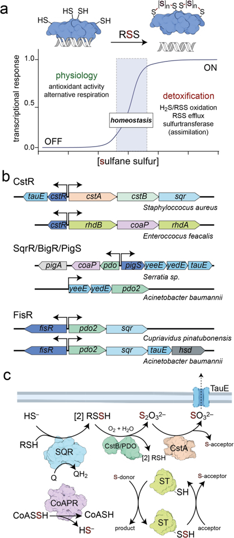
Best characterized bacterial RSS-sensing transcriptional regulators engage in persulfidation chemistry that leads to allosteric modulation of DNA binding or transcriptional activation (Figure 1a), and ultimately a change in the cellular abundance of enzymes encoded by downstream genes that oxidize H2S and reestablish H2S and RSS homeostasis (Figure 3a–b). We designate these RSS-sensing regulators as primary sensors of RSS, the action of which allows the cell to tightly regulate the intracellular (cytoplasmic) concentrations of these specific effector molecules [11,44,45]. Primary RSS sensor-regulated gene products include SQR [46], mononuclear, non-heme iron persulfide dioxygenase (PDO) [47,48], flavin-dependent coenzyme A persulfide reductase [30,49], various sulfurtransferases (ST) [39], and one or a number of membrane transporters, including the candidate sulfite exporter TauE [50] and thiosulfate importers YedE/YeeE [51,52] (Figure 3b–c). In some cases, these genes are regulated by more than one sensor that may or may not be responsive exclusively to RSS. For example, in A. baumannii pdo and tauE are regulated by a distinct regulator relative to genes encoding the importers YedE/YeeE [12] and in Bacillus licheniformis pdo and sqr are regulated by a two-component regulatory system, nreBC, while the expression of genes encoding the sulfur carriers seem to be regulated by another RSS-sensing CstR-family regulator (see below) [53].
Figure 3.
(a) Concept of RSS homeostasis under the transcriptional control of a primary RSS-sensing repressor, adapted from [86]. (b) A selection of operons known to be controlled by the indicated primary RSS sensor. coaP encodes coenzyme A persulfide reductase (CoAPR), while cstA and rhdA/rhdB are multidomain or single domain sulfurtransferases (ST), respectively. (c) Illustration of the chemical transformations carried out by the enzymes encoded by genes in panel (b). YeeE and YedE transporters, reported to bring thiosulfate into cells, are not shown [52].
In other cases, the sensing and detoxification of RSS by prototypical RSS-sensing regulators is linked to the production of secondary metabolites, including pigments and antibiotics in developmentally complex organisms. These include prodigiosin in Serratia spp. [51] and actinorhodin in Streptomyces coelicolor [45] (Figure 3b). The extent to which these regulators also contribute to RSS homeostasis or solely regulate other adaptive responses to increased RSS levels is not yet clear, thus their classification as primary sensors is based on the high level sequence and structural similarity to other primary RSS-sensing sensors. Moreover, why cellular RSS is linked to antibiotic production is not yet understood; however, it is well-established that bacterial resistance against antibiotics is enhanced (and can be selected for) by increasing endogenous H2S or thiol persulfide production [40,54]. Cysteine persulfide (CSSH) is, in fact, capable of ring-opening β-lactam (penicillin and carbapenem class) antibiotics to form carbothioic S-acids, but is unreactive toward other non-β-lactam classes [55], thus providing a compelling chemical rationale for linking RSS homeostasis to β-lactam resistance [56].
Secondary RSS sensors, in contrast, have a primary role distinct from H2S/RSS homeostasis, e.g., in ROS sensing and detoxification, or in virulence gene regulation, and generally tend to be global regulators that drive changes in complex developmental and morphogenesis programs. Secondary RSS sensors may have another specific, well-characterized input exemplified by the ubiquitous H2O2 sensors OxyR and PerR, where cysteine persulfidation is likely an acute phase (over) response to what is effectively a minor or even non-physiological stressor [57,58]. However, evidence continues to emerge that historically classified ROS-regulated enzymes, including peroxiredoxins and glutaredoxins, are capable of clearing excess H2S or RSS [34,59]; further, H2O2 can induce the upregulation of H2S and RSS in some bacteria [13], which leverages RSS as an effective scavenger of H2O2 [2].
Other secondary RSS sensors likely have multiple primary inputs; here, RSS sensing operates as a rheostat to augment or otherwise integrate a complex cellular response to a primary input or a range of inputs. One such example is the global regulator ScAdpA, which when persulfidated at a single conserved Cys in cells, upregulates adpA and AdpA target gene expression, including those required for actinorhodin biosynthesis and morphological differentiation [60]. Like ScAdpA, other global regulators can be detected as persulfidated in sulfide- or RSS-stressed cells and include the global virulence gene regulator MgrA in Staphylococcus aureus, the master biofilm regulator BfmR in Acinetobacter baumannii [12,25] and MexR and LasR, which regulate multidrug efflux [61–63] and quorum sensing [64], respectively, in Pseudomonas aeruginosa. These secondary sensors are not necessarily exclusively thiol-based sensors as implicated recently for candidate heme-based sensors in M. tuberculosis and B. licheniformis [53,65].
Structural classification and transpersulfidation chemistry of primary RSS sensors
All bacterial primary RSS-sensing regulators characterized thus far belong to one of three structurally unrelated protein families, consistent with the idea that adaptation to H2S toxicity arose at least three independent times during the course of evolution. They belong to the copper-sensitive operon repressor (CsoR), arsenic repressor (ArsR) and Fis superfamilies, with one or more sometimes encoded in a bacterial genome [16,66–68]. Each exploits dithiol chemistry to form either disulfide or polysulfide bridges between reactive cysteine residues.
CstR.
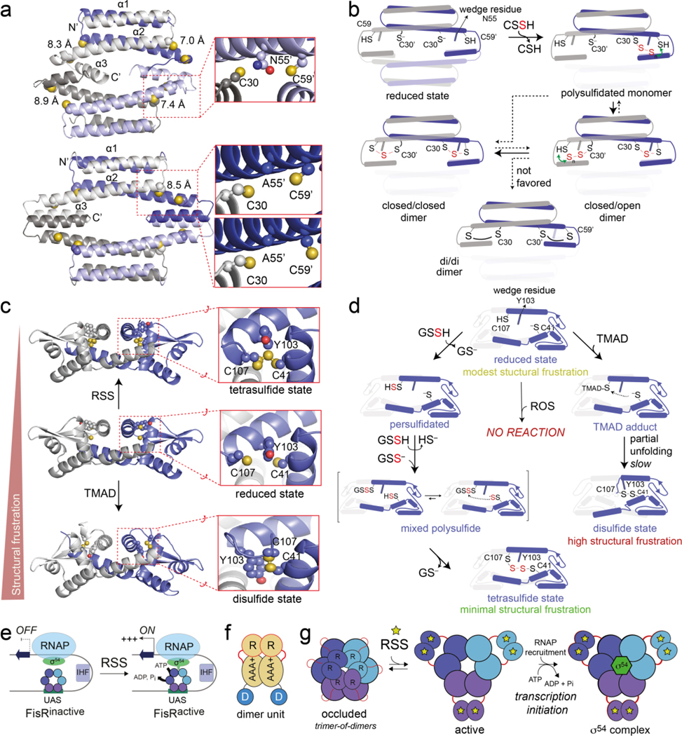
CstR (CsoR-like sulfurtransferase repressor), initially discovered in Staphylococcus aureus, is found largely in Gram-positive organisms (Firmicutes) [14,53,69]. The structure of pneumococcal CstR reveals an all-α-helical dimer-of-dimers quaternary structure, with the two Cys found on opposite subunits thus creating four peripheral dithiol sensing sites on the tetramer (Figure 4a). The two Cys in S. pneumoniae CstR (C30, C59’) are characterized by long intersubunit Sγ-Sγ distances (7–9 Å), mediated in part by N55, which is wedged between them (Figure 4a). This structure enhances the nucleophilicity of the N-terminal Cys (C30) relative to the structurally related copper sensor CsoR [69]. A mass spectrometry-based kinetic profiling method performed with a variety of oxidants, including CSSH [70], reveals a striking asymmetry of transpersulfidation within each CstR dimer unit, with one side of the dimer reacting and ultimately closing to a crosslinked product far faster than the opposite side. This asymmetry of reactivity is lost in a “wedge” mutant (N55A), as is much of the structural asymmetry in the tetramer itself [69] (Figure 4a). Although reactivity profiles of even closely related CstRs are distinct from one another, a per- or polysulfidated monomer is formed rapidly in all cases, which interconverts to the “singly-closed” (closed/open) and “doubly-closed” (closed/closed) dimers at various rates (Figure 4b). In no case does the “doubly-closed” disulfide product (di/di) accumulate when CSSH is the transpersulfidation donor, consistent with a general tendency of CstRs to form polysulfide-crosslinked linkages in vitro [69] and persulfidated products in sulfide-stressed cells [30]. CstR also reacts rapidly with H2O2 in vitro to form the di/di species, but sluggishly with GSSG, while retaining a strong asymmetry of reactivity with these non-native oxidants [69]. The lack of an H2O2-specific CsoR-family sensor prevents a detailed evaluation of the oxidant specificity of a bona fide RSS and ROS sensor in this structural class.
Figure 4.
(a) X-ray crystal structures of C9A (upper) and C9A/N55A S. pneumoniae CstRs, with dithiol sensing sites highlighted in the inset panels [69]. (b) A summary of the results of kinetically resolved native mass spectrometry-based profiling of reaction products when reduced CstR is incubated with a molar excess of CSSH. Only one of the dimer units of the CstR tetramer are shown in the foreground for clarity. These five states shown are not representative of discrete intermediates, but instead capture collections of structures that conform to the indicated trivial designation, e.g., “closed/closed” represents species that harbor tri- or tetrasulfide linkages on both sides of the dimer, not just the doubly trisulfidated species as shown [69,70]. (c) X-ray crystal structures of three distinct states of the RcSqrR dimer, with one of the two dithiol sensing sites highlighted in the expanded view [17]. (d) A cartoon summary of kinetically resolved native mass spectrometry-based reaction products when reduced SqrR is reacted with a molar excess of GSSH [17]. Reactivity of only one of the protomers of the SqrR dimer are shown in the foreground for clarity. (e) Generic cartoon of the regulatory mechanism of hexameric AAA+ σ54-dependent transcriptional activators like FisR. UAS, upstream activation sequence; IHF, integration host factor; RNAP, RNA polymerase. (f) The fundamental functional unit of FisR is a dimer, where R, AAA+ and D correspond to the N-terminal regulatory domain, the catalytic ATPase domain and the DNA binding domain, which engages the UAS, respectively. (g) One of a number of possible regulatory models for a FisR, with the nature of the RSS-sensing mechanism not broadly established, but in one case appears to involve transpersulfidation of the R domain directly [16].
SqrR and related ArsR-family sensors.
The prototypical ArsR-family persulfide sensor in many Gram-negative organisms is Rhodobacter capsulatus SqrR (sulfide:quinone reductase repressor), the master regulator of sulfide-dependent photosynthesis in this purple sulfur bacterium [15]. SqrR is representative of a family of very closely related ArsR subfamily members, now known to include Xylella fastidiosa and A. baumannii BigR (biofilm repressor), E. coli YgaV, Vibrio spp. HlyU and likely Serratia PigS [12,13,51,71,72]. It is interesting to note that unlike CstR-like repressors, many members of the RSS-sensing ArsR subfamily regulate a wider variety of genes related to exotoxin expression [13], and biofilm regulation [12]. ArsR proteins are characterized by a core α1-α2-α3-α4-β1-β2-α5 secondary structure, with the α3-α4 segment engaging successive major grooves in the DNA-bound state [73]. RSS-sensing ArsRs harbor a characteristic pair of cysteines in the α2 and α5 helices (C41, C107 in RcSqrR) that form an intraprotomer tetrasulfide bridge when presented with sulfane sulfur transpersulfidation donors, while showing no reaction with oxidants like H2O2 [15,17] (Figure 4c–d).
Crystallographic structures of RcSqrR in five functionally distinct states, coupled with kinetically-resolved reactivity profiling experiments, provide unprecedented insights into the mechanism of allosteric inhibition of DNA binding upon installation of a tetrasulfide crosslink and how these linkages are formed [17] (Figure 4c–d). As in CstRs, the two sensing Cys are quite far apart, mediated here by a “wedge” aromatic residue (Y103); unlike CstRs, the transpersulfidation reaction proceeds smoothly to the tetrasulfide product in RcSqrR, with some formation of a pentasulfide linkage in other RSS-sensing ArsR-family repressors, e.g., AbBigR and V. cholerae HylU [13,17]. Like in CstR, the disulfide-crosslinked species does not accumulate, nor is it a major on-pathway intermediate to the polysulfide product [17]. Two distinct RcSqrR structures obtained upon incubation with the disulfide-inducing electrophile diamide suggest a possible rationale for this. The ability to trap a monothiol S-N adduct between C107 and diamide suggests an energy barrier that slows closure to the disulfide, while inspection of the disulfide-crosslinked structure reveals a high degree of structural frustration, consistent with a higher global energy relative to the thiol-reduced and polysulfide-crosslinked states [17] (Figure 4d).
Remarkably, the global structures of the SqrR dimer in the DNA-binding-competent reduced and DNA-binding-inhibited tetrasulfide states are virtually identical, consistent with a dynamics-driven allosteric model [74]. Further, quantitative DNA binding experiments reveal that while formation of the disulfide is inhibitory to DNA binding, the tetrasulfide is inhibited to a greater degree [17]. Most importantly, SqrR and related repressors show no reaction with a conventional thiol disulfide or H2O2 and no evidence of even a transiently populated sulfenylated intermediate in the latter case, collectively highlighting the exquisite specificity of SqrR-like repressors for per- and polysulfide species, cognate oxidants that can only give rise to polysulfide products observed (Figure 4d) [13,17]. This behavior contrasts sharply with that of CstR [69]. Recent work suggests that CSSH is a more efficacious transpersulfidation donor than glutathione persulfide, GSSH, and more rapidly induces dissociation of RcSqrR from the DNA both in vitro and in cells [44]. The general significance of this finding is unknown but suggests that some degree of ligand specificity can be incorporated into even simple transpersulfidation reactions.
FisR.
The third major class of RSS sensor is exemplified by Cupriavidas spp. FisR [16]. FisR is a σ54-dependent transcriptional activator or bacterial enhancer-binding protein (bEBP), which activates transcription initiation via DNA looping by engaging σ54 thereby relieving the strong inhibition by σ 54 tightly bound to the promoter (Figure 4e–g). The basic functional unit of FisR is a dimer (Figure 4f), which is equilibrium with the hexamer, the functional assembly state (Figure 4g). FisRs are also reported to employ thiol transpersufidation chemistry in the N-terminal regulatory (R) domain to activate ATP hydrolysis and drive RNAP open complex formation from these stress responsive, σ54-dependent promoters [16]. The extent to which this persulfidation model characterizes other FisR activators is unknown, particularly given that the persulfidated cysteines in Cupriavidas FisR are not generally conserved; in fact, FisR is the primary RSS sensor in A. baumannii and lacks cysteines in the regulatory domain altogether and is not found to be persulfidated in cells, under conditions where SQR, a FisR-regulated PDO and the master regulator of biofilm formation, BfmR, are persulfidated [12,26]. The precise nature of the transcription activation signal in AbFisR remains unknown (Figure 4g).
Conclusions and open questions
The extent to which H2S/RSS homeostasis, polysulfide chemistry, sensing and signaling discussed here is harnessed by bacteria, to sustain an infection is not yet known. Our current understanding, however, points towards an important role of this chemistry for both for pathogens and commensal bacteria during infection, where specialized antioxidants like ergothioneine are now known to be deployed [75,76], and may be particularly relevant in the gastrointestinal tract (GIT). In this sulfur-rich, generally anaerobic chemosphere, taurocholic acid (TCA) accumulates in the gut upon infection [77]. Bile salt hydrolases cleave TCA, regenerating cholic acid (to induce bacterial membrane stress [78]) and taurine, which is metabolized by commensal microbiota to make hydrogen sulfide (H2S) [79]. H2S, in turn, limits re-colonization and minimizes inflammation associated with subsequent infections caused by enteric bacteria, including Klebsiella pneumoniae and Enterococcus faecalis [80].
For pathogens, studies on the beneficial aspects of the biogenesis of H2S and RSS are focused on antibiotic defenses and more recently, resistance to immune system killing and oxidative stress [54,81,82]. Here, H2S/polysulfide chemistry is viewed as complementary to other strategies that bacteria use to combat host oxidative stressors, as shown by the measurable virulence phenotypes obtained with ΔcstR strains in Staphylococcus aureus and Enterococcus faecalis [49,83]. Indeed, the development of inhibitors of 3-MST and CSE, in efforts to blunt bacterial H2S biogenesis, has emerged as a strategy to attenuate antibiotic resistance and biofilm formation, which may well involve the intermediacy of RSS sensing and signaling.
The three distinct regulatory strategies discussed here (Figure 4) appear to have evolved independently, with each RSS sensor a member of large superfamily of regulators that have evolved collectively to respond to a wide range of diverse signals [68]. While reactivity studies have focused on the transpersulfidation chemistry in vitro using a variety of small molecule sulfane sulfur donors, an important unanswered question is the nature of the transpersulfidation donor in cells, which might be protein-catalyzed by a persulfidase. This is suggested by the fact that these reaction rates are slow, even with a large molar excess of RSS, although not strongly attenuated in the DNA-bound state [13,17,44]. In addition, the response of changes in intracellular H2S and RSS speciation in cells by an RSS does not necessarily track with the longer-term elevation of cellular RSS, particularly when exogenous sulfide is used to “turn on” the regulon [25,44,49]. Thus, a protein catalyst might be responsible for reducing per- and polysulfide linkages on the regulator itself and might implicate a housekeeping or a specialized thioredoxin in this depersulfidase activity [24,43,84]. A better understanding of how persulfides are dynamically trafficked within and between cells as well as their regulatory potential are important areas for future study.
Acknowledgements
This work was supported by the National Institutes of Health (R35 GM118157) to DPG and MinCyT Argentina (PICT 2019-0011, 2019-3805) to DAC.
Footnotes
Declaration of competing interests
The authors declare no competing financial interests.
Declaration of interests
The authors declare that they have no known competing financial interests or personal relationships that could have appeared to influence the work reported in this paper.
This review comes from a themed issue on The Chemical biology and Sulfur and Selenium
Publisher's Disclaimer: This is a PDF file of an unedited manuscript that has been accepted for publication. As a service to our customers we are providing this early version of the manuscript. The manuscript will undergo copyediting, typesetting, and review of the resulting proof before it is published in its final form. Please note that during the production process errors may be discovered which could affect the content, and all legal disclaimers that apply to the journal pertain.
Data availability
Data will be made available upon request.
References
Papers of particular interest, published within the period of review, have been highlighted as: *, of special interest
**, of outstanding interest.
- 1.Lin VS, Chen W, Xian M, Chang CJ: Chemical probes for molecular imaging and detection of hydrogen sulfide and reactive sulfur species in biological systems. Chem Soc Rev 2014, 44:4596–4618. [DOI] [PMC free article] [PubMed] [Google Scholar]
- 2.Ida T, Sawa T, Ihara H, Tsuchiya Y, Watanabe Y, Kumagai Y, Suematsu M, Motohashi H, Fujii S, Matsunaga T, et al. : Reactive cysteine persulfides and S-polythiolation regulate oxidative stress and redox signaling. Proc Natl Acad Sci U S A 2014, 111:7606–7611. [DOI] [PMC free article] [PubMed] [Google Scholar]
- 3.Paulsen CE, Carroll KS: Cysteine-mediated redox signaling: Chemistry, biology, and tools for discovery. Chem Rev 2013, 113:4633–4679. [DOI] [PMC free article] [PubMed] [Google Scholar]
- 4.Filipovic MR, Zivanovic J, Alvarez B, Banerjee R: Chemical biology of H2S signaling through persulfidation. Chem Rev 2018, 118:1253–1337. [DOI] [PMC free article] [PubMed] [Google Scholar]
- 5.Iciek M, Bilska-Wilkosz A, Gorny M: Sulfane sulfur - new findings on an old topic. Acta Biochim Pol 2019, 66:533–544. [DOI] [PubMed] [Google Scholar]
- 6.Benchoam D, Semelak JA, Cuevasanta E, Mastrogiovanni M, Grassano JS, Ferrer-Sueta G, Zeida A, Trujillo M, Moller MN, Estrin DA, et al. : Acidity and nucleophilic reactivity of glutathione persulfide. J Biol Chem 2020, 295:15466–15481. [DOI] [PMC free article] [PubMed] [Google Scholar]
- 7.Cuevasanta E, Benchoam D, Semelak JA, Moller MN, Zeida A, Trujillo M, Alvarez B, Estrin DA: Possible molecular basis of the biochemical effects of cysteine-derived persulfides. Front Mol Biosci 2022, 9:975988. [DOI] [PMC free article] [PubMed] [Google Scholar]
- 8.Greiner R, Palinkas Z, Basell K, Becher D, Antelmann H, Nagy P, Dick TP: Polysulfides link H2S to protein thiol oxidation. Antioxid Redox Signal 2013, 19:1749–1765. [DOI] [PMC free article] [PubMed] [Google Scholar]
- 9. Barayeu U, Schilling D, Eid M, Xavier da Silva TN, Schlicker L, Mitreska N, Zapp C, Grater F, Miller AK, Kappl R, et al. : Hydropersulfides inhibit lipid peroxidation and ferroptosis by scavenging radicals. Nat Chem Biol 2023, 19:28–37. *One of two papers that connects the chemistry of hydropersulfides to inhibition of membrane lipid peroxidation.
- 10. Wu Z, Khodade VS, Chauvin JR, Rodriguez D, Toscano JP, Pratt DA: Hydropersulfides inhibit lipid peroxidation and protect cells from ferroptosis. J Am Chem Soc 2022, 144:15825–15837. *One of two papers that links the radical chemistry of thiol hydropersulfides with inhibition of membrane lipid peroxidation associated with ferroptosis.
- 11.Peng H, Shen J, Edmonds KA, Luebke JL, Hickey AK, Palmer LD, Chang FJ, Bruce KA, Kehl-Fie TE, Skaar EP, et al. : Sulfide homeostasis and nitroxyl intersect via formation of reactive sulfur species in Staphylococcus aureus. mSphere 2017, 2:e00082–00017. [DOI] [PMC free article] [PubMed] [Google Scholar]
- 12.Walsh BJC, Wang J, Edmonds KA, Palmer LD, Zhang Y, Trinidad JC, Skaar EP, Giedroc DP: The response of Acinetobacter baumannii to hydrogen sulfide reveals two independent persulfide-sensing systems and a connection to biofilm regulation. mBio 2020, 11:e01254–01220. [DOI] [PMC free article] [PubMed] [Google Scholar]
- 13. Pis Diez CM, Antelo GT, Dalia TN, Dalia AB, Giedroc DP, Capdevila DA: Increased intracellular persulfide levels attenuate HlyU-mediated hemolysin transcriptional activation in Vibrio cholerae. bioRxiv 2023, 10.1101/2023.03.13.532278. *First evidence of RSS dependent regulation of exotoxin expression in a gut pathogen.
- 14.Luebke JL, Shen J, Bruce KE, Kehl-Fie TE, Peng H, Skaar EP, Giedroc DP: The CsoR-like sulfurtransferase repressor (CstR) is a persulfide sensor in Staphylococcus aureus. Mol Microbiol 2014, 94:1343–1360. [DOI] [PMC free article] [PubMed] [Google Scholar]
- 15.Shimizu T, Shen J, Fang M, Zhang Y, Hori K, Trinidad JC, Bauer CE, Giedroc DP, Masuda S: Sulfide-responsive transcriptional repressor SqrR functions as a master regulator of sulfide-dependent photosynthesis. Proc Natl Acad Sci U S A 2017, 114:2355–2360. [DOI] [PMC free article] [PubMed] [Google Scholar]
- 16.Li H, Li J, Lu C, Xia Y, Xin Y, Liu H, Xun L, Liu H: FisR activates sigma54 -dependent transcription of sulfide-oxidizing genes in Cupriavidus pinatubonensis JMP134. Mol Microbiol 2017, 105:373–384. [DOI] [PubMed] [Google Scholar]
- 17. Capdevila DA, Walsh BJC, Zhang Y, Dietrich C, Gonzalez-Gutierrez G, Giedroc DP: Structural basis for persulfide-sensing specificity in a transcriptional regulator. Nat Chem Biol 2021, 17:65–70. **First detailed structural description of the mechanism of persulfide specificity in any RSS-sensing transcriptional regulator.
- 18.Sawa T, Ono K, Tsutsuki H, Zhang T, Ida T, Nishida M, Akaike T: Reactive cysteine persulphides: Occurrence, biosynthesis, antioxidant activity, methodologies, and bacterial persulphide signalling. Adv Microb Physiol 2018, 72:1–28. [DOI] [PubMed] [Google Scholar]
- 19.Sawa T, Takata T, Matsunaga T, Ihara H, Motohashi H, Akaike T: Chemical biology of reactive sulfur species: Hydrolysis-driven equilibrium of polysulfides as a determinant of physiological functions. Antioxid Redox Signal 2022, 36:327–336. [DOI] [PMC free article] [PubMed] [Google Scholar]
- 20.Zhang T, Ono K, Tsutsuki H, Ihara H, Islam W, Akaike T, Sawa T: Enhanced Cellular Polysulfides negatively regulate TLR4 signaling and mitigate lethal endotoxin shock. Cell Chem Biol 2019, 26:686–698. [DOI] [PubMed] [Google Scholar]
- 21.Landry AP, Ballou DP, Banerjee R: Hydrogen sulfide oxidation by sulfide quinone oxidoreductase. Chembiochem 2021, 22:949–960. [DOI] [PMC free article] [PubMed] [Google Scholar]
- 22.Nelp MT, Zheng V, Davis KM, Stiefel KJE, Groves JT: Potent activation of indoleamine 2,3-dioxygenase by polysulfides. J Am Chem Soc 2019, 141:15288–15300. [DOI] [PubMed] [Google Scholar]
- 23.Vitvitsky V, Miljkovic JL, Bostelaar T, Adhikari B, Yadav PK, Steiger AK, Torregrossa R, Pluth MD, Whiteman M, Banerjee R, et al. : Cytochrome c reduction by H2S potentiates sulfide signaling. ACS Chem Biol 2018, 13:2300–2307. [DOI] [PMC free article] [PubMed] [Google Scholar]
- 24.Dóka E, Pader I, Biro A, Johansson K, Cheng Q, Ballago K, Prigge JR, Pastor-Flores D, Dick TP, Schmidt EE, et al. : A novel persulfide detection method reveals protein persulfide- and polysulfide-reducing functions of thioredoxin and glutathione systems. Sci Adv 2016, 2:e1500968. [DOI] [PMC free article] [PubMed] [Google Scholar]
- 25.Peng H, Zhang Y, Palmer LD, Kehl-Fie TE, Skaar EP, Trinidad JC, Giedroc DP: Hydrogen Sulfide and reactive sulfur species impact proteome S-sulfhydration and global virulence regulation in Staphylococcus aureus. ACS Infect Dis 2017, 3:744–755. [DOI] [PMC free article] [PubMed] [Google Scholar]
- 26.Walsh BJC, Giedroc DP: Proteomics profiling of S-sulfurated proteins in Acinetobacter baumannii. Bio Protoc 2021, 11:e4000. [DOI] [PMC free article] [PubMed] [Google Scholar]
- 27.Wedmann R, Onderka C, Wei S, Szijarto IA, Miljkovic JL, Mitrovic A, Lange M, Savitsky S, Yadav PK, Torregrossa R, et al. : Improved tag-switch method reveals that thioredoxin acts as depersulfidase and controls the intracellular levels of protein persulfidation. Chem Sci 2016, 7:3414–3426. [DOI] [PMC free article] [PubMed] [Google Scholar]
- 28.Zivanovic J, Kouroussis E, Kohl JB, Adhikari B, Bursac B, Schott-Roux S, Petrovic D, Miljkovic JL, Thomas-Lopez D, Jung Y, et al. : Selective Persulfide detection reveals evolutionarily conserved antiaging effects of S-sulfhydration. Cell Metab 2019, 30:1152–1170 e1113. [DOI] [PMC free article] [PubMed] [Google Scholar]
- 29.Yadav V, Gao XH, Willard B, Hatzoglou M, Banerjee R, Kabil O: Hydrogen sulfide modulates eukaryotic translation initiation factor 2alpha (eIF2alpha) phosphorylation status in the integrated stress-response pathway. J Biol Chem 2017, 292:13143–13153. [DOI] [PMC free article] [PubMed] [Google Scholar]
- 30.Walsh BJC, Costa SS, Edmonds KA, Trinidad JC, Issoglio FM, Brito JA, Giedroc DP: Metabolic and structural insights into hydrogen sulfide mis-regulation in Enterococcus faecalis. Antioxidants 2022, 11: 1607. [DOI] [PMC free article] [PubMed] [Google Scholar]
- 31.Anand P, Hausladen A, Wang YJ, Zhang GF, Stomberski C, Brunengraber H, Hess DT, Stamler JS: Identification of S-nitroso-CoA reductases that regulate protein S-nitrosylation. Proc Natl Acad Sci U S A 2014, 111:18572–18577. [DOI] [PMC free article] [PubMed] [Google Scholar]
- 32.Seim GL, John SV, Arp NL, Fang Z, Pagliarini DJ, Fan J: Nitric oxide-driven modifications of lipoic arm inhibit alpha-ketoacid dehydrogenases. Nat Chem Biol 2023, 19:265–274. [DOI] [PMC free article] [PubMed] [Google Scholar]
- 33.Schilling D, Barayeu U, Steimbach RR, Talwar D, Miller AK, Dick TP: Commonly used alkylating agents limit persulfide detection by converting protein persulfides into thioethers. Angew Chem Int Ed Engl 2022, 61:e202203684. [DOI] [PMC free article] [PubMed] [Google Scholar]
- 34.Cuevasanta E, Reyes AM, Zeida A, Mastrogiovanni M, De Armas MI, Radi R, Alvarez B, Trujillo M: Kinetics of formation and reactivity of the persulfide in the one-cysteine peroxiredoxin from Mycobacterium tuberculosis. J Biol Chem 2019, 294:13593–13605. [DOI] [PMC free article] [PubMed] [Google Scholar]
- 35. Pedre B, Talwar D, Barayeu U, Schilling D, Luzarowski M, Sokolowski M, Glatt S, Dick TP: 3-Mercaptopyruvate sulfur transferase is a protein persulfidase. Nat Chem Biol 2023. *First report to demonstrate that a persulfidated protein functions as global proteome persulfidation donor in cells.
- 36.Moseler A, Dhalleine T, Rouhier N, Couturier J: Arabidopsis thaliana 3-mercaptopyruvate sulfurtransferases interact with and are protected by reducing systems. J Biol Chem 2021, 296:100429. [DOI] [PMC free article] [PubMed] [Google Scholar]
- 37.Higgins KA, Peng H, Luebke JL, Chang FM, Giedroc DP: Conformational analysis and chemical reactivity of the multidomain sulfurtransferase, Staphylococcus aureus CstA. Biochemistry 2015, 54:2385–2398. [DOI] [PubMed] [Google Scholar]
- 38.Selles B, Moseler A, Caubriere D, Sun SK, Ziesel M, Dhalleine T, Heriche M, Wirtz M,Rouhier N, Couturier J: The cytosolic Arabidopsis thaliana cysteine desulfurase ABA3 delivers sulfur to the sulfurtransferase STR18. J Biol Chem 2022:101749. [DOI] [PMC free article] [PubMed] [Google Scholar]
- 39. Ran M, Li Q, Xin Y, Ma S, Zhao R, Wang M, Xun L, Xia Y: Rhodaneses minimize the accumulation of cellular sulfane sulfur to avoid disulfide stress during sulfide oxidation in bacteria. Redox Biol 2022, 53:102345. *First direct demonstration that rhodanases reduce intracellular sulfane sulfur levels in E. coli.
- 40.Luhachack L, Rasouly A, Shamovsky I, Nudler E: Transcription factor YcjW controls the emergency H2S production in E. coli. Nat Commun 2019, 10:2868. [DOI] [PMC free article] [PubMed] [Google Scholar]
- 41.Libiad M, Motl N, Akey DL, Sakamoto N, Fearon ER, Smith JL, Banerjee R: Thiosulfate sulfurtransferase-like domain-containing 1 protein interacts with thioredoxin. J Biol Chem 2018, 293:2675–2686. [DOI] [PMC free article] [PubMed] [Google Scholar]
- 42.Zheng C, Guo S, Tennant WG, Pradhan PK, Black KA, Dos Santos PC: The thioredoxin system reduces protein persulfide intermediates formed during the synthesis of thiocofactors in Bacillus subtilis. Biochemistry 2019, 58:1892–1904. [DOI] [PubMed] [Google Scholar]
- 43.Peng H, Zhang Y, Trinidad JC, Giedroc DP: Thioredoxin profiling of multiple thioredoxin-like proteins in Staphylococcus aureus. Front Microbiol 2018, 9:2385. [DOI] [PMC free article] [PubMed] [Google Scholar]
- 44.Shimizu T, Ida T, Antelo GT, Ihara Y, Fakhoury JN, Masuda S, Giedroc DP, Akaike T,Capdevila DA, Masuda T: Polysulfide metabolizing enzymes influence SqrR-mediated sulfide-induced transcription by impacting intracellular polysulfide dynamics. PNAS Nexus 2023, 2:pgad048. [DOI] [PMC free article] [PubMed] [Google Scholar]
- 45.Lu T, Cao Q, Pang X, Xia Y, Xun L, Liu H: Sulfane sulfur-activated actinorhodin production and sporulation is maintained by a natural gene circuit in Streptomyces coelicolor. Microb Biotechnol 2020, 13:1917–1932. [DOI] [PMC free article] [PubMed] [Google Scholar]
- 46.Zhang X, Xin Y, Chen Z, Xia Y, Xun L, Liu H: Sulfide-quinone oxidoreductase is required for cysteine synthesis and indispensable to mitochondrial health. Redox Biol 2021, 47:102169. [DOI] [PMC free article] [PubMed] [Google Scholar]
- 47.Motl N, Skiba MA, Kabil O, Smith JL, Banerjee R: Structural and biochemical analyses indicate that a bacterial persulfide dioxygenase-rhodanese fusion protein functions in sulfur assimilation. J Biol Chem 2017, 292:14026–14038. [DOI] [PMC free article] [PubMed] [Google Scholar]
- 48.Pettinati I, Brem J, McDonough MA, Schofield CJ: Crystal structure of human persulfide dioxygenase: Structural basis of ethylmalonic encephalopathy. Hum Mol Genet 2015, 24:2458–2469. [DOI] [PMC free article] [PubMed] [Google Scholar]
- 49.Shen J, Walsh BJC, Flores-Mireles AL, Peng H, Zhang Y, Zhang Y, Trinidad JC, Hultgren SJ, Giedroc DP: Hydrogen sulfide sensing through reactive sulfur species (RSS) and nitroxyl (HNO) in Enterococcus faecalis. ACS Chem Biol 2018, 13:1610–1620. [DOI] [PMC free article] [PubMed] [Google Scholar]
- 50.Weinitschke S, Denger K, Cook AM, Smits TH: The DUF81 protein TauE in Cupriavidus necator H16, a sulfite exporter in the metabolism of C2 sulfonates. Microbiology 2007, 153:3055–3060. [DOI] [PubMed] [Google Scholar]
- 51.Gristwood T, McNeil MB, Clulow JS, Salmond GP, Fineran PC: PigS and PigP regulate prodigiosin biosynthesis in Serratia via differential control of divergent operons, which include predicted transporters of sulfur-containing molecules. J Bacteriol 2011, 193:1076–1085. [DOI] [PMC free article] [PubMed] [Google Scholar]
- 52.Tanaka Y, Yoshikaie K, Takeuchi A, Ichikawa M, Mori T, Uchino S, Sugano Y, Hakoshima T, Takagi H, Nonaka G, et al.: Crystal structure of a YeeE/YedE family protein engaged in thiosulfate uptake. Sci Adv 2020, 6:eaba7637. [DOI] [PMC free article] [PubMed] [Google Scholar]
- 53.Tang C, Li J, Shen Y, Liu M, Liu H, Liu H, Xun L, Xia Y: A sulfide-sensor and a sulfane sulfur-sensor collectively regulate sulfur-oxidation for feather degradation by Bacillus licheniformis. Commun Biol 2023, 6:167. [DOI] [PMC free article] [PubMed] [Google Scholar]
- 54. Shatalin K, Nuthanakanti A, Kaushik A, Shishov D, Peselis A, Shamovsky I, Pani B, Lechpammer M, Vasilyev N, Shatalina E, et al. : Inhibitors of bacterial H2S biogenesis targeting antibiotic resistance and tolerance. Science 2021, 372:1169–1175. **A recent report that provides support for the idea that targeting of bacterial hydrogen sulfide biogenesis can be used as a strategy to attenuate widespread antimicrobial resistance.
- 55. Ono K, Kitamura Y, Zhang T, Tsutsuki H, Rahman A, Ihara T, Akaike T, Sawa T: Cysteine hydropersulfide inactivates beta-lactam antibiotics with formation of ring-opened carbothioic S-acids in bacteria. ACS Chem Biol 2021, 16:731–739. *First direct evidence of the chemical rationale linking RSS homeostasis to β-lactam resistance.
- 56.Shen J, Keithly ME, Armstrong RN, Higgins KA, Edmonds KA, Giedroc DP: Staphylococcus aureus CstB Is a novel multidomain persulfide dioxygenase-sulfurtransferase involved in hydrogen sulfide detoxification. Biochemistry 2015, 54:4542–4554. [DOI] [PMC free article] [PubMed] [Google Scholar]
- 57.Hou N, Yan Z, Fan K, Li H, Zhao R, Xia Y, Xun L, Liu H: OxyR senses sulfane sulfur and activates the genes for its removal in Escherichia coli. Redox Biol 2019, 26:101293. [DOI] [PMC free article] [PubMed] [Google Scholar]
- 58.Liu D, Song H, Li Y, Huang R, Liu H, Tang K, Jiao N, Liu J: The transcriptional repressor PerR senses sulfane sulfur by cysteine persulfidation at the structural Zn(2+) site in Synechococcus sp. PCC7002. Antioxidants 2023, 12: 423. [DOI] [PMC free article] [PubMed] [Google Scholar]
- 59.Liu D, Chen J, Wang Y, Meng Y, Li Y, Huang R, Xia Y, Liu H, Jiao N, Xun L, et al. : Synechococcus sp. PCC7002 uses peroxiredoxin to cope with reactive sulfur species stress. mBio 2022, 13:e0103922. [DOI] [PMC free article] [PubMed] [Google Scholar]
- 60.Lu T, Wu X, Cao Q, Xia Y, Xun L, Liu H: Sulfane sulfur posttranslationally modifies the global regulator AdpA to influence actinorhodin production and morphological differentiation of Streptomyces coelicolor. mBio 2022, 13:e0386221. [DOI] [PMC free article] [PubMed] [Google Scholar]
- 61.Chen H, Hu J, Chen PR, Lan L, Li Z, Hicks LM, Dinner AR, He C: The Pseudomonas aeruginosa multidrug efflux regulator MexR uses an oxidation-sensing mechanism. Proc Natl Acad Sci U S A 2008, 105:13586–13591. [DOI] [PMC free article] [PubMed] [Google Scholar]
- 62.Chen H, Yi C, Zhang J, Zhang W, Ge Z, Yang CG, He C: Structural insight into the oxidation-sensing mechanism of the antibiotic resistance of regulator MexR. EMBO Rep 2010, 11:685–690. [DOI] [PMC free article] [PubMed] [Google Scholar]
- 63.Xuan G, Lu C, Xu H, Chen Z, Li K, Liu H, Xia Y, Xun L: Sulfane sulfur is an intrinsic signal activating MexR-regulated antibiotic resistance in Pseudomonas aeruginosa. Mol Microbiol 2020, 114:1038–1048. [DOI] [PubMed] [Google Scholar]
- 64.Xuan G, Lv C, Xu H, Li K, Liu H, Xia Y, Xun L: Sulfane sulfur regulates LasR-mediated quorum sensing and virulence in Pseudomonas aeruginosa PAO1. Antioxidants 2021, 10: 1498 [DOI] [PMC free article] [PubMed] [Google Scholar]
- 65.Sevalkar RR, Glasgow JN, Pettinati M, Marti MA, Reddy VP, Basu S, Alipour E, Kim-Shapiro DB, Estrin DA, Lancaster JR Jr., et al. : Mycobacterium tuberculosis DosS binds H2S through its Fe3+ heme iron to regulate the DosR dormancy regulon. Redox Biol 2022, 52:102316. [DOI] [PMC free article] [PubMed] [Google Scholar]
- 66.Higgins KA, Giedroc D: Insights into protein Aalostery in the CsoR/RcnR family of transcriptional repressors. Chem Lett 2014, 43:20–25. [DOI] [PMC free article] [PubMed] [Google Scholar]
- 67.Liu T, Ramesh A, Ma Z, Ward SK, Zhang L, George GN, Talaat AM, Sacchettini JC, Giedroc DP: CsoR is a novel Mycobacterium tuberculosis copper-sensing transcriptional regulator. Nat Chem Biol 2007, 3:60–68. [DOI] [PubMed] [Google Scholar]
- 68.Capdevila DA, Edmonds KA, Giedroc DP: Metallochaperones and metalloregulation in bacteria. Essays Biochem 2017, 61:177–200. [DOI] [PMC free article] [PubMed] [Google Scholar]
- 69. Fakhoury JN, Zhang Y, Edmonds KA, Bringas M, Luebke JL, Gonzalez-Gutierrez G, Capdevila DA, Giedroc DP: Functional asymmetry and chemical reactivity of CsoR family persulfide sensors. Nucleic Acids Res 2021, 49:12556–12576. **First structural analysis of the CsoR-family persulfide sensor CstR placed into the broader context of evolutionarily-related CsoR-family sensors.
- 70.Fakhoury JN, Capdevila DA, Giedroc DP: Protocol for using organic persulfides to measure the chemical reactivity of persulfide sensors. STAR Protoc 2022, 3:101424. [DOI] [PMC free article] [PubMed] [Google Scholar]
- 71.Guimarães BG, Barbosa RL, Soprano AS, Campos BM, De Souza Ta, Tonoli CCC, Leme AFP, Murakami MT, Benedetti CE: Plant pathogenic bacteria utilize biofilm growth-associated repressor (BigR), a novel winged-helix redox switch, to control hydrogen sulfide detoxification under hypoxia. J Biol Chem 2011, 286:26148–26157. [DOI] [PMC free article] [PubMed] [Google Scholar]
- 72.Balasubramanian R, Hori K, Shimizu T, Kasamatsu S, Okamura K, Tanaka K, Ihara H, Masuda S: The sulfide-responsive SqrR/BigR homologous regulator YgaV of Escherichia coli controls expression of anaerobic respiratory genes and antibiotic tolerance. Antioxidants 2022, 11: 2359. [DOI] [PMC free article] [PubMed] [Google Scholar]
- 73.Arunkumar AI, Campanello GC, Giedroc DP: Solution structure of a paradigm ArsR family zinc sensor in the DNA-bound state. Proc Natl Acad Sci U S A 2009, 106:18177–18182. [DOI] [PMC free article] [PubMed] [Google Scholar]
- 74.Capdevila DA, Braymer JJ, Edmonds KA, Wu H, Giedroc DP: Entropy redistribution controls allostery in a metalloregulatory protein. Proc Natl Acad Sci U S A 2017, 114:4424–4429. [DOI] [PMC free article] [PubMed] [Google Scholar]
- 75.Zhang Y, Gonzalez-Gutierrez G, Legg KA, Walsh BJC, Pis Diez CM, Edmonds KA, Giedroc DP: Discovery and structure of a widespread bacterial ABC transporter specific for ergothioneine. Nat Commun 2022, 13:7586. [DOI] [PMC free article] [PubMed] [Google Scholar]
- 76.Dumitrescu DG, Gordon EM, Kovalyova Y, Seminara AB, Duncan-Lowey B, Forster ER, Zhou W, Booth CJ, Shen A, Kranzusch PJ, et al. : A microbial transporter of the dietary antioxidant ergothioneine. Cell 2022, 185:4526–4540. [DOI] [PMC free article] [PubMed] [Google Scholar]
- 77.Wexler AG, Guiberson ER, Beavers WN, Shupe JA, Washington MK, Lacy DB, Caprioli RM, Spraggins JM, Skaar EP: Clostridioides difficile infection induces a rapid influx of bile acids into the gut during colonization of the host. Cell Rep 2021, 36:109683. [DOI] [PMC free article] [PubMed] [Google Scholar]
- 78.Singla P, Kaur M, Kumari A, Kumari L, Pawar SV, Singh R, Salunke DB: Facially amphiphilic cholic acid-lysine conjugates as promising antimicrobials. ACS Omega 2020, 5:3952–3963. [DOI] [PMC free article] [PubMed] [Google Scholar]
- 79.Peck SC, Denger K, Burrichter A, Irwin SM, Balskus EP, Schleheck D: A glycyl radical enzyme enables hydrogen sulfide production by the human intestinal bacterium Bilophila wadsworthia. Proc Natl Acad Sci U S A 2019, 116:3171–3176. [DOI] [PMC free article] [PubMed] [Google Scholar]
- 80. Stacy A, Andrade-Oliveira V, McCulloch JA, Hild B, Oh JH, Perez-Chaparro PJ, Sim CK, Lim AI, Link VM, Enamorado M, et al. : Infection trains the host for microbiota-enhanced resistance to pathogens. Cell 2021, 184:615–627. *This report reveals that an infected host of prior infections enhances resistance to subsequent infections by utilizing the sulfonic acid taurine to provide this resistance.
- 81.Toliver-Kinsky T, Cui W, Toro G, Lee SJ, Shatalin K, Nudler E, Szabo C: H2S, a Bacterial Defense Mechanism against the Host Immune Response. Infect Immun 2019, 87:e00272–00218. [DOI] [PMC free article] [PubMed] [Google Scholar]
- 82.Mironov A, Seregina T, Nagornykh M, Luhachack LG, Korolkova N, Lopes LE, Kotova V, Zavilgelsky G, Shakulov R, Shatalin K, et al. : Mechanism of H2S-mediated protection against oxidative stress in Escherichia coli. Proc Natl Acad Sci U S A 2017, 114:6022–6027. [DOI] [PMC free article] [PubMed] [Google Scholar]
- 83.Grossoehme N, Kehl-Fie TE, Ma Z, Adams KW, Cowart DM, Scott RA, Skaar EP, Giedroc DP: Control of copper resistance and inorganic sulfur metabolism by paralogous regulators in Staphylococcus aureus. J Biol Chem 2011, 286:13522–13531. [DOI] [PMC free article] [PubMed] [Google Scholar]
- 84.Shimizu T, Hashimoto M, Masuda T: Thioredoxin-2 Regulates SqrR-Mediated Polysulfide-Responsive Transcription via Reduction of a Polysulfide Link in SqrR. Antioxidants 2023, 12:699. [DOI] [PMC free article] [PubMed] [Google Scholar]
- 85.Hamid HA, Tanaka A, Ida T, Nishimura A, Matsunaga T, Fujii S, Morita M, Sawa T, Fukuto JM, Nagy P, et al. : Polysulfide stabilization by tyrosine and hydroxyphenyl-containing derivatives that is important for a reactive sulfur metabolomics analysis. Redox Biol 2019, 21:101096. [DOI] [PMC free article] [PubMed] [Google Scholar]
- 86.Walsh BJC, Giedroc DP: H2S and reactive sulfur signaling at the host-bacterial pathogen interface. J Biol Chem 2020, 295:13150–13168. [DOI] [PMC free article] [PubMed] [Google Scholar]
Associated Data
This section collects any data citations, data availability statements, or supplementary materials included in this article.
Data Availability Statement
Data will be made available upon request.


