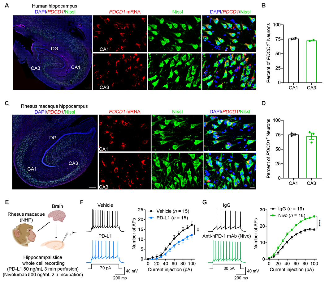
Figure 8. PDCD1 is widely expressed by human and monkey hippocampal neurons and regulates neuronal excitability in NHP slices.

(A) PDCD1 mRNA expression in human hippocampal neurons as shown by in situ hybridization. Left, representative image of the whole hippocampus (Scale bar, 500 μm). Right panels, high magnification images showing PDCD1 mRNA expression in Nissl-labeled neurons (Scale bar, 20 μm).
(B) Quantification of PDCD1+ neurons in the CA1 and CA3 regions of humans.
(C) PDCD1 mRNA expression in NHP hippocampal neurons as shown by in situ hybridization. Left, representative image of the whole hippocampus. Scale bar, 500 μm. Right panels, high magnification images showing PDCD1 mRNA expression in Nissl-labeled neurons. Scale bar, 20 μm.
(D) Quantification of PDCD1+ neurons in the CA1 and CA3 regions of NHPs.
(E) Schematic of experimental design for whole-cell patch clamp recordings in NHP brain slices.
(F) Representative AP traces (left) and quantification of AP firing rate (right) before and after treatment of recombinant monkey PD-L1.
(G) Representative AP traces (left) and quantification of current-evoked APs (right) in NHP hippocampal neurons treated with control IgG and anti-human PD-1 mAb (nivolumab).
Data are represented as mean ± SEM. Also see Figure S11. Sample size and statistical tests are reported in detail in Tables S1 and S5.
