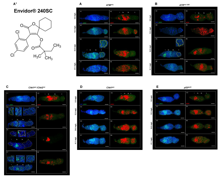
Figure 5.
(A–E) Germarium of DDR mutant females immunostained against anti-γH2AV after 72 h of exposure to concentrations of 0.0, 12.3, 24.6, and 41.1 mg/L to Envidor® 240SC. Composite image in blue DAPI marking cell nuclei and in green γH2AV, red immunolocalization of γH2AV in the germarium, scale bar represents 10 μm. (A1) Chemical structure of Spirodiclofen (a.i. Envidor® 240SC). (A) ATMtefu. (A1,A2) expression of γH2AV in regions 1, 2a, 2b of the germarium. (A3,A4) expression of γH2AV in all regions of the germarium. (A5,A6) expression of γH2AV in regions 1, 2a and 2b of the germarium and absence of nuclei in region 3 (yellow-dotted line). (A7,A8) expression of γH2AV in all regions of the germarium. (B) ATRmei−29D. (B1–B4) expression of γH2AV in all regions of the germarium. (B5,B6) expression of γH2AV in regions 1, 2a and 2b of the germarium, absence of nuclei in region 3 (yellow-dotted line) and morphological changes. (B7,B8) expression of γH2AV in all regions of the germarium. (C) Chk1grp/Chk2lok. (C1,C2) expression of γH2AV in regions 2a and 2b of the germarium. (C3,C4) expression of γH2AV in regions 1 and 2a of the germarium and absence of nuclei in regions 1, 2a and 3 (yellow-dotted line). (C5,C6) expression of γH2AV in region 2a of the germarium, absence of nuclei in regions 1, 2a and 3 (yellow-dotted line) and morphological changes. (C7,C8) expression of γH2AV in all regions of the germarium and absence of nuclei in regions 1, 2b and 3 (yellow-dotted line). (D) Chk1grp. (D1,D2) expression of γH2AV in regions 1, 2a and 2b of the germarium. (D3–D8) expression of γH2AV in all regions of the germarium. (E) p53dp53. (E1,E2) expression of γH2AV in regions 2a and 3 of the germarium. (E3–E6) expression of γH2AV in all regions of the germarium. (E7,E8) expression of γH2AV in all regions of the germarium, morphological changes and reduction in the size of the germarium.

