Abstract
Acute hepatopancreatic necrosis disease (AHPND) is a serious bacterial disease affecting shrimp aquaculture worldwide. In this study, natural microbes were used in disease prevention and control. Probiotics derived from Bacillus spp. were isolated from the stomachs of AHPND-surviving Pacific white shrimp Litopenaeus vannamei (22 isolates) and mangrove forest soil near the shrimp farms (10 isolates). Bacillus spp. were genetically identified and characterized based on the availability of antimicrobial peptide (AMP)-related genes. The phenotypic characterization of all Bacillus spp. was determined based on their capability to inhibit AHPND-causing strains of Vibrio parahaemolyticus (VPAHPND). The results showed that Bacillus spp. without AMP-related genes were incapable of inhibiting VPAHPND in vitro, while other Bacillus spp. harboring at least two AMP-related genes exhibited diverse inhibition activities. Interestingly, K3 [B. subtilis (srfAA+ and bacA+)], isolated from shrimp, exerted remarkable inhibition against VPAHPND (80% survival) in Pacific white shrimp and maintained a reduction in shrimp mortality within different ranges of salinity (75–95% survival). Moreover, with different strains of VPAHPND, B. subtilis (K3) showed outstanding protection, and the survival rate of shrimp remained stable among the tested groups (80–95% survival). Thus, B. subtilis (K3) was further used to determine its efficiency in shrimp farms in different locations of Vietnam. Lower disease occurrences (2 ponds out of 30 ponds) and greater production efficiency were noticeable in the B. subtilis (K3)-treated farms. Taking the results of this study together, the heat-shock isolation and genotypic-phenotypic characterization of Bacillus spp. enable the selection of probiotics that control AHPND in Pacific white shrimp. Consequently, greater disease prevention and growth performance were affirmed to be beneficial in the use of these probiotics in shrimp cultivation, which will sustain shrimp aquaculture and be environmentally friendly.
Keywords: shrimp, probiotics, aquaculture, acute hepatopancreatic necrosis disease (AHPND), Bacillus spp., nursery trial, antimicrobial peptide
1. Introduction
Acute hepatopancreatic necrosis disease (AHPND) is one of the most serious threats to shrimp farming [1]. AHPND is caused mainly by Vibrio parahaemolyticus (VPAHPND), which carries plasmid encoding-specific toxin genes [2]. This pathogen affects penaeid shrimp, including the Pacific white shrimp Litopenaeus vannamei and black tiger shrimp Penaeus monodon, with mortality up to 100% within 20 to 30 days of cultivation, resulting in significant economic losses in shrimp aquaculture [1,3]. Although the use of antibiotics is effective against this bacterial disease, the risk of resistance against drugs among bacteria in the environment requires alternative strategies to address this disease.
In general, bacteria, which are promising contributors to shrimp health, are widely applied in shrimp farms for health management as an alternative strategy to reduce the risk of diseases [4]. Probiotics, ‘live microorganisms which when administered in adequate amounts confer a health benefit on the host’ [5], and biological control agents [6] have been studied in the research field of aquaculture. Previous studies suggest that oral administration of exogenous bacteria stimulates shrimp immune reactions [7,8], and bacterial antagonism occurs in the environment, including the shrimp gut and pond water [9,10]. However, the exact mechanism of the probiotic effects of bacteria on shrimp and the appropriate application are uncertain.
Bacillus spp. is one of the most studied and used bacteria as a probiotic or biocontrol agent in aquaculture [11]. In shrimp, oral administration of spores or vegetative cells of specific strains of Bacillus spp. reduces the mortality of shrimp caused by bacterial infections, with induction of the host immune system and/or antagonism between the bacteria as possible mechanisms [7,8,12,13,14,15]. Antimicrobial peptides (AMPs) secreted by Bacillus spp., such as bacillomycin, fengycin, iturin, surfactin, bacilysin, and subtilin, inhibit the growth of other microorganisms, especially bacteria and fungi [16,17]. The risk of resistance among bacteria against those AMPs is thought to be small [18]; thus, the application of AMP-producing Bacillus to aquaculture fields is promising.
The stomach microbiota of penaeid shrimp may play a crucial role in protecting against bacterial infections. Vibrio bacteria, such as VPAHPND and Vibrio penaeicida, colonize the shrimp’s stomach during the initial stages of infection [19,20]. Moreover, in contrast to mammalian animals, shrimp stomachs host a diverse range of bacteria [21,22], with variations observed in the presence or absence of AHPND development [1,23]. This knowledge suggests that the bacterial community in the stomach microbiota of shrimp, similar to the intestinal microbiota of mammals, might confer beneficial effects on shrimp.
Southeast Asian countries have been heavily impacted by AHPND [24]. However, interestingly, not all shrimp farms or shrimp ponds were affected by the disease. Thus, we expected that shrimp may have specific factors that reduce the risk of AHPND. In this study, Bacillus spp. were isolated from the shrimp stomach and environment, and their potential as beneficial bacteria for shrimp was evaluated.
2. Materials and Methods
2.1. Bacillus spp. Isolation
Bacillus spp. in this study were isolated from two sources: (1) from the stomachs of surviving shrimp from ponds that were positive for AHPND outbreaks on local farms in Samut Songkhram Province and (2) from soils in mangrove forests in Thailand. The isolation of Bacillus spp. followed the heat-cold shock method [25]. Briefly, stomachs of AHPND-surviving shrimp and soils from mangroves were ground and diluted in 0.85% Normal saline solution (NSS) and then heated at 80 °C for 20 min before rapidly chilling on ice for 1 to 2 min. Suspensions were serially diluted in 0.85% NSS and spread on tryptic soy agar (TSA). After incubation at 30 °C for 20 h, Bacillus-like colonies were selected on the basis of their morphology and kept at −80 °C until use.
2.2. VPAHPND Isolation and Identification
To obtain the V. parahaemolyticus (VP) AHPND strain (VPAHPND), the stomachs of diseased shrimp from different areas in the eastern and southern areas of Thailand were collected, including Chanthaburi (CT), Rayong (RY), Trat (TR), Nakhon Si Thammarat (NK), Surat Thani (SR), Chumphon (CP), and Songkhla (SK). The shrimp stomachs were aseptically dissected, minced, and serially diluted 10-fold in 0.85% NaCl (normal saline; NSS), spread on thiosulfate citrate bile salt (TCBS) agar, and incubated at 30 °C overnight [26]. Individual green colonies were chosen and identified using specific primers for VP [27] and VPAHPND [28]. One VP isolate from each province that was positive with both primer sets was examined for pathogenicity in a shrimp challenge assay. VP isolates that caused AHPND were designated VPAHPND hereafter. VPAHPND isolates were collected in glycerol stocks and stored at −80 °C.
2.3. Phenotypic Characterization of Bacillus spp.
2.3.1. In Vitro Inhibition Assay: Solid Medium
The soft agar overlay technique was used to determine inhibition on a solid medium [29]. Soft agar was prepared by mixing melted TSA and TSB at a ratio of 1:2. Then, 3 mL of soft agar was mixed with 50 µL of overnight cultured VPAHPND. The mixture was poured onto the surface of solidified TSA in 87 mm diameter Petri dishes and left for 20 to 30 min to solidify. Then, blank antimicrobial susceptibility disks (Oxoid) were placed on the surface of the agar, and 10 µL of 1 × 10⁸ CFU/mL of each Bacillus culture was dropped on the blank disks. After incubation at 30 °C for 16 h, the diameters of the clear zones surrounding the disks were measured.
2.3.2. In Vitro Inhibition Assay: Liquid Medium
Overnight cultures of different Bacillus spp. were diluted to 1 × 10⁵ CFU·mL−1 in 10 mL of TSB. The overnight culture of VPAHPND was prepared as a 100-fold dilution with Bacillus culture and shaken at 200 rpm 30 °C for 20 h. TSB was used as the control (without Bacillus). For the negative control, TSB was not inoculated with either Bacillus or VPAHPND. Samples with different Bacillus isolates were 10-fold serially diluted in 0.85% NSS and spread on TCBS to count VPAHPND and TSB to count Bacillus spp. All assays were performed in triplicate for each Bacillus isolate.
2.3.3. Antibiotic Susceptibility of Bacillus spp.
The soft agar overlay technique was used to examine antibiotic susceptibility inhibition on a solid medium [29]. The preparation of soft agar followed the method described above. A mixture of soft agar containing Bacillus isolates was poured on the surface of solidified TSA and allowed to solidify. Once solidified, 11 antibiotic discs (Oxoid), including amoxicillin (10 µg), oxytetracycline (30 µg), sulfa-trimethoprim (25 µg), doxycycline (30 µg), erythromycin (15 µg), gentamycin (10 µg), enrofloxacin (5 µg), tetracycline (30 µg), ceftriaxone (30 µg), streptomycin (10 µg) and norfloxacin (10 µg), were placed on the surface of solid agar. After incubation at 30 °C for 16 h, the diameter of the clear zone surrounding the antibiotic discs was measured and interpreted as susceptible (S), intermediate (I), and resistant (R) following the standards of the Clinical and Laboratory Standards Institute [30].
2.4. Genotypic Characterization of Bacillus spp.
2.4.1. Species Identification
The species identification of isolated Bacillus spp. was based on the entire sequence of 16S rRNA gene (1500 bp) using universal primer 8f and 1490r as described below. The isolated Bacillus-like colonies were cultured in tryptic soy broth (TSB) at 30 °C for 20 h. Bacterial DNA was extracted for species identification following the standard phenol–chloroform extraction method. Primers 8f (3′GAGTTTGATCCTGTGCTCAG5′) and 1490r (5′GACTTACCAGGGTATCTAATCC-3′) were used as 16S rRNA universal primers for the bacteria. PCR products were purified with a GeneJET PCR Purification Kit (Thermo Fisher Scientific, Waltham, MA, USA) and sequenced using Sanger sequencing using a 3730XL DNA Analyzer (Applied Biosystems, Waltham, MA, USA). Bacterial identification was performed using NCBI BLAST (https://blast.ncbi.nlm.nih.gov/Blast.cgi, 20 April 2020).
2.4.2. Antimicrobial Peptide (AMP)-Related Gene Determination
Genomic DNA of Bacillus spp. was used as a template for examining the presence of AMP genes, including bmyB (bacillomycin L synthetase B), fenD (fengycin synthetase), ituC (iturin A synthetase C), srfAA (surfactin synthetase subunit 1), bacA (bacilysin biosynthesis protein), and spaS (subtilin) [31](Mora et al., 2011). All PCR amplifications were performed in 25 μL reactions containing 100 μg of genomic DNA template, 2.5 μL of 10x DreamTaq Buffer, 0.2 mM dNTP (Thermo Scientific, Hong Kong, China), 0.5 μM of each primer (Table 1), and 0.6 U of DreamTaq DNA Polymerase (Thermo Scientific, Hong Kong, China). All reactions were run on a MyCycler (Bio-Rad, Hong Kong, China). The cycling conditions for the amplification of all targets were as follows: initial denaturation at 95 °C for 2 min; 35 cycles of 95 °C for 30 s, 55 °C for 30 s and 72 °C for 40 s; and a final extension at 72 °C for 5 min. The PCR amplicons were analyzed in a 2% (w/v) agarose gel.
Table 1.
Primers used to detect the AMP-related genes: bmyB (bacillomycin L synthetase B), fenD (fengycin synthetase), ituC (iturin A synthetase C), srfAA (surfactin synthetase subunit 1), bacA (bacilysin biosynthesis protein) and spaS (subtilin).
| Gene | AMP | Primer | Sequence (5′ → 3′) | Product Size (bp) |
|---|---|---|---|---|
| fenD | Fengycin | fenD_F | GGCCCGTTCTCTAAATCCAT | 269 |
| fenD_R | GTCATGCTGACGAGAGCAAA | |||
| bmyB | Bacillomycin | bmyB_F | GAATCCCGTTGTTCTCCAAA | 370 |
| bmyB_R | GCGGGTATTGAATGCTTGTT | |||
| ituC | Iturin | ituC_F | GGCTGCTGCAGATGCTTTAT | 423 |
| ituC_R | TCGCAGATAATCGCAGTGAG | |||
| srfAA | Surfactin | srfAA_F | TCGGGACAGGAAGACATCAT | 201 |
| srfAA_R | CCACTCAAACGGATAATCCTGA | |||
| bacA | Bacilysin | bacA_F | CAGCTCATGGGAATGCTTTT | 498 |
| bacA_R | CTCGGTCCTGAAGGGACAAG | |||
| spaS | Subtilin | spaS_F | GGTTTGTTGGATGGAGCTGT | 375 |
| spaS_R | GCAAGGAGTCAGAGCAAGGT |
2.5. In Vivo AHPND Challenge Test and Efficacy Analysis
2.5.1. Experimental Shrimp
Healthy Pacific white shrimp were kindly provided by Aquatic Animal Research Mae Klong, Charoen Pokphand Foods Public Co., Ltd. (CP), Bangkok, Thailand. Shrimp were maintained in aerated aquaculture tanks at Kasetsart University until the challenge test. The shrimp were randomly screened for the presence of AHPND, EHP, WSSV, IHHNV, TSV, and YHV using PCR based on a previous method [28,32,33,34,35,36]. Shrimp conditions were maintained during the experiment as follows: pH 7.8–8.2, temperature 28–32 °C, salinity 20 ppt, alkalinity 170–190 mg, TAN less than 1 ppm, and NO2− less than 1 ppm.
2.5.2. Preparation of Bacillus spp. and VPAHPND
Bacillus isolates and VPAHPND in glycerol stocks were streaked on TSA and TCBS agar, respectively. After incubation at 30 °C overnight, a single colony was inoculated in TSB and shaken at 250 rpm and 30 °C for 20 h. The bacterial amount was quantified using the plate count method on TSA and TCBS agar.
2.5.3. Pathogenicity Analysis of Isolated VPAHPND in Shrimp
Three hundred sixty shrimp, 0.5 ± 0.03 g, were acclimated in aerated experimental 200-L tanks for 3 days. The shrimp were separated into two groups: VPAHPND challenge and control group, 60 shrimp in each group with triplicate. In the challenge groups, shrimp were inoculated with isolated VPAHPND at 104 CFU·mL−1 using the immersion method [37]. In the control group, shrimp were inoculated with 100 mL of TSB. The hepatopancreas of moribund shrimps were examined for the presence of VPAHPND using PCR [28], and histomorphology was determined using H&E staining. The number of dead shrimp was observed every 24 h after the challenge until 10 days after the challenge. All assays were performed in triplicate for each VPAHPND isolate.
2.5.4. Efficiency of Isolated Bacillus in Controlling AHPND: Laboratory Level
Three hundred sixty shrimp, 0.5 g ± 0.03 g, were kept in a 400-L container with aeration, and the diseased contaminant was determined prior to testing. To test the disease control, 60 shrimps in each group were directly immersed in different Bacillus isolates (including K3, K5, K6, K11, K12, K13, K19, P4, and P6) at 1 × 10⁵ CFU·mL−1 for 10 h. After that, VPAHPND at 1 × 10⁴ CFU·mL−1 was immediately added to the aquarium. For the control group, 60 shrimp were treated with TSB instead of Bacillus spp. All experiments were performed in triplication. Moribund shrimp were examined for the presence of VPAHPND using PCR. The number of dead shrimp was observed every 24 h. All assays were performed in triplicate for each Bacillus isolate.
2.5.5. Evaluation of AHPND Control Efficiency at Different Salinities
Shrimp were kept in tanks with salinities of 5, 20, and 40 ppt. Shrimp were immersed in Bacillus spp. at 1 × 10⁵ CFU·mL−1 for 10 h and then challenged with VPAHPND at 1 × 10⁴ CFU·mL−1 using the immersion method. The other methodology of this experiment was the same as that mentioned above. All assays were performed in triplicate for each salinity.
2.5.6. Validation of Disease Control Efficiency against Different Strains of VPAHPND
The efficacy of Bacillus sp. against various strains of VPAHPND was evaluated. Shrimp were immersed in 1 × 10⁵ CFU·mL−1 Bacillus sp. for 10 h and then challenged with 1 × 10⁴ CFU·mL−1 of different VPAHPND strains (strains CT, RY, TR NK, SR, CP, and SK) in a salinity of 20 ppt. The other methodology of this experiment was the same as that mentioned above. All assays were performed in triplicate for each VPAHPND isolate.
2.5.7. Efficiency of Isolated Bacillus in Controlling AHPND: Field Level
Field trials were conducted in nursery farms in Quang Binh, Binh Dinh, Ninh Thuan, Ben Tre, Bac Lieu, and Kien Giang at local farms in Vietnam (Supplementary date S1). Healthy postlarvae were reared in 250 m3 aerated aquaculture tanks at a density of 1600 (pcs/m3) for 35 days. Shrimp were initially randomly screened for the presence of AHPND, EHP, WSSV, IHHNV, TSV, and YHV using PCR based on previously described methods. Culture conditions for shrimp were maintained during the whole experiment as follows: pH at 7.8–8.2, temperature at 28–32 °C, salinity at 20 ppt, alkalinity at 170–190 mg, TAN less than 1 ppm and NO2− less than 1 ppm. In the treatment group (30 tanks), Bacillus spp. was added directly to the water at a dose of 105 CFU·mL−1 every 3 days. In the control group (30 tanks), postlarvae did not receive any probiotics. The number of final shrimp (pcs), total weight (kg), final size (pcs·kg−1), total feed (kg), FCR, % survival, and VPAHPND infection were recorded at the end of the experiment.
2.5.8. Statistical Analysis
In bacterial inhibition analysis, Dunnett’s multiple comparisons test was performed to compare the bacterial number in the test groups and the control. In challenge tests, the survival of shrimp was analyzed using the log-rank test. These statistical analyses and figure preparations were conducted using GraphPad Prism v.6 (GraphPad).
3. Results
3.1. Bacillus spp. Isolation
To collect the Bacillus spp., the stomachs of AHPND-surviving shrimp and the soil from the AHPND outbreak area were targeted for Bacillus spp. isolation. After heat and cold shock treatment, Bacillus spp. were entered into the sporulation stage, while other bacteria were killed. Thus, the spore suspension was spread on solid agar to allow the Bacillus spp. to re-enter the vegetative stage. The bacteria showing different morphological characteristics of the colonies were collected, and their species were determined based on 16S rRNA analysis. A total of 22 isolates, designated K1–K22, were isolated from AHPND-surviving shrimp stomachs, whereas 10 isolates, named P1–P10, were isolated from the soil in mangrove forests (Table 1).
3.2. VPAHPND Isolation, Identification, and Pathogenicity of Isolated VPAHPND
VPAHPND was isolated from the stomachs of naturally AHPND-infected shrimp in commercial shrimp farms. Seven strains were collected from different provinces in Thailand: Chanthaburi (CT), Rayong (RY), Trat (TR), Nakhon Si Thammarat (NK), Surat Thani (SR), Chumphon (CP), and Songkhla (SK). Those strains were confirmed using PCR using specific primers for V. parahaemolyticus and VPAHPND. The pathogenicity of these isolates demonstrated acute mortality, with survival rates ranging from 40.56–67.78% (Supplementary date S2). The mortality of AHPND-challenged shrimp ceased at 6–8 days after infection. The presence of VPAHPND in the hepatopancreas of moribund shrimp was confirmed using PCR, and the AHPND histomorphology was confirmed using H&E staining (Supplement data S1 and S2). Pathogenic bacteria were reisolated from dead shrimp, demonstrating authentic infection using the tested VPAHPND.
3.3. Phenotypic Characterization of Bacillus spp.
3.3.1. In Vitro Inhibition Assay: Solid Medium and Liquid Medium
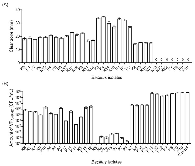
To assess the inhibition activity of Bacillus isolates against VPAHPND in vitro, the width of the inhibition zone on agar plates and the growth of bacteria in liquid media were evaluated. The twenty-five Bacillus isolates exhibited inhibition zones ranging from 14.33–34.67 mm against VPAHPND (strain RY, topic 3.4). Isolates K3, K4, K14, K15, P1, P2, and P3 displayed outstanding inhibition with clear zone diameters ranging from 27.00–34.67 mm (Figure 1A).
Figure 1.
Growth inhibition analysis of Bacillus spp. against VPAHPND. (A) Solid agar; isolated Bacillus spp. were grown on the lawn of VPAHPND (strain RY) on solid agar plates. The inhibition zone was measured and demonstrated as the diameter of the clear zone (mm). (B) Liquid medium; Bacillus spp. were cocultured overnight with VPAHPND (strain RY). The control is the culture without Bacillus. The order corresponds to Table 2, which shows the presence of AMP-related genes.
In liquid media, Bacillus isolates showed significant (Dunnett’s multiple comparisons test, p < 0.001) inhibition against the growth of VPAHPND, except isolates K13, P9, and P10 (Figure 1B). These results concurred with the inhibition assay on a solid medium.
3.3.2. Antibiotic Susceptibility of B. subtilis (K3)
B. subtilis (K3) was susceptible to almost all the tested antibiotics except for amoxicillin (intermediate), oxytetracycline (intermediate), and streptomycin (intermediate) (Table 3).
Table 3.
Presence of AMP genes in Bacillus isolates.
| Isolate | Origin of Isolation | 16S rRNA | BacA | srfAA | ituC | fenD | spaS | bmyB |
|---|---|---|---|---|---|---|---|---|
| K1 | Shrimp | B. methylotrophicus | + | + | − | + | − | + |
| K2 | Shrimp | B. licheniformis | − | + | − | − | − | − |
| K3 | Shrimp | B. subtilis | + | + | − | − | − | − |
| K4 | Shrimp | B. subtilis | + | + | − | − | − | − |
| K5 | Shrimp | B. licheniformis | − | + | − | − | − | − |
| K6 | Shrimp | B. amyloliquefaciens | + | + | + | + | − | + |
| K7 | Shrimp | B. amyloliquefaciens | + | + | − | + | − | + |
| K8 | Shrimp | B. subtilis | + | + | − | − | − | + |
| K9 | Shrimp | B. amyloliquefaciens | + | + | − | + | − | + |
| K10 | Shrimp | B. subtilis | + | + | − | + | − | + |
| K11 | Shrimp | B. vallismortis | − | + | − | − | − | + |
| K12 | Shrimp | B. subtilis | − | + | − | + | − | − |
| K13 | Shrimp | B. subtilis | − | − | − | − | − | − |
| K14 | Shrimp | B. subtilis | + | + | − | − | − | − |
| K15 | Shrimp | B. subtilis | + | + | − | − | − | − |
| K16 | Shrimp | B. licheniformis | − | + | − | − | − | − |
| K17 | Shrimp | B. subtilis | + | + | − | + | − | + |
| K18 | Shrimp | B. subtilis | + | + | − | − | + | − |
| K19 | Shrimp | B. subtilis | + | + | − | − | − | + |
| K20 | Shrimp | B. cereus | − | − | − | − | − | − |
| K21 | Shrimp | B. licheniformis | − | + | − | − | − | − |
| K22 | Shrimp | B. flexus | − | − | − | − | − | − |
| P1 | Mangrove | B. tequilensis | + | + | − | − | − | − |
| P2 | Mangrove | B. amyloliquefaciens | + | + | − | − | − | − |
| P3 | Mangrove | B. tequilensis | + | + | − | − | − | − |
| P4 | Mangrove | B. amyloliquefaciens | + | + | − | + | − | − |
| P5 | Mangrove | B. velezensis | + | + | − | + | − | + |
| P6 | Mangrove | B. velezensis | + | + | − | + | − | + |
| P7 | Mangrove | B. methylotrophicus | − | − | − | − | − | − |
| P8 | Mangrove | B. velezensis | − | − | − | − | − | − |
| P9 | Mangrove | B. firmus | − | − | − | − | − | − |
| P10 | Mangrove | B. velezensis | − | − | − | − | − | − |
3.4. Genotypic Characterization of Bacillus spp.
3.4.1. Species Identification
Upon 16S rRNA identification, the nucleotide sequences (1500 bp) from the isolated Bacillus spp. were compared with references sequenced in the NCBI database to identify their species. In conclusion, the populations of isolated Bacillus species were B. subtilis (11 isolates), B. amyloliquefaciens (5 isolates), B. velezensis (4 isolates), and B. licheniformis (4 isolates) (Table 3).
3.4.2. Antimicrobial Peptide (AMP)-Related Gene Determination
To determine the inhibition activity of isolated Bacillus spp. against VPAHPND, six different AMP genes were tested. Analysis based on gene-specific PCR detection showed diverse distributions of AMP genes in different Bacillus isolates (Table 2). From a total of 32 isolates, the srfAA gene was most frequently found (25 isolates), followed by bacA (19 isolates), bmyB (11 isolates), fenD (11 isolates), and ituC and spaS (1 isolate). More than half of the isolates (21 isolates) harbored at least two of the tested AMP genes, while seven isolates had none of the tested genes. The most frequent patterns of AMP genes were srfAA+-bacA+ (7 isolates) and srfAA+-bacA+-bmyB+-fenD+ (7 isolates). Bacillus isolates carrying AMP genes contained at least srfAA+.
Table 2.
Susceptibility of Bacillus isolates (K3) against antibiotics, including amoxicillin, oxytetracycline, sulfa-trimethoprim, doxycycline, erythromycin, gentamycin, enrofloxacin, tetracycline, ceftriaxone, streptomycin and norfloxacin.
| Antibiotic | Disc Potency (µg) | Bacillus sp. Isolate K3 | |
|---|---|---|---|
| Zone Diameter (mm) | Interpretation | ||
| Amoxicillin | 10 | 15.0 | I |
| Ceftriaxone | 30 | 38.5 | S |
| Doxycycline | 30 | 27.0 | S |
| Gentamycin | 10 | 16.5 | S |
| Enrofloxacin | 5 | 27.5 | S |
| Erythromycin | 15 | 21.0 | S |
| Norfloxacin | 10 | 31.0 | S |
| Oxytetracycline | 30 | 26.5 | I |
| Streptomycin | 10 | 14.0 | I |
| Sulfa-trimethoprim | 25 | 28.5 | S |
| Tetracycline | 30 | 28.0 | S |
3.5. In Vivo Efficacy Analysis of Bacillus against AHPND
3.5.1. Efficiency of Isolated Bacillus in Controlling AHPND: Laboratory Level
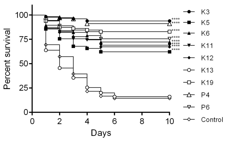
Challenge tests were performed to evaluate the capability of Bacillus isolates P4, P6, K3, K5, K6, K11, K12, K13, and K19 to reduce shrimp mortality during an artificial challenge by VPAHPND (strain RY). Shrimp were treated with different Bacillus isolates (isolates P4, P6, K3, K5, K6, K11, K12, and K19) and showed significantly higher survival rates than the control (p < 0.0001). However, K3 (B. subtilis) exerted the highest effectiveness in reducing shrimp mortality. Isolate K13 (B. subtilis), which lacks the tested AMP genes, did not show a significant difference compared to the control (Figure 2).
Figure 2.
Evaluation of AHPND disease control by Bacillus spp. Shrimp were treated with different Bacillus spp. isolates (isolates K3, K5, K6, K11, K12, K13, K19, P4, and P6) for 10 h following challenge by immersion with 104 CFU·mL−1 VPAHPND (strain RY). **** p < 0.0001 compared with the control.
3.5.2. Evaluation of AHPND Control Efficiency at Different Salinities
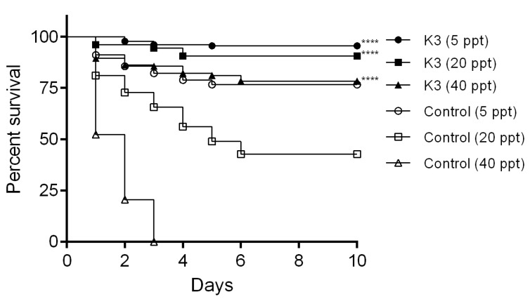
To ensure that B. subtilis (K3) had the highest AHPND control effectiveness, whether it remained effective at different salinities was evaluated. The challenge test was monitored after treatment with B. subtilis (K3). Survival rates of shrimp 76.67% (5 ppt), 42.78% (20 ppt), and 0% (40 ppt) were observed in control shrimp after immersion with VPAHPND (strain RY) without pretreatment with B. subtilis (K3) (Figure 3). This result indicated that the virulence of VPAHPND (strain RY) was salinity-dependent. However, after treatment with B. subtilis (K3), a significantly higher survival rate than the control groups for each salinity (p < 0.0001) was observed (Figure 3).
Figure 3.
Effect of water salinity on B. subtilis (K3) efficiency in controlling AHPND. Shrimp were treated with B. subtilis (K3) for 10 h following the challenge with VPAHPND (strain RY) at 104 CFU·mL−1 by the immersion method. Shrimp were reared at different salinities, including 5 ppt, 20 ppt, and 40 ppt. **** p < 0.0001 compared with the control for each salinity.
3.5.3. Validation of Disease Control Efficiency against Different Strains of VPAHPND
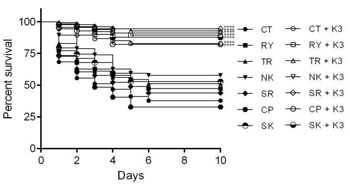
To determine the efficiency of B. subtilis (K3) in protection from different VPAHPND strains, the survival rates of shrimp challenged with different VPAHPND strains were observed in shrimp treated with 105 CFU·mL−1 B. subtilis (K3) at 20 ppt salinity. Similar survival rates among different strains of VPAHPND were observed as follows: CP (82.22%), CT (82.78%), SK (87.78%), RY (89.44%), NK (91.11%), TR (92.78%) and SR (94.44%); all tested groups exhibited significantly greater survival rates than the controls (32.78%, 37.78%, 52.78%, 47.22%, 57.78%, 51.11% and 43.89%, respectively) (Figure 4). The PCR determination of VPAHPND and histomorphology of diseased shrimp confirmed the pathogenicity caused by AHPND disease (Supplementary Figures S3 and S4).
Figure 4.
Efficiency of B. subtilis (K3) in controlling different strains of VPAHPND. Shrimp treated with B. subtilis (K3) for 10 h following challenge with different strains of VPAHPND (strains CP, CT, SK, RY, NK, TR, and SR) at 104 CFU·mL−1. **** p < 0.0001 compared with the control without Bacillus.
3.5.4. Efficiency of Isolated Bacillus in Controlling AHPND: Field Level
Field trials were performed in a local nursery farm in Vietnam to evaluate the protection efficacy of B. subtilis (K3). Shrimp ponds that were treated with B. subtilis (K3) showed a low number of AHPND occurrences, and if eventually positive for AHPND, those ponds can be continually cultivated until harvested. Moreover, with B. subtilis (K3) threat, higher weight with lower FCR compared to the control was observed, which led to greater shrimp production (Table 4).
Table 4.
Results of the field trial. Number of AHPND infection ponds, number of drain ponds, % average survival, total shrimp weight (kg), total feed (kg), and FCR of shrimp receiving B. subtilis (K3) and the control group.
| Parameters | Control | B. subtilis (K3) |
|---|---|---|
| No. of ponds | 30 | 30 |
| No. of shrimp stocked/pond | 600,000 | 600,000 |
| No. of AHPND-positive ponds | 10 | 2 |
| Average survival rate (%) | 68.1 | 94.6 |
| Average FCR | 1.67 | 1.45 |
| Number of drain ponds | 5 | 0 |
| Final size (pcs/kg) | 942.48 | 783.53 |
| Total shrimp weight (kg)/pond | 433.75 | 724.53 |
| Total feed (kg)/pond | 725.75 | 1053.90 |
| No. of surviving shrimp/pond | 408,801 | 567,604 |
4. Discussion
Probiotics in aquaculture are a well-known method to improve the health status of aquatic animals; thus, the use of probiotics to control disease has been widely discussed. However, the validity of their use should be confirmed using rational probiotic screening and selection, as well as the elucidation of their efficiency in laboratory and field trials.
Upon AHPND devastation, we noticed that some shrimp survived in the AHPND-positive ponds. Hence, the ecosystem of the microbiome in the shrimp digestive system might reflect the regulation of the population of pathogenic bacteria by healthy bacteria. Therefore, probiotics, Bacillus spp., were isolated from two different sources: (1) from the surviving shrimp from the AHPND outbreak pond and (2) from the soils in mangrove forests in Thailand.
By using the heat-cold shock method, Bacillus spp. can be isolated from other bacterial species [25]. Based on genotypic and phenotypic identification, AMP identification was used to screen and group all Bacillus spp. AMP synthesis-related genes tested in this study have long been known to be responsible for the inhibitory activity of Bacillus spp. against other microorganisms [31], and it is not surprising that various Bacillus isolates possess these genes. However, a diverse pattern of AMPs containing Bacillus spp. was found, and it is not appropriate to classify them regarding the presentation of AMPs. In addition, the phenotypic determination of their capability to inhibit and control VPAHPND growth and pathogenesis in vitro and in vivo facilitated the selection of candidate Bacillus spp. for further analysis.
B. subtilis (K3) markedly reduced shrimp mortality both at the laboratory level and at the farm level. The bacteria harbor srfAA and bacA genes. The BacA gene is known to contribute to the synthesis of bacilysin, but insufficient reports on controlling or inhibiting Gram-negative bacteria, including Vibrio spp., have been noted. Conversely, many inhibitory effects of srfAA-mediated surfactin against Vibrio were reported [38,39,40]. Therefore, it is expected that surfactin secreted by B. subtilis (K3) would inhibit the growth of VPAHPND in vitro, and a similar mechanism would be proposed in vivo. However, the effect of secreted AMPs or bacterial components on the activation of the shrimp immune system, which contributes to retained survival, should be further elucidated.
Despite the apparent differences in environmental conditions between shrimp stomachs and mangrove forest soil, there were no clear patterns in the distributions of the tested AMP-related genes among Bacillus isolates from each origin. It is expected that the Bacillus present in the shrimp stomach is not specifically adapted to the shrimp stomach environment but rather that Bacillus that could be present in the external environment is ingested orally and colonizes the shrimp stomach. While some previous studies claimed that bacteria administered by feeding colonized shrimp [15,41], it is not clear whether supplied Bacillus in water can stably colonize the digestive tract of shrimp. A previous report showed very low colonization rates of Bacillus bacteria in the digestive tracts of shrimp, especially in earthen ponds [42]. For this reason, the field trial in this study was conducted with continuous administration of the test bacterium, but the optimization of dosing methods is needed.
In general, strains of VPAHPND are halophilic, and the salinity in the water affects their virulence [43,44]. Experimental infections in this study also showed different mortality rates of shrimp in a salinity-dependent manner. However, in shrimp hatcheries, it may be difficult to reduce the salinity [45]. In contrast, inland water aquaculture of Pacific white shrimp uses low-salinity water for cultivation [46,47]. In this study, at each salinity, mortality during VPAHPND challenge was lower in the experimental group using B. subtilis (K3). This indicates that the effects can be expected under a variety of environmental salinity conditions.
Given the definition of the term probiotic [5], it may not be appropriate to use the designation probiotic for the use of Bacillus in this study (exposure via immersion). However, shrimp are expected to take up bacteria in the water, as evidenced by the fact that oral infection of Vibrio is experimentally established using immersion [26]. Further research is needed on the dynamics of the supplied bacteria during immersion, but it might become possible to use the term probiotics for this strategy of the use of beneficial bacteria for shrimp in this method.
Although Bacillus bacteria have been isolated from the shrimp gut [14,15,48], previous analyses of the 16S rDNA-based microbiome show that the genus Bacillus is rarely the dominant genus in the shrimp gut [49], despite its strong inhibitory activity against other bacteria. In the environment of the shrimp digestive tract, it is likely that the balance of microbes is maintained among many bacterial species via bacterial competition or interrelationships with the host. Human studies and subsequent mouse model studies have shown that rare Bacillus in the gastrointestinal tract reduces the risk of infectious disease outbreaks [50]. Surely, the findings in mammals cannot be easily applied to shrimp, but there are phenomena that cannot be fully elucidated by sequencing microbiome analysis alone, and isolation of bacterial strains and subsequent in vitro and in vivo analysis remain useful.
This study was based on selected AMP-related genes for genetic analysis rather than whole genome analysis. We cannot deny that the isolates might harbor novel or overlooked AMP genes that contribute to bacterial inhibition. In addition, it remains unclear whether the differences in inhibitory activity between isolates in vitro are dependent on the amount of AMP secreted or the activity of each peptide molecule. Because of the limitations of this study, it is not certain that similar results can be obtained with other Bacillus isolates.
Importantly, selected B. subtilis (K3) isolate do not possess antibiotic resistance properties. It is suggested that the use of B. subtilis (K3) as a probiotic is safe for aquatic animal cultivation and suitable for producing fishery products for human consumption. In the future, the replacement of antibiotics with probiotics not only reduces the use of chemicals in agriculture, which leave residues in the environment, but also reduces bacteria harboring AMR, which reduces the transfer of antimicrobial-resistant bacteria to humans.
In conclusion, an isolate of Bacillus spp., which was obtained in this study, decreased the mortality of shrimp challenged with VPAHPND. The criterion to screen potential beneficial Bacillus spp. is useful for the search for probiotics for shrimp. This study also shows one of the possible mechanisms of the beneficial effects of probiotics on shrimp.
Supplementary Materials
The following supporting information can be downloaded at: https://www.mdpi.com/article/10.3390/microorganisms11092176/s1.
Author Contributions
P.P.—conducting the experiment, collection and assembly of data, data analysis, figure preparation, and writing of the draft. R.M. and I.H.—conception and design, supervision, provide experimental resources. K.I.—data analysis, figure preparation, and writing of the draft. S.U. administration, funding acquisition, supervision, writing, figure preparation, reviewing articles, and editing. All authors have read and agreed to the published version of the manuscript.
Institutional Review Board Statement
The study was conducted in accordance with guidelines approved by the National Research Council of Thailand.
Informed Consent Statement
Not applicable.
Data Availability Statement
Data will be made available on request.
Conflicts of Interest
The authors declare no conflict of interest.
Funding Statement
This work was financed by Kasetsart University Research and Development Institute (KURDI) under grant number FFKU17.64.
Footnotes
Disclaimer/Publisher’s Note: The statements, opinions and data contained in all publications are solely those of the individual author(s) and contributor(s) and not of MDPI and/or the editor(s). MDPI and/or the editor(s) disclaim responsibility for any injury to people or property resulting from any ideas, methods, instructions or products referred to in the content.
References
- 1.Kumar R., Ng T.H., Wang H.C. Acute hepatopancreatic necrosis disease in penaeid shrimp. Rev. Aquac. 2020;12:1867–1880. doi: 10.1111/raq.12414. [DOI] [Google Scholar]
- 2.Lee C.T., Chen I.T., Yang Y.T., Ko T.P., Huang Y.T., Huang J.Y., Huang M.H., Lin S.J., Chen C.Y., Lin S.S., et al. The opportunistic marine pathogen Vibrio parahaemolyticus becomes virulent by acquiring a plasmid that expresses a deadly toxin. Proc. Natl. Acad. Sci. USA. 2015;112:10798–10803. doi: 10.1073/pnas.1503129112. [DOI] [PMC free article] [PubMed] [Google Scholar]
- 3.Schryver P.D., Defoirdt T., Sorgeloos P. Early mortality syndrome outbreaks: A microbial management issue in shrimp farming. PLoS Pathog. 2014;10:e1003919. doi: 10.1371/journal.ppat.1003919. [DOI] [PMC free article] [PubMed] [Google Scholar]
- 4.Decamp O., Moriarty D.J., Lavens P. Probiotics for shrimp larviculture: Review of field data from Asia and Latin America. Aquac. Res. 2008;39:334–338. doi: 10.1111/j.1365-2109.2007.01664.x. [DOI] [Google Scholar]
- 5.Evaluations of Health and Nutritional Properties of Probiotics in Food Including Powder Milk and Live Lactic Acid Bacteria. FAO/WHO; Cordoba, Argentina: 2001. FAO/WHO Joint Expert Consultation Report. [Google Scholar]
- 6.Verschuere L., Rombaut G., Sorgeloos P., Verstraete W. Probiotic bacteria as biological control agents in aquaculture. Microbiol. Mol. Biol. Rev. 2000;64:655–671. doi: 10.1128/MMBR.64.4.655-671.2000. [DOI] [PMC free article] [PubMed] [Google Scholar]
- 7.Tseng D.Y., Ho P.L., Huang S.Y., Cheng S.C., Shiu Y.L., Chiu C.S., Liu C.H. Enhancement of immunity and disease resistance in the white shrimp, Litopenaeus vannamei, by the probiotic, Bacillus subtilis E20. Fish ShellFish Immunol. 2009;26:339–344. doi: 10.1016/j.fsi.2008.12.003. [DOI] [PubMed] [Google Scholar]
- 8.Liu K.F., Chiu C.H., Shiu Y.L., Cheng W., Liu C.H. Effects of the probiotic, Bacillus subtilis E20, on the survival, development, stress tolerance, and immune status of white shrimp, Litopenaeus vannamei larvae. Fish ShellFish Immunol. 2010;28:837–844. doi: 10.1016/j.fsi.2010.01.012. [DOI] [PubMed] [Google Scholar]
- 9.Knipe H., Temperton B., Lange A., Bass D., Tyler C.R. Probiotics and competitive exclusion of pathogens in shrimp aquaculture. Rev. Aquac. 2021;13:324–352. doi: 10.1111/raq.12477. [DOI] [Google Scholar]
- 10.Vaseeharan B.A.R.P., Ramasamy P. Control of pathogenic Vibrio spp. by Bacillus subtilis BT23, a possible probiotic treatment for black tiger shrimp Penaeus monodon. Lett. Appl. Microbiol. 2003;36:83–87. doi: 10.1046/j.1472-765X.2003.01255.x. [DOI] [PubMed] [Google Scholar]
- 11.Kuebutornye F.K., Abarike E.D., Lu Y. A review on the application of Bacillus as probiotics in aquaculture. Fish ShellFish Immunol. 2019;87:820–828. doi: 10.1016/j.fsi.2019.02.010. [DOI] [PubMed] [Google Scholar]
- 12.Balcázar J.L., Rojas-Luna T. Inhibitory activity of probiotic Bacillus subtilis UTM 126 against Vibrio species confers protection against vibriosis in juvenile shrimp (Litopenaeus vannamei) Curr. Microbiol. 2007;55:409–412. doi: 10.1007/s00284-007-9000-0. [DOI] [PubMed] [Google Scholar]
- 13.Balcázar J.L., Rojas-Luna T., Cunningham D.P. Effect of the addition of four potential probiotic strains on the survival of pacific white shrimp (Litopenaeus vannamei) following immersion challenge with Vibrio parahaemolyticus. J. Invertebr. Pathol. 2007;96:147–150. doi: 10.1016/j.jip.2007.04.008. [DOI] [PubMed] [Google Scholar]
- 14.Hill J.E., Baiano J.C.F., Barnes A.C. Isolation of a novel strain of Bacillus pumilus from penaeid shrimp that is inhibitory against marine pathogens. J. Fish Dis. 2009;32:1007–1016. doi: 10.1111/j.1365-2761.2009.01084.x. [DOI] [PubMed] [Google Scholar]
- 15.Kewcharoen W., Srisapoome P. Probiotic effects of Bacillus spp. from Pacific white shrimp (Litopenaeus vannamei) on water quality and shrimp growth, immune responses, and resistance to Vibrio parahaemolyticus (AHPND strains) Fish ShellFish Immunol. 2019;94:175–189. doi: 10.1016/j.fsi.2019.09.013. [DOI] [PubMed] [Google Scholar]
- 16.Stein T. Bacillus subtilis antibiotics: Structures, syntheses and specific functions: Bacillus subtilis antibiotics. Mol. Microbiol. 2005;56:845–857. doi: 10.1111/j.1365-2958.2005.04587.x. [DOI] [PubMed] [Google Scholar]
- 17.Sumi C.D., Yang B.W., Yeo In-Cheol Hahm Y.T. Antimicrobial peptides of the genus Bacillus: A new era for antibiotics. Can. J. Microbiol. 2015;61:93–103. doi: 10.1139/cjm-2014-0613. [DOI] [PubMed] [Google Scholar]
- 18.Sang Y., Blecha F. Antimicrobial peptides and bacteriocins: Alternatives to traditional antibiotics. Anim. Health Res. Rev. 2008;9:227–235. doi: 10.1017/S1466252308001497. [DOI] [PubMed] [Google Scholar]
- 19.Lai H.C., Ng T.H., Ando M., Lee C.T., Chen I.T., Chuang J.C., Mavichak R., Chang S.H., Yeh M.D., Chiang Y.A., et al. Pathogenesis of acute hepatopancreatic necrosis disease (AHPND) in shrimp. Fish ShellFish Immunol. 2015;47:1006–1014. doi: 10.1016/j.fsi.2015.11.008. [DOI] [PubMed] [Google Scholar]
- 20.de la Peña L.D., Nakai T., Muroga K. Dynamics of Vibrio sp. PJ in organs of orally infected kuruma prawn, Penaeus japonicus. Fish Pathol. 1995;30:39–45. doi: 10.3147/jsfp.30.39. [DOI] [Google Scholar]
- 21.Imaizumi K., Tinwongger S., Kondo H., Hirono I. Analysis of microbiota in the stomach and midgut of two penaeid shrimps during probiotic feeding. Sci. Rep. 2021;11:9936. doi: 10.1038/s41598-021-89415-w. [DOI] [PMC free article] [PubMed] [Google Scholar]
- 22.Imaizumi K., Molex W., Jitnavee C., Direkbusarakom S., Kondo H., Hirono I. Bacterial and eukaryotic communities in pond water of whiteleg shrimp Litopenaeus vannamei and the bacterial communities of their stomach and midgut. Aquaculture. 2022;554:738139. doi: 10.1016/j.aquaculture.2022.738139. [DOI] [Google Scholar]
- 23.Restrepo L., Domínguez-Borbor C., Bajaña L., Betancourt I., Rodríguez J., Bayot B., Reyes A. Microbial community characterization of shrimp survivors to AHPND challenge test treated with an effective shrimp probiotic (Vibrio diabolicus) Microbiome. 2021;9:88. doi: 10.1186/s40168-021-01043-8. [DOI] [PMC free article] [PubMed] [Google Scholar]
- 24.Thitamadee S., Prachumwat A., Srisala J., Jaroenlak P., Salachan P.V., Sritunyalucksana K., Flegel T.W., Itsathitphaisarn O. Review of current disease threats for cultivated penaeid shrimp in Asia. Aquaculture. 2016;452:69–87. doi: 10.1016/j.aquaculture.2015.10.028. [DOI] [Google Scholar]
- 25.Sookchaiyaporn N., Srisapoome P., Unajak S., Areechon N. Efficacy of Bacillus spp. isolated from Nile tilapia Oreochromis niloticus Linn. on its growth and immunity, and control of pathogenic bacteria. Fish Sci. 2020;86:353–365. doi: 10.1007/s12562-019-01394-0. [DOI] [Google Scholar]
- 26.Tran L., Nunan L., Redman R.M., Mohney L.L., Pantoja C.R., Fitzsimmons K., Lightner D.V. Determination of the infectious nature of the agent of acute hepatopancreatic necrosis syndrome affecting penaeid shrimp. Dis. Aquat. Organ. 2013;105:45–55. doi: 10.3354/dao02621. [DOI] [PubMed] [Google Scholar]
- 27.Prompamorn P., Sithigorngul P., Rukpratanporn S., Longyant S., Sridulyakul P., Chaivisuthangkura P. The development of loop-mediated isothermal amplification combined with lateral flow dipstick for detection of Vibrio parahaemolyticus. Lett. Appl. Microbiol. 2011;52:344–351. doi: 10.1111/j.1472-765X.2011.03007.x. [DOI] [PubMed] [Google Scholar]
- 28.Tinwongger S., Proespraiwong P., Thawonsuwan J., Sriwanayos P., Kongkumnerd J., Chaweepack T., Mavichak R., Unajak S., Nozaki R., Kondo H., et al. Development of PCR diagnosis for shrimp acute hepatopancreatic necrosis disease (AHPND) strain of Vibrio parahaemolyticus. Fish Pathol. 2014;49:159–164. doi: 10.3147/jsfp.49.159. [DOI] [Google Scholar]
- 29.Hockett K.L., Baltrus D.A. Use of the soft-agar overlay technique to screen for bacterially produced inhibitory compounds. J. Vis. Exp. 2017;119:55064. doi: 10.3791/55064. [DOI] [PMC free article] [PubMed] [Google Scholar]
- 30.Weinstein M.P. Performance Standards for Antimicrobial Susceptibility Testing. 31st ed. Clinical and Laboratory Standards Institute; Wayne, PA, USA: 2021. [Google Scholar]
- 31.Mora I., Cabrefiga J., Montesinos E. Antimicrobial peptide genes in Bacillus strains from plant environments. Int. Microbiol. 2011;14:213–223. doi: 10.2436/20.1501.01.151. [DOI] [PubMed] [Google Scholar]
- 32.Jaroenlak P., Sanguanrut P., Williams B.A.P., Stentiford G.D., Flegel T.W., Sritunyalucksana K., Itsathitphaisarn O. A nested PCR assay to avoid false positive detection of the microsporidian Enterocytozoon hepatopenaei (EHP) in environmental samples in shrimp farms. PLoS ONE. 2016;11:e0166320. doi: 10.1371/journal.pone.0166320. [DOI] [PMC free article] [PubMed] [Google Scholar]
- 33.Durand S.V., Lightner D.V. Quantitative real time PCR for the measurement of white spot syndrome virus in shrimp. J. Fish Dis. 2002;25:381–389. doi: 10.1046/j.1365-2761.2002.00367.x. [DOI] [Google Scholar]
- 34.Tang K.F., Navarro S.A., Lightner D.V. PCR assay for discriminating between infectious hypodermal and hematopoietic necrosis virus (IHHNV) and virus-related sequences in the genome of Penaeus monodon. Dis. Aquat. Org. 2007;74:165–170. doi: 10.3354/dao074165. [DOI] [PubMed] [Google Scholar]
- 35.Tang K.F., Wang J., Lightner D.V. Quantitation of Taura syndrome virus by real-time RT-PCR with a TaqMan assay. J. Virol. Methods. 2004;115:109–114. doi: 10.1016/j.jviromet.2003.09.021. [DOI] [PubMed] [Google Scholar]
- 36.Wijegoonawardane P.K., Cowley J.A., Walker P.J. A consensus real-time RT-PCR for detection of all genotypic variants of yellow head virus of penaeid shrimp. J. Virol. Methods. 2010;167:5–9. doi: 10.1016/j.jviromet.2010.02.024. [DOI] [PubMed] [Google Scholar]
- 37.Dangtip S., Sirikharin R., Sanguanrut P., Thitamadee S., Sritunyalucksana K., Taengchaiyaphum S., Mavichak R., Proespraiwong P., Flegel T.W. AP4 method for two-tube nested PCR detection of AHPND isolates of Vibrio parahaemolyticus. Aquac. Rep. 2015;2:158–162. doi: 10.1016/j.aqrep.2015.10.002. [DOI] [Google Scholar]
- 38.Kim K.M., Lee J.Y., Kim C.K., Kang J.S. Isolation and characterization of surfactin produced by Bacillus polyfermenticus KJS-2. Arch. Pharm. Res. 2009;32:711–715. doi: 10.1007/s12272-009-1509-2. [DOI] [PubMed] [Google Scholar]
- 39.Xu H.M., Rong Y.J., Zhao M.X., Song B., Chi Z.M. Antibacterial activity of the lipopetides produced by Bacillus amyloliquefaciens M1 against multidrug-resistant Vibrio spp. isolated from diseased marine animals. Appl. Microbiol. Biotechnol. 2014;98:127–136. doi: 10.1007/s00253-013-5291-1. [DOI] [PubMed] [Google Scholar]
- 40.Chen Y., Liu S.A., Mou H., Ma Y., Li M., Hu X. Characterization of lipopeptide biosurfactants produced by Bacillus licheniformis MB01 from marine sediments. Front. Microbiol. 2017;8:871. doi: 10.3389/fmicb.2017.00871. [DOI] [PMC free article] [PubMed] [Google Scholar]
- 41.Liu C.H., Chiu C.S., Ho P.L., Wang S.W. Improvement in the growth performance of white shrimp, Litopenaeus vannamei, by a protease-producing probiotic, Bacillus subtilis E20, from natto. J. Appl. Microbiol. 2009;107:1031–1041. doi: 10.1111/j.1365-2672.2009.04284.x. [DOI] [PubMed] [Google Scholar]
- 42.Ziaei-Nejad S., Rezaei M.H., Takami G.A., Lovett D.L., Mirvaghefi A.R., Shakouri M. The effect of Bacillus spp. bacteria used as probiotics on digestive enzyme activity, survival and growth in the Indian white shrimp Fenneropenaeus indicus. Aquaculture. 2006;252:516–524.4444. doi: 10.1016/j.aquaculture.2005.07.021. [DOI] [Google Scholar]
- 43.López-Cervantes G., Álvarez-Ruiz P., Luna-Suárez S., Luna-González A., Esparza-Leal H.M., Castro-Martínez C., Gámez-Jiménez C., Soto-Alcalá J. Temperature and salinity modulate virulence and PirA gene expression of Vibrio parahaemolyticus, the causative agent of AHPND. Aquac. Int. 2021;29:743–756. doi: 10.1007/s10499-021-00654-0. [DOI] [Google Scholar]
- 44.Schofield P.J., Noble B.L., Caro L.F.A., Mai H.N., Padilla T.J., Millabas J., Dhar A.K. Pathogenicity of acute hepatopancreatic necrosis disease (AHPND) on the freshwater prawn, Macrobrachium rosenbergii, and Pacific white shrimp, Penaeus vannamei, at various salinities. Aquac. Res. 2021;52:1480–1489. doi: 10.1111/are.15001. [DOI] [Google Scholar]
- 45.Charmantier G., Charmantier-Daures M., Bouaricha N., Thuet P., Trilles J.P., Aiken D.E. Ontogeny of osmoregulation and salinity tolerance in two decapod crustaceans: Homarus americanus and Penaeus japonicus. Biol. Bull. 1988;175:102–110. doi: 10.2307/1541897. [DOI] [Google Scholar]
- 46.Roy L.A., Davis D.A., Saoud I.P., Boyd C.A., Pine H.J., Boyd C.E. Shrimp culture in inland low salinity waters. Rev. Aquac. 2010;2:191–208. doi: 10.1111/j.1753-5131.2010.01036.x. [DOI] [Google Scholar]
- 47.Li E., Wang X., Chen K., Xu C., Qin J.G., Chen L. Physiological change and nutritional requirement of Pacific white shrimp Litopenaeus vannamei at low salinity. Rev. Aquac. 2017;9:57–75. doi: 10.1111/raq.12104. [DOI] [Google Scholar]
- 48.Chai P.C., Song X.L., Chen G.F., Xu H., Huang J. Dietary supplementation of probiotic Bacillus PC465 isolated from the gut of Fenneropenaeus chinensis improves the health status and resistance of Litopenaeus vannamei against white spot syndrome virus. Fish ShellFish Immunol. 2016;54:602–611. doi: 10.1016/j.fsi.2016.05.011. [DOI] [PubMed] [Google Scholar]
- 49.Holt C.C., Bass D., Stentiford G.D., van der Giezen M. Understanding the role of the shrimp gut microbiome in health and disease. J. Invertebr. Pathol. 2021;186:107387. doi: 10.1016/j.jip.2020.107387. [DOI] [PubMed] [Google Scholar]
- 50.Piewngam P., Zheng Y., Nguyen T.H., Dickey S.W., Joo H.S., Villaruz A.E., Glose K.A., Fisher E.L., Hunt R.L., Li B., et al. Pathogen elimination by probiotic Bacillus via signalling interference. Nature. 2018;562:532–537. doi: 10.1038/s41586-018-0616-y. [DOI] [PMC free article] [PubMed] [Google Scholar]
Associated Data
This section collects any data citations, data availability statements, or supplementary materials included in this article.
Supplementary Materials
Data Availability Statement
Data will be made available on request.


