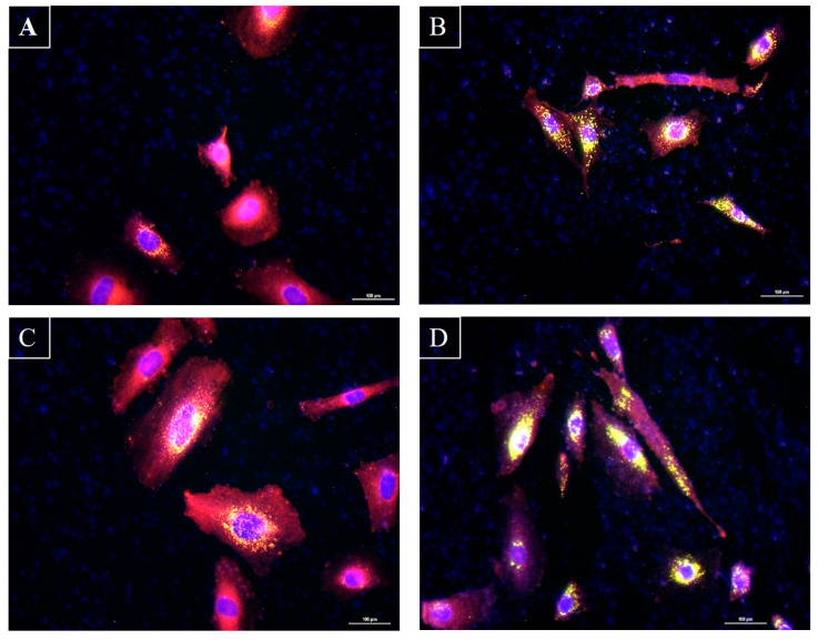
Figure 6.
Microscopic observation of sebocytes cultured after fixation and labeling with Nile Red and Hoechst. Panel (A): Sebocytes alone. Panel (B): Sebocytes with S. epidermidis. Panel (C): Sebocytes with C. acnes. Panel (D): Sebocytes with S. epidermidis and C. acnes. Pictures were taken via an epifluorescence microscope. Hoechst marks the nuclei in blue, while Nile Red reveals neutral lipids in green and total lipids in red. Superimposing the two colors highlights the lipid droplets in yellow. Scale bar: 100 µm.

