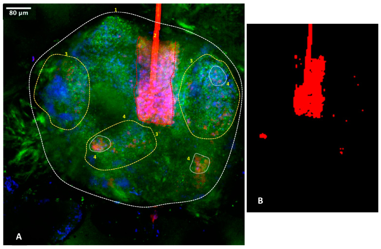
Figure 8.
Representative 3D bioimaging of C. acnes on ex vivo sebaceous gland via confocal microscopy. Orientation has been corrected to the surface direction of the skin. Blue represents cell nuclei, green represents MC5R-positive sebocytes and red represents for C. acnes bacteria. Area 1: overall location of the gland. Area 2: hair follicle and hair sheath passing through gland with red autofluorescence of the follicle material and other C. acnes red labelling in the lower bulb area. Area 3: active areas of the gland. Area 4: examples of location of C. acnes. Panel (A): full gland. Panel (B): C. acnes areas labelled red. Localization of the C. acnes bacteria to active areas of the glands, including the hair follicles and gland lobes, is noted at areas 3 and 4. Supplementary Videos S1 and S2 available from the authors show further examples.

