Abstract
The key factors contributing to radiculopathy caused by lumbar disc herniation include mechanical compression. It was commonly believed that the disc herniation causes the compression on the nerve root exiting under the pedicle of the vertebral body at the adjacent inferior level. However, a disc herniation might occasionally result in non-adjacent, isolated radicular symptoms. We report the case of a 74-year-old female who presented with a 2-years history of progressive low back pain associated with L5 radiculopathy and reduced quality of life. The patient had undergone a magnetic resonance image showing a large L2/3 disc herniation. Symptoms had progressively worsened and failed to respond to conservative treatments including pain medication, exercise rehabilitation, and acupuncture at the lower lumbar region. The patient was diagnosed with L5 radiculopathy caused by L2/3 disc herniation. Consequently, her symptoms improved with chiropractic rehabilitation which involved spinal manipulative therapy and intermittent motorized traction at the L2/3 level to reduce herniated disc. Therefore, an L2/3 Disc herniation-related L5 radiculopathy should be considered in the differential diagnosis of cases of inconsistency of level of disc herniation and nerve root pattern.
Keywords: Chiropractic , lumbar disc herniation , manipulative therapy , spine surgery , lumbar radiculopathy
Introduction
Over the past few decades, lower back pain has been the primary musculoskeletal disorder negatively impacting people's quality of life [1].
The key factor contributing to low back pain and radiculopathy caused by lumbar disc herniation include mechanical compression [2].
It was commonly believed that the disc herniation causes the compression on the nerve root exiting under the pedicle of the vertebral body at the adjacent inferior level.
However, the lumbar herniated disc presents contradictory symptoms that are challenging to explain by a straightforward mechanical defect.
The inflammatory mediators and autoimmune response was found to be actively involved in the pathophysiology of radiculopathy.
A disc herniation might occasionally result in non-adjacent, isolated radicular symptoms.
Herein, we report an atypical case of L5 nerve root radiculopathy caused by a herniated intervertebral disc at the L2/3 segment.
L2/3 disc herniation was eventually confirmed as the etiology of L5 radiculopathy resulting from the magnetic resonance image and positive response from the spinal manipulative therapy at the L2/3 level.
We reviewed the literature for similar cases and briefly described the diagnosis and management of L5 nerve root radiculopathy caused by upper lumbar disc herniation.
Case History
A 74-year-old female presented complaining of low back pain and radicular symptoms prominent in the left L5 nerve root territory, with pain in the anterolateral aspect of the distal extremity for 2 years.
She first experienced the symptoms in early 2020 with a mild gradual numbness sensation to her left foot.
The magnetic resonance imaging (MRI) performed for the orthopedic workup revealed a large L2/3 disc herniation.
The patient has been treated with NSAID (diclofenac), 6 months exercise rehabilitation, and acupuncture for pain relief with minimum improvement.
The symptoms were aggravated by prolonged sleep, walk, and exercise.
She also had a past medical history of early-stage colon cancer in 1991, which was treated by polypectomy; severe knee osteoarthritis diagnosed in 2015; and 10-years history of cardiovascular and diabetes.
In January 2022, she was infected with Covid-19 with basic flu symptoms.
10 days after her recovery, her low back pain symptoms suddenly worsened, accompanied by radiating burning pain down the left foot, which she had not experienced previously.
She was required to use crutches for walking and up and down stairs for her daily living.
She was unable to stand/walk for over 5 minutes and unable to take public transportation.
Her orthopedic surgeon recommended her for surgery and she sought a chiropractor for a second opinion.
Low back soreness could be induced by passive lumbar flexion.
Spinal palpation revealed intersegmental restriction at T4-T7; and L2-L3 vertebral levels.
Hypertonicity was palpable in the right quadratus lumborum, bilateral gluteal muscles, and paraspinal muscles.
The neurological examination showed a motor weakness and graded at 4/5 in the left extensor hallucis longus, tibialis posterior, and anterior tibialis, and a weakness in foot inversion.
Sensory examination included reduced pin-prick sensation at the L5 dermatome.
The orthopedic examination was positive at left straight-leg raise test at 25 degrees.
She was diagnosed with lumbar disc herniation on the basis of imaging findings and clinical history.
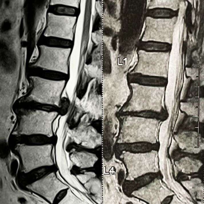
Her MRI of the lumbar spine identified severe circumferential L2/3 disc bulging with right-sided eccentricity.
Right-sided eccentricity with protruded components measures 7mm x 15.5mm x 18.4mm in size (Figure 1 left).
Figure 1.
MRI of the lumbar spine identified severe circumferential L2/3 disc bulging. (Left) The protruded disc measures 7mm x 15.5mm x 18.4mm in size. Moderate central canal stenosis with anterior-posterior diameter of spinal canal measures 0.78cm. (Right) At the 4th month follow-up visit, the size of her disc extrusion had significantly reduced by 60% at the MRI.
Posterior annular tear seen.
Moderate central canal stenosis with AP diameter of spinal canal measures 0.78cm.
She had to walk with a cane and all of her previous symptoms became much worse.
Her WHOQOL score was reduced to 52%.
She was immediately scheduled with the therapeutic plans three times a week for 4 weeks and her symptoms recovered slowly.
By the end of the 4th week, her WHOQOL score had returned to 86% and she had resumed participation in her daily taichi exercise.
Following 6 consecutive days of spinal manipulative therapy at L2/3 level to release intersegmental restriction combined with therapeutic ultrasound and instrument assisted soft tissue manipulation (IASTM) to provide deep heating and relax soft tissues, the patient’s pain complaints were substantially reduced.
Subsequent treatment sessions consisted of intermittent motorized lumbar traction (Spine Decompression Device, SpineMT, Korea), therapeutic ultrasound, instrument assisted soft tissue mobilization, and spinal manipulation of the upper lumbar spine.
The spinal decompression device was aimed specifically at L2/3 level to restore his neurological dysfunction.
Treatment frequency was reduced to three times weekly for a period of 3 weeks.
Low back pain and radiating leg pain were reduced from 7/10 to 1/10 and the burning sensation reduced by 50%.
The World Health Organization Quality of Life (WHOQOL) score improved from 56% to 86% (100% being the highest possible well-being) after the therapy.
Her treatment frequency was reduced to two times a week for a period of 3 weeks.
By the end of the treatment, her lumbar range of motions, neurological and orthopedic exams had returned to normal.
The burning sensation transformed into a warm feeling with minimal paresthesias; and the symptoms may mildly relapsed due to her housework but recovered quickly with chiropractic treatment.
The size of her disc extrusion had significantly reduced by 60% at the 4th month follow-up MRI (Figure 1 Right).
She maintained her treatment once a month to ensure her nerve integrity for an additional 3 months and remained stable.
The patient enrolled in the study was included only after signing the informed consent and her participation was voluntary.
Discussions
Radiculopathy in the lower extremity with dermatomal distribution is the most traditional presentation of lumbar disc herniation with nerve root compression.
Since the anterior tibialis, extensor hallucis longus, extensor digitorum brevis, and lateral gastrocnemius muscles receive their major motor innervation from the L5 nerve root, these muscles are frequently affected by the symptoms of L5 nerve root compression.
A L2/3 disc herniation typically pinches the L2 nerve root, and causes motor deficiency in an iliopsoas, quadriceps femoris, hip adductors muscles, and/or sensory alteration that radiates into the anterior and anterolateral thigh [3].
In this case, the L3 nerve root was not implicated clinically; there were only L5 clinical symptoms.
If the passing nerves were also squeezed within the thecal sac, a sufficiently substantial disc herniation would also result in a polyradiculopathy, which would affect the primary level and caudal level components.
The peculiarity of this patient is that L3 and L4 were unaffected, and only the 5th lumbar dermatome and myotome was.
As the L5 nerve root travels inferiorly in the spinal canal as part of the cauda equina, the lumbar disc herniation could compress the L5 nerve root at the L2-3 lumbar level.
The more distant sacral nerve roots are located dorsally within the thecal sac, while the lumbar nerve roots travel ventrally within the thecal sac before leaving the spinal canal through the specified intervertebral foramina.
The third lumbar nerve was able to leave and the intradural roots of the L4 nerve were able to pass through due to sufficient laxity in the sacral and contralateral roots and sufficient space lateral to the herniation.
A comparable example of L5 radiculopathy brought on by an L2/3 disc herniation was documented by Stien et al.
The case also shares similar clinical presentation with MRI findings that did not correlate, and the L2/3 disc herniation was identified to be the only compressive lesion [4].
Unlike our case management with conservative treatment, the patient was recovered by microsurgical discectomy.
For a complete knowledge of the pathogenesis of L5 radiculopathy caused by an L2/3 disc herniation, more research is required.
MRI is the gold standard to assess the association between the lumbar herniated intervertebral disc and changes in the angle of the lumbar spine's facet joints [5].
Anatomical and morphological analysis of the lumbar spine provides the foundation for the early detection and detailed diagnosis of lumbar disc herniation [5].
Although electrophysiological studies in peripheral nerves and muscles of lower limbs in prolapsed lumbar intervertebral discs are efficacious methods in the diagnosis and predicting the prognosis of radiculopathies [6], we did not prescribe the studies.
The patient’s radiological finding was clear and obvious, and the additional invasive examination will not change the treatment plan of the large disc herniation.
Furthermore, initial spinal manipulative therapy at L2/3 can also help in confirming the diagnosis of L2/3 disc herniation by observing the prognosis.
Surgical intervention can always be considered when the conservative treatment fails.
The majority of individuals with lumbar disc herniation should have conservative treatment as their first line of defense [7, 8].
Mechanical traction and spinal manipulative therapy are the common conservative treatments to treat radiculopathy caused by lumbar disc herniation [9].
In a single-arm clinical experiment, patients with acute low back pain caused by lumbar disc herniation were assessed for the effects of segmental traction therapy on lumbar disc herniation, pain, lumbar range of motion (ROM), and back extensor muscle endurance [10].
The size of the herniated disc and the patients' pain were greatly reduced after following the therapy plan.
Additionally, the range of motion in the lumbar region improved significantly [10].
A review also concluded that oblique pulling spinal manipulation is more effective than acupuncture and lumbar traction in managing lumbar disc herniation [11].
Spinal manipulative therapy has biomechanical consequences on symptom reduction, such as relaxing hypertonic muscles, releasing pinched nerves, rupturing periarticular adhesions, and restoring spinal alignment and lumbar traction, increases the intervertebral space's volume and decreases its pressure [12].
Large study also found that the incidence of severe adverse events in chiropractic therapy is very rare [13], the spinal manipulative therapy and intermittent motorized lumbar traction were both utilized at the L2/3 level, which also demonstrated reduction of protruded disc size and improvement of quality of life.
Although the spine surgeon faces additional challenges in treating upper disc herniations because of their low incidence and slow detection as a result of the lack of recognizable clinical symptoms [3], the upper lumbar disc herniation causing non-adjacent and isolated radiculopathy is a rare finding.
According to a search of Google Scholar, PubMed, Medline, and the Index to Chiropractic Literature on September 1st, 2022, we reviewed 7 published cases of patients with a lumbar disc herniation causing non-adjacent radiculopathy.
2 cases are directly related to L2/3 disc herniation; 4 cases are related to L1/2 disc herniation; and 1 case is involved with L1/2/3 stenosis.
All past cases were successfully managed by operations.
To prove a proper diagnosis of the condition, a complete medical history, orthopedic tests, neurological assessments, and MRI scans are required.
Accurate diagnosis and treatment of L5 radiculopathy secondary to L2/3 disc herniation can avoid unnecessary investigations, operation, and surgical adverse effects.
Conclusions
Rare lumbar disc herniations may also cause isolated, non-adjacent radiculopathy.
Low back pain and radiculopathy is often multifactorial and the cause of the symptoms sometimes cannot easily be diagnosed.
Recognition of rare lumbar disc herniation and its referral pattern is essential for the management of atypical clinical presentation.
As the majority of individuals with lumbar disc herniation should have conservative treatment as their first line of defense, spinal manipulative therapy and mechanical traction should always be utilized as the first options even with atypical cases.
Conflict of interests
None to declare
References
- 1.Vos TP, Flaxman ADP, Naghavi MP, Lozano RP, Michaud CMD, Ezzati MP, Shibuya KP, Salomon JAP, Abdalla SM, Aboyans VP, et al. Years lived with disability (YLDs) for 1160 sequelae of 289 diseases and injuries 1990-2010: A systematic analysis for the Global Burden of Disease Study 2010. Lancet. 2012;380(9859):2163–2196. doi: 10.1016/S0140-6736(12)61729-2. [DOI] [PMC free article] [PubMed] [Google Scholar]
- 2.Meng Z, Zheng J, Fu K, Kang Y, Wang L. Curative Effect of Foraminal Endoscopic Surgery and Efficacy of the Wearable Lumbar Spine Protection Equipment in the Treatment of Lumbar Disc Herniation. J Healthc Eng. 2022;2022:6463863–6463863. doi: 10.1155/2022/6463863. [DOI] [PMC free article] [PubMed] [Google Scholar] [Retracted]
- 3.Kim DS, Lee JK, Jang JW, Ko BS, Lee JH, Kim SH. Clinical features and treatments of upper lumbar disc herniations. J Korean Neurosurg Soc. 2010;48(2):119–124. doi: 10.3340/jkns.2010.48.2.119. [DOI] [PMC free article] [PubMed] [Google Scholar]
- 4.Stein AA, Vrionis F, Espinosa PS, Moskowitz S. Report of an Isolated L5 Radiculopathy Caused by an L2-3 Disc Herniation and Review of the Literature. Cureus. 2018;30;10(4):e2552–e2552. doi: 10.7759/cureus.2552. [DOI] [PMC free article] [PubMed] [Google Scholar]
- 5.Zheng K, Wen Z, Li D. The Clinical Diagnostic Value of Lumbar Intervertebral Disc Herniation Based on MRI Images. J Healthc Eng. 2021;2021:5594920–5594920. doi: 10.1155/2021/5594920. [DOI] [PMC free article] [PubMed] [Google Scholar] [Retracted]
- 6.Sarmast AH, Kirmani AR, Bhat AR. Evaluation of Role of Electrophysiological Studies in Patients with Lumbar Disc Disease. Asian J Neurosurg. 2018;13(3):585–589. doi: 10.4103/ajns.AJNS_341_16. [DOI] [PMC free article] [PubMed] [Google Scholar]
- 7.Benzakour T, Igoumenou V, Mavrogenis AF, Benzakour A. Current concepts for lumbar disc herniation. Int Orthop. 2019;43(4):841–851. doi: 10.1007/s00264-018-4247-6. [DOI] [PubMed] [Google Scholar]
- 8.Chu ECP, Wong AYL. Chronic Orchialgia Stemming From Lumbar Disc Herniation: A Case Report and Brief Review. Am J Mens Health. 2021;15(3):15579883211018431–15579883211018431. doi: 10.1177/15579883211018431. [DOI] [PMC free article] [PubMed] [Google Scholar]
- 9.Chu ECP, Chan AKC, Lin AFC. Pitting oedema in a polio survivor with lumbar radiculopathy complicated disc herniation. J Family Med Prim Care. 2019;8(5):1765–1768. doi: 10.4103/jfmpc.jfmpc_254_19. [DOI] [PMC free article] [PubMed] [Google Scholar]
- 10.Karimi N, Akbarov P, Rahnama L. Effects of segmental traction therapy on lumbar disc herniation in patients with acute low back pain measured by magnetic resonance imaging: A single arm clinical trial. J Back Musculoskelet Rehabil. 2017;30(2):247–253. doi: 10.3233/BMR-160741. [DOI] [PubMed] [Google Scholar]
- 11.Mo Z, Zhang R, Chen J, Shu X, Shujie T. Comparison Between Oblique Pulling Spinal Manipulation and Other Treatments for Lumbar Disc Herniation: A Systematic Review and Meta-Analysis. J Manipulative Physiol Ther. 2018;41(9):771–779. doi: 10.1016/j.jmpt.2018.04.005. [DOI] [PubMed] [Google Scholar]
- 12.Chu ECP. Taming of the Testicular Pain Complicating Lumbar Disc Herniation with Spinal Manipulation. Am J Mens Health. 2020;14(4):1557988320949358–1557988320949358. doi: 10.1177/1557988320949358. [DOI] [PMC free article] [PubMed] [Google Scholar]
- 13.Chu EC, Trager RJ, Lee LY, Niazi IK. A retrospective analysis of the incidence of severe adverse events among recipients of chiropractic spinal manipulative therapy. Sci Rep. 2023;13(1):1254–1254. doi: 10.1038/s41598-023-28520-4. [DOI] [PMC free article] [PubMed] [Google Scholar]


