Point-of-care ultrasound (POCUS) is an imaging technique performed at the patient’s bedside by a treating medical doctor. It assists in the diagnosis, monitors changes in clinical conditions, and guides invasive procedures.1 In critically ill patients, POCUS allows for rapid, focused, and integrated assessments of ultrasound images, technical skills, and clinical knowledge. Lately, the use of POCUS has expanded across many pediatric disciplines.2 Bladder and renal ultrasound imaging analyses to identify hydronephrosis and the volume of urine are relevant applications to pediatric patients.1
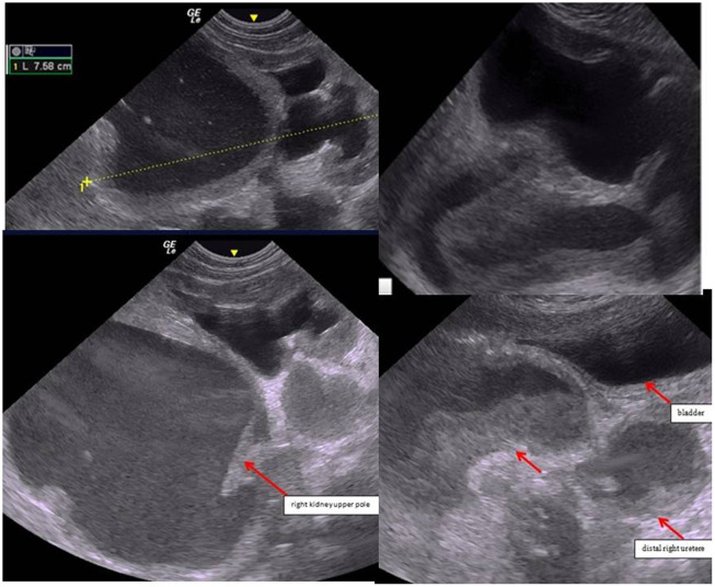
We describe a case of a 50-day-old female infant born at term, a normal pregnancy, and correct birth weight. She had a prenatal diagnosis of right renal pelvis dilatation, not otherwise specified. On admission to our emergency room she had a poor clinical condition and presented with fever, refusal to feed, and lethargy. Clinical laboratory test results showed elevated serum levels of C-reactive protein (211 mg/dL, n = 0-5) and procalcitonin (2.26 mg/dL, n <0.5). A urine test revealed an elevated white blood cell count (3+). The others laboratory findings (creatinine, etc.) were normal. The POCUS study of the kidney and urinary tract showed a markedly enlarged right kidney, severe thinning of the cortical parenchyma, dilation of the renal calyces (hydronephrosis IV grade),3 and a dilated and tortuous right ureter (Figure 1). Ultrasound findings of the left kidney and bladder were normal. Then, empirical antimicrobial therapy was started with amoxicillin/clavulanate (50 mg/kg/day in 2 doses intravenously) and amikacin (15 mg/kg/day single dose intravenously). After 24 hours, we modified the antimicrobial therapy with meropenem because urine culture revealed the growth of Pseudomonas colonies. Despite clinical improvement, follow-up with POCUS showed enlargement of the hydronephrotic sac of the right kidney upper pole, enlargement of the right ureter, and increased purulent content in both, and diffuse thickening of the bladder wall. Moreover, it seemed evident that the hydronephrotic sac of the right kidney upper pole did not communicate with the remaining renal parenchyma (Figure 1). Therefore, we suspected a complete duplex pelvis with malformation of the ureter. Then, the child was transferred to pediatric surgery, where she underwent cutaneous ureterostomy, with drainage of 100 mL of purulent fluid and intraoperative confirmation of complete duplex system and ectopic obstructive insertion of the ureter of the right kidney upper pole moiety into the bladder neck. Magnetic resonance imaging confirmed the diagnosis. After 2 months, cystography showed non-refluxing ureters, and renal scintigraphy showed that the right kidney function was normal. To date, the child has undergone regular follow-up after surgical correction.
Figure 1.
Point-of-care ultrasound (POCUS) performed at the time of hospitalization in the pediatric ward showed severe hydronephrosis of the right kidney, with a large hydronephrotic sac on the upper pole, proximal right ureterectasis, distal right ureterectasis, and POCUS performed after 36 hours showed enlargement of hydronephrotic sac of right kidney upper pole, enlargement of distal right ureterectasis in cross-section with increased purulent content in both.
Duplex pelvis is an urinary malformation with an incidence rate of 0.8%-1%.4 In the complete type, there are 2 pelvises and 2 ureters, which open separately. The sites of insertion of ectopic ureters are usually the bladder neck/urethra and rarely the vagina.5-7 This malformation can be complicated by ureterocele, vesicoureteral reflux, obstruction of the urinary tract, and renal dysplasia. Duplex collecting systems are present in 80% of ectopic ureter cases and in 80% of duplicated systems, the ureterocele is associated with the upper pole.8 The presence of a double pelvis and malformation of the ureter with clinical manifestations require surgical intervention.8,9 Urinary incontinence and abdominal pain are the main symptoms of an ectopic ureter. However, obstructive uropathy may present precociously because of recurring urinary tract infections.9 Therefore, in early childhood, it is a congenital malformation complex and potentially fatal due to the high risk of acute pyelonephritis, urosepsis, and pyonephrosis.
In our case, the ureter came from the right kidney upper pole moiety opened with an ectopic obstructive insertion into the bladder neck and caused an obstruction of the urinary tract and accumulation of purulent material. Furthermore, the absence of communication between the hydronephrotic sac and the remaining renal parenchyma hindered the effects of the antibiotics. Therefore ultrasound performed at the patient’s bedside by the treating medical doctor, in emergency conditions and when is not possible radiologist assessment, allowes saves a considerable amount of time in the management of the patient. In fact, in this case, despite apparent clinical improvement, POCUS monitoring, by the treating medical doctor, allowed early diagnosis of a worsening suppurative process within the kidney and adequate management in the surgical department.
Footnotes
Ethics Committee Approval: The study was conducted ethically in accordance with the ethical standards of the institutional research committee and with the 1964 Helsinki Declaration and its later amendments or comparable ethical standards and was approved by the ethics committee of University of Catania, Italy.
Informed Consent: Informed consent was obtained from the parents of the proband regarding publishing. The authors affirm that parents provided informed consent for the publication of the images.
Peer-review: Externally peer-reviewed.
Author Contributions: Concept – M.E.C., F.B., V.A.D.S.; Design – M.E.C., F.B., V.A.D.S.; Data Collection and/or Processing – M.E.C., F.B.; Analysis and/or Interpretation – M.E.C., F.B.; Writing Manuscript – M.E.C.; Critical Review – V.A.D.S.
Acknowledgments: The authors thank the little patient and her family. They thank American Journal Experts (AJE) to revise manuscript’s English (code 59ED-B732-29CF-5095-B432).
Declaration of Interests: The authors have no conflict of interest to declare.
Funding: This study received no funding.
References
- 1. Vieira RL, Hsu D, Nagler J, et al. Pediatric emergency medicine fellow training in ultrasound: consensus educational guidelines. Acad Emerg Med. 2013;20(3):300 306. ( 10.1111/acem.12087) [DOI] [PubMed] [Google Scholar]
- 2. Singh Yogen, Tissot C, Fraga MV, et al. International evidence-based guidelines on Point of Care Ultrasound (POCUS) for critically ill neonates and children issued by the POCUS Working Group of the European Society of Paediatric and Neonatal Intensive Care (ESPNIC). Crit Care. 2020;24(1):65. ( 10.1186/s13054-020-2787-9) [DOI] [PMC free article] [PubMed] [Google Scholar]
- 3. Fernbach SK, Maizels M, Conway JJ. Ultrasound grading of hydronephrosis: introduction to the system used by the Society for Fetal Urology. Pediatr Radiol. 1993;23(6):478 480. ( 10.1007/BF02012459) [DOI] [PubMed] [Google Scholar]
- 4. Chertin L, Neeman BB, Stav K, et al. Robotic versus laparoscopic ipsilateral uretero-ureterostomy for upper urinary tract duplications in the pediatric population: a multi-institutional review of outcomes and complications. J Pediatr Surg. 2021;56(12):2377 2380. ( 10.1016/j.jpedsurg.2020.12.022) [DOI] [PubMed] [Google Scholar]
- 5. Singh S, Dahal S, Kayastha A, Thapa B, Thapa A. Duplex collecting system with ectopic ureters opening into vagina: a case report. JNMA J Nepal Med Assoc. 2022;60(246):204 206. ( 10.31729/jnma.6570) [DOI] [PMC free article] [PubMed] [Google Scholar]
- 6. Chu H, Zhang XS, Cao Y-S, Deng QF. A single-center study of two types of upper kidney preservation surgery for complete duplicated kidney in children. Front Pediatr. 2022;10(10):1056349. ( 10.3389/fped.2022.1056349) [DOI] [PMC free article] [PubMed] [Google Scholar]
- 7. Liu W, Du G, Wu X, et al. Peadiatric transvesicoscopic dismembered ureteric reimplantation for ectopic upper ureter in duplication anomalies. J Pediatr Urol. 2021;17(3):412.e1 412.e5. ( 10.1016/j.jpurol.2021.01.021) [DOI] [PubMed] [Google Scholar]
- 8. Li Z, Psooy K, Morris M, Dharamsi N, Retrosi G. Laparoscopic ligation of ectopic ureter in pediatric patients: a safe surgical option for the management of urinary incontinence due to ectopic ureters. Pediatr Surg Int. 2021;37(5):667 671. ( 10.1007/s00383-020-04852-4) [DOI] [PubMed] [Google Scholar]
- 9. Liu X, Sun J, Liu F. Ultrasonography of complete duplex ureter with paraurethralectopic opening of the upper kidney ureter. The International Urogynecological. 2021;10:1007. [Google Scholar]


 Content of this journal is licensed under a