Abstract
Purpose:
The EORTC-26101 study was a randomized phase II and III clinical trial of bevacizumab in combination with lomustine versus lomustine alone in progressive glioblastoma. Other than for progression-free survival (PFS), there was no benefit from addition of bevacizumab for overall survival (OS). However, molecular data allow for the rare opportunity to assess prognostic biomarkers from primary surgery for their impact in progressive glioblastoma.
Experimental Design:
We analyzed DNA methylation array data and panel sequencing from 170 genes of 380 tumor samples of the EORTC-26101 study. These patients were comparable with the overall study cohort in regard to baseline characteristics, study treatment, and survival.
Results:
Of patients' samples, 295/380 (78%) were classified into one of the main glioblastoma groups, receptor tyrosine kinase (RTK)1, RTK2 and mesenchymal. There were 10 patients (2.6%) with isocitrate dehydrogenase mutant tumors in the biomarker cohort. Patients with RTK1 and RTK2 classified tumors had lower median OS compared with mesenchymal (7.6 vs. 9.2 vs. 10.5 months). O6-methylguanine DNA-methyltransferase (MGMT) promoter methylation was prognostic for PFS and OS. Neurofibromin (NF)1 mutations were predictive of response to bevacizumab treatment.
Conclusions:
Thorough molecular classification is important for brain tumor clinical trial inclusion and evaluation. MGMT promoter methylation and RTK1 classifier assignment were prognostic in progressive glioblastoma. NF1 mutation may be a predictive biomarker for bevacizumab treatment.
Translational Relevance.
There are few molecular markers predicting survival and therapy response in patients with glioblastoma that help decision-making, especially in the progressive situation. Even the EORTC-26101 trial comparing bevacizumab and lomustine versus lomustine alone failed to meet its overall survival (OS) endpoint; thus prognostic and predictive markers may define subgroups predicting benefit. Here, we present comprehensive molecular data of this large, controlled glioblastoma clinical trial that emphasizes the importance of thorough molecular workup based on methylation profiling and next-generation sequencing for clinical trial inclusion. We established O6-methylguanine DNA-methyltransferase promoter methylation and receptor tyrosine kinase 1 methylation phenotype as prognostic biomarkers for patient OS upon tumor progression after a lomustine-based treatment. In addition, with neurofibromin 1 mutation we describe a probable alteration predicting response to bevacizumab in addition to lomustine treatment. Despite its limitations of a single clinical trial and tissue derived from the primary operation, this study could enable further research on molecularly guided decision-making for progressive glioblastoma.
Introduction
The European Organization for Research and Treatment of Cancer (EORTC)-26101 study was a randomized phase II/III clinical trial evaluating the benefit of the VEGF antibody bevacizumab in combination with lomustine versus lomustine alone in patients with progressive glioblastoma after standard radiochemotherapy with temozolomide (1). This trial failed to show a benefit of the addition of bevacizumab to lomustine for overall survival (OS), despite a longer time to progression for patients in the combined treatment group, but analysis of the molecular data opened the opportunity to analyze the diagnostic and prognostic value of methylation profiling and sequencing in clinical trials for progressive glioblastoma. This seems of value even though molecular information is derived from pretreatment samples, because the information assessed does not relevantly alter during treatment (2, 3).
Methylation profiling is an increasingly used tool to classify brain tumors (4). In the recent 5th World Health Organization classification of brain tumors (5), methylation classification is mentioned as a tool to support and refine the diagnosis in complicated or equivocal cases. Copy-number variation (CNV) data is furthermore derived from methylation array analysis and allows identification of structural aberrations of which some are typical for glioblastoma (e.g., EGFR amplification, chromosome seven gain and ten loss) and allow together with telomerase (TERT) promoter mutation status the diagnosis of glioblastoma in the presence of an isocitrate dehydrogenase (IDH) wild-type diffuse glioma (5). As the prognosis of patients can effectively be predicted on the basis of molecular markers (6–8), correct diagnosis and identification of patients prior to study inclusion is of importance to minimize bias attributed to naturally different prognosis. Here, we present the analysis of methylation and next-generation sequencing (NGS) data from the EORTC-26101 trial and particularly assess the prognostic value of methylation classification and O6-methylguanine DNA-methyltransferase (MGMT) promoter methylation in the progressive situation.
Materials and Methods
Study cohort
The study cohort consisted of 380 patients from the EORTC-26101 phase II and III clinical trials from which sufficient tissue for DNA methylation profiling and panel sequencing was available (Supplementary Fig. S1). Of note, bias only resulted from availability of sufficient material and data, not any exclusion based on sites or clinical courses. There was no specific power analysis for the biomarker cohort. The original study cohorts of the combined EORTC-26101 phase II and III trial included 596 patients. Written informed consent was obtained from all patients included for the clinical trial and use of tumor tissue material for molecular analysis. The study was conducted in accordance with the Declaration of Helsinki and approved by the local Heidelberg ethics committee (S-130/2022) and additionally through the original approval of the EORTC-26101 study (1). The representativeness of those patients included in the study is presented in Supplementary Table S1.
DNA methylation profiling
The Illumina Infinium HumanMethylation450 (450k) bead chip kit was used to obtain the DNA methylation status at >450,000 CpG sites (Illumina), according to the manufacturer's instructions at the Genomics and Proteomics Core Facility of the German Cancer Research Center (DKFZ) in Heidelberg, Germany, from paraffin-embedded tissue of samples from the EORTC-26101 biomarker cohort.
IDH mutations were assessed with panel sequencing (see below) and on the basis of the glioma CpG island methylator phenotype (6), and MGMT promoter methylation was assessed with the use of Illumina 450k methylation arrays based on the MGMT-STP27 model (9). Classification of tumors was performed with the Heidelberg classifier (www.molecularneuropathology.org) using the versions v11b4 and v12.5 (4).
Samples were analyzed using the R (www.r-project.org) based methylation pipeline “ChAMP” (version 2.24.0, RRID:SCR_012891). In brief, filtering was done for multihit sites, SNPs and XY chromosome related CpGs; then data were normalized with a BMIQ-based method.
Custom scripts based on the R packages “minfi” (version 1.26.2) and “conumee” (version 1.14.0) were implemented for CNV profiling and visualization.
DNA panel sequencing
DNA sequencing was conducted as described previously (10). In brief, an adapted version of the original panel consisting of 170 genes recurrently altered in brain tumors was used from paraffin-embedded tissue samples of the EORTC-26101 cohort.
DNA was extracted on the Promega Maxwell device (Promega) following the manufacturer's instructions. Sequencing was performed on a NovaSeq instrument (Illumina, RRID:SCR_016387).
For data processing, raw data were de-multiplexed and converted into fastq format with subsequent alignment to the reference genome. For single-nucleotide variant (SNV) calling, we used SAMtools mpileup (version 1.17, RRID:SCR_002105), and for InDel calling Platypus (11) was used. Common Seq artifacts were removed. Filtering was done for snp138 variants and exonic SNV were included.
Statistical analysis
All statistical analysis was performed in R (version 4.1.2, RRID:SCR_001905). Statistical significance for comparison between the biomarker cohort and the full study cohort was assessed with Fisher exact test or t test as appropriate. For the survival analysis, the R packages survminer (version 0.4.9, RRID:SCR_021094) and survival (version 3.2–13, RRID:SCR_021137) were used. Survival dates were calculated from study entry (first progression after chemoradiotherapy) until progression or death. Patients lost to follow-up were censored on the last day when a clinical visit was recorded. Graphics were created with the ggplot2 package (version 3.3.5, RRID:SCR_014601). Circos plotting of classifier versions v11b4 and v12.5 was performed with the circlize R package (version 0.4.13, RRID:SCR_002141). P < 0.05 was used to indicate statistical significance.
Data availability
Methylation raw and processed data are available via the Gene Expression Omnibus (GEO) database (https://www.ncbi.nlm.nih.gov/geo/; RRID:SCR_005012) under the GEO accession number GSE237103. Sequence data have been deposited at the European Genome-phenome Archive (EGA), which is hosted by the EBI and the CRG, under accession number EGAS00001007421 (https://ega-archive.org; RRID:SCR_004944). Source code can be made available upon reasonable request.
Results
Trial cohort
Samples with sufficient tissue material available were subjected to molecular analysis. Successful methylation profiling and panel sequencing were performed for samples from 380 of 596 patients from the EORTC-26101 study (63.8%; Supplementary Fig. S1). Characteristics of these patients are shown in Table 1. The baseline characteristics were similar between the patients in the full EORTC-26101 study and the subgroup for molecular analysis. Furthermore, there were no significant differences in OS and progression-free survival (PFS) between the two groups (Supplementary Fig. S2). From the 380 patients in the biomarker cohort, 99 (26.1%) solely received lomustine after progression, 189 (49.7%) a combination of lomustine and bevacizumab, and 92 (24.2%) a sequential treatment. These numbers are as well comparable with the full study cohort. Tissue for DNA methylation and panel sequencing analysis was mainly derived from the primary tumor (n = 245, 64.5%). From 2 (0.5%) patients, tissue was taken from a reoperation prior to study entry, whereas no documentation was available for the remaining patients. In ten patients (2.6%) of the biomarker cohort, an IDH-mutant tumor was identified by panel sequencing. MGMT promoter methylation was present in tumors of 167 patients (43.9%), 146 patients (38.4%) had tumors without MGMT promoter methylation, and in 67 patients (17.6%) the MGMT promotor methylation status was not determinable from methylation array data.
Table 1.
Characteristics of the EORTC-26101 clinical study and biomarker cohort.
| EORTC-26101 study cohort | EORTC-26101 biomarker cohort | P value | |
|---|---|---|---|
| N (%) | 596 (100) | 380 (63.8) | |
| Sex | |||
| Female | 223 (37.4) | 153 (40.3) | 0.38 |
| Male | 373 (62.6) | 227 (59.7) | |
| OS [median (95% CI)] | 8.9 (8.3–9.6) | 9.2 (8.4–10.0) | 0.57 |
| PFS [median (95% CI)] | 2.9 (2.8–3.0) | 2.9 (2.8–3.4) | 0.48 |
| Age [mean ± SD] | 56.8 ± 10.7 | 57.0 ± 10.7 | 0.76 |
| Steroid use | |||
| yes | 295 (49.5) | 181 (47.6) | 0.60 |
| no | 301 (50.5) | 199 (52.4) | |
| ECOG performance status | |||
| 0 | 204 (34.2) | 139 (36.6) | 0.49 |
| >0 | 392 (65.8) | 241 (63.4) | |
| Tumor diameter | |||
| Equal or smaller than 40 mm | 323 (54.2) | 210 (55.3) | 0.79 |
| Larger than 40 mm | 273 (45.8) | 170 (44.7) | |
| Origin of tissue for molecular analysis | |||
| Primary tumor | NA* | 245 (64.5) | NA* |
| Progressive tumor | 2 (0.5) | ||
| No information available | 133 (35.0) | ||
| Treatment | |||
| Lomustine | 149 (25.0) | 99 (26.1) | 0.87 |
| Lomustine + bevacizumab | 288 (48.3) | 189 (49.7) | |
| Sequence | 159 (26.7) | 92 (24.2) | |
| MGMT | |||
| Methylated | NA* | 167 (43.9) | NA* |
| Unmethylated | 146 (38.4) | ||
| Not determinable | 67 (17.6) | ||
| IDH | |||
| Wild-type | NA* | 370 (97.4) | NA* |
| Mutated | 10 (2.6) | ||
Clinical characteristics did not differ significantly between the clinical study and biomarker cohort. *NA, not available; inherent discrepancy between the whole cohort, which contains patients with and without biomarker data available, and the biomarker cohort.
Abbreviation: ECOG, Eastern Cooperative Oncology Group.
Methylation classes in the trial cohort
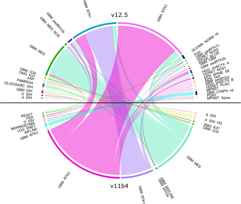
Methylation classification using the Heidelberg classifier in the 11b4 version (4) showed that 89.7% of tumor samples from patients in the EORTC-26101 biomarker cohort had the highest classifier score for the three main glioblastoma groups, receptor tyrosine kinase (RTK)1 (63, 16.6%), RTK2 (174, 45.8%) and mesenchymal (MES, 104, 27.4%). Thirteen tumors (3.4%) were classified as IDH-mutant gliomas, three tumors as H3 G34-mutant glioblastoma and glioblastoma subclass midline, two as diffuse midline glioma, H3 K27-mutant. Seven tumors classified most consistently with other glioma groups and in 11 patients (3%) the classification was most similar to inflammatory or reactive tissue. Of these 11 tumors, the TERT promoter mutation was found in three samples; in one of them the copy-number profile identified a 7+/10- signature with cyclin-dependent kinase inhibitor N2A/B deletion, allowing the diagnosis of glioblastoma. Analysis of these samples with the new v12.5 version of the classifier was able to resolve a diagnosis of glioblastoma in all patients. In 10 of 11 tumors, v12.5 classification established the prediction of an infiltration zone of glioblastoma and the remaining tumor was classified as mesenchymal glioblastoma. Analysis of all samples with the v12.5 version of the classifier identified a shift in the subclassification for 119/380 (31%) tumor samples (Fig. 1). Of note, 44/119 (37%) tumors with a shift in subclassification reached a class specific score of >0.9 in the v12.5 version. However, the largest groups remained RTK1 (89, 23.4%), RTK2 (140, 36.8%), and MES (66, 17.4%), but with a substantial increase in rare tumor entities. We used the current v12.5 version of the classifier to assess the prognostic value of methylation groups in glioblastoma in the progressive situation.
Figure 1.
Methylation classes in progressive glioblastoma of the EORTC-26101 study. Circos blot showing methylation classification according to the v11b4 and v12.5 version of the methylation classifier (n = 380).
Prognostic value of methylation classes and MGMT promoter methylation
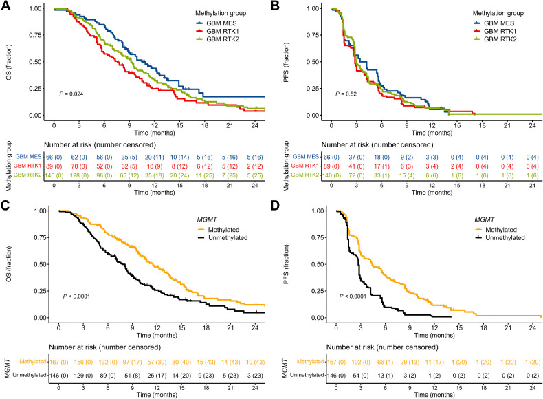
Patients with RTK1 tumors classified by the v12.5 classifier showed a worse prognosis for OS in comparison with the other two main glioblastoma methylation groups RTK2 and MES [median {95% confidence interval (CI)}: RTK1, 7.6 {5.9–9.6}; RTK2, 9.2 {8.1–10.7}; MES, 10.5 {8.8–13.5}; P = 0.024; Fig. 2A]. However, there was no difference regarding PFS (Fig. 2B). The same OS disadvantage of patients with RTK1 tumors was previously shown in the Neurooncology Working Group of the German Cancer Society (NOA)-08 cohort for patients with primary glioblastoma (12).
Figure 2.
Prognostic effect of the main glioblastoma methylation classes and MGMT promoter methylation status. A, OS of patients with the three main glioblastoma groups (RTK1, RTK2, and MES, n = 295) identified with classifier v12.5. B, PFS of the same patient cohort as in A (n = 295). C, OS according to MGMT promoter methylation (n = 313). Patients with MGMT promoter methylation status of “undeterminable” were excluded from the analysis. D, PFS according to MGMT promoter methylation (n = 313).
In most patients, MGMT promoter methylation is retained at tumor progression (13). We previously reported longer PFS and OS of patients with MGMT promoter methylation in a progressive situation in a subgroup of these EORTC-26101 patients (1). In the present full biomarker cohort, MGMT promoter methylation was prognostic for both prolonged OS [median 11.5 (10.2–13.4) vs. 7.8 (6.9–8.7) months; P < 0.0001] and PFS [median 4.4 (3.4–5.7) vs. 2.7 (2.5–2.8); P < 0.0001; Fig. 2C and D]. MGMT promoter methylation was more prevalent in the RTK2 methylation class (Supplementary Fig. S3A). However, in a multivariate analysis taking the methylation subgroup and MGMT promoter methylation status into account, there was still an OS disadvantage of patients with RTK1 tumors (Supplementary Fig. S4). Clinical characteristics did not differ between patients with RTK1 tumors and RTK2/MES tumors (Supplementary Table S2). However, RTK1 tumors had more tumor protein (TP)53 mutations compared with RTK2 tumors (24% vs. 9%; P = 0.0094; Supplementary Fig. S3B and S3C) and neurofibromin (NF)1 mutations were the most frequent mutations in MES tumors, but rare in RTK1 (21% vs. 4%; P = 0.0015; Supplementary Fig. S3B and S3D). Copy-number analysis identified a higher rate of platelet-derived growth factor receptor (PDGFR)A amplifications in RTK1 tumors and gains of chromosomes 19 and 20 in RTK2 tumors (Supplementary Fig. S5).
Prognostic effect of recurrent mutations in glioma
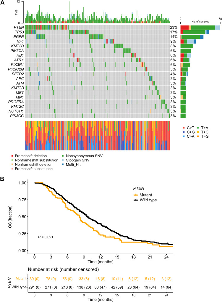
We identified five genes with most frequent mutations in panel sequencing analysis and assessed the impact on OS in the progressive situation in the full EORTC-26101 biomarker cohort (n = 380). We applied stringent filtering for sequencing artefacts and common nonpathogenic synonymous variants. Finally, exonic SNVs and indels likely to be pathogenic were included in the analysis. The most prevalent mutations were found in phosphatase and tensin homolog (PTEN, 23%), TP53 (17%), EGFR (14%), NF1 (9%) and lysine methyltransferase 2D (KMT2D, 8%). Mutations in these genes were mainly SNV (Fig. 3A). Of these, only PTEN mutations were prognostic for OS (P = 0.021; Fig. 3B). To exclude a bias potentially introduced by prognostically favorable IDH-mutant tumors, we defined a subgroup of 362/380 (95.3%) patients with IDH–wild-type glioblastoma. In this subgroup, PTEN mutations were still prognostic (P = 0.047; Supplementary Fig. S6), while all other above-mentioned genes were not.
Figure 3.
Impact of somatic mutations in the EORTC-26101 cohort. A, Oncoplot with SNVs and indels in the 20 most frequently affected genes in the complete EORTC-26101 biomarker cohort (n = 380). B, OS according to PTEN mutation status (n = 380).
NF1 mutation predicts response to bevacizumab treatment
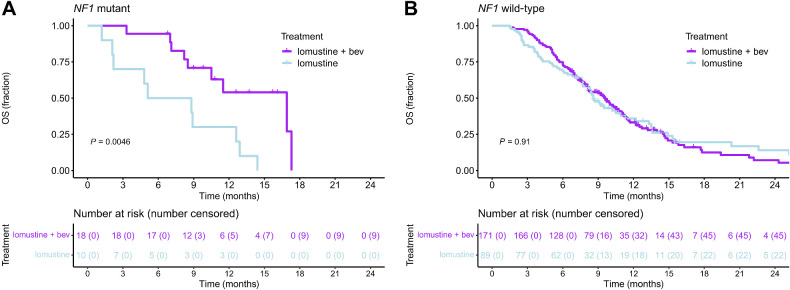
We used the methylation subgroups and the genes with recurrent mutations to identify markers that are prognostic for bevacizumab response. The cohort was restricted to patients receiving lomustine or lomustine + bevacizumab (n = 288); patients with sequential treatments were excluded (n = 92). We evaluated the five above mentioned most frequent mutations (PTEN, TP53, EGFR, NF1, and KMT2D) as well as MGMT promoter methylations status. Patients with NF1 mutation had longer OS in the lomustine + bevacizumab arm compared with the lomustine alone arm (P = 0.0046; FDR = 0.0276; Fig. 4A). Of note, all patients with NF1 mutations (28/28) had IDH–wild-type glioblastomas. No difference was seen between the two treatments in the NF1 wild-type subgroup (Fig. 4B). The other factors were not significant.
Figure 4.
NF1 mutation is a prognostic factor for response to bevacizumab therapy. A, OS of patients with NF1 mutations according to treatment (n = 28). B, OS of patients with NF1 wild-type according to treatment (n = 260). Bev, bevacizumab.
Exceptional survivors
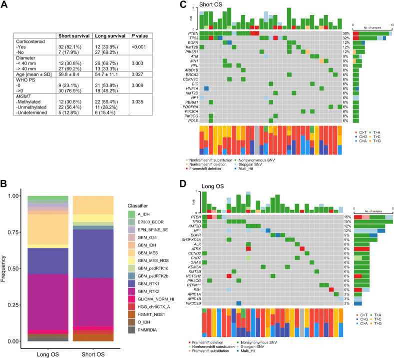
Patients with progression of glioblastoma after standard therapy usually have an unfavorable prognosis with a median OS of only 8.9 (8.3–9.6) months in the EORTC-26101 study (Table 1). We therefore did an exploratory analysis of patients with the 10% shortest (n = 39) and 10% longest (n = 39) OS times in the EORTC-26101 biomarker cohort to identify specific molecular profiles associated with survival in a progressive glioblastoma cohort. Median OS was 2.3 (2.1–2.6) months in the short survival group and 22.7 (21–27.4) months in the long survival group (Supplementary Fig. S7). As expected, general risk factors such as corticosteroid intake, large tumor size at study entry, age, performance status >0 and an unmethylated MGMT promoter were more frequent in the short survival group (Fig. 5A). There were more patients with RTK1 tumors in the short survival group compared with long surviving patients [short: 7/39 (17.9%), long: 13/39 (33.3%); Fig. 5B]. In patients with short survival PTEN [short: 13/39 (33.3%), long 5/39 (12.8%)] and TP53 [short: 11/39 (28.2%), long: 5/39 (12.8%)] mutations were more prevalent (Fig. 5C). In patients with long survival, there was no specific enrichment of mutations found (Fig. 5D).
Figure 5.
Features of patients with long and short OS. A, Clinical characteristics of patients with the 10% shortest (n = 39) and longest (n = 39) OS. B, Distribution of methylation classification in patients with short (n = 39) and long (n = 39) OS. C, Oncoplot of patients with the 10% shortest OS (n = 39). D, Oncoplot of patients with the 10% longest OS (n = 39).
Discussion
Patients with progression of glioblastoma usually have a limited prognosis with few therapeutic options. In the EORTC-26101 study, bevacizumab failed to show a clinical benefit in terms of OS when given in addition to lomustine over lomustine alone in patients with progressive glioblastoma. However, methylation profiling and NGS allow stratification into diagnostic and prognostic groups to optimize targeted treatment.
This study was able to show that patients with the RTK1 methylation phenotype have a worse prognosis compared with the other two main glioblastoma phenotypes, RTK2 and mesenchymal here for the first time, especially in a progressive situation. We have previously shown this as a result of the NOA-08 biomarker analysis in a study cohort of elderly patients treated with either temozolomide or radiotherapy for primary glioblastoma (12). Here, this OS disadvantage was valid for a progressive situation; however, we did not identify differences in PFS in these three methylation groups. Nonetheless, despite the fact that PDGFRA amplification and TP53 mutation were found to be more prevalent in RTK1 tumors, the exact reason for the survival disadvantage of these patients remains unclear and may be subject to further functional evaluation. MGMT promoter methylation is a well-defined biomarker for response to temozolomide treatment in primary glioblastoma (13, 14) based on its ability to reverse methylation of the O6 position of guanine introduced by temozolomide (15). The EORTC-26101 trial established MGMT promoter methylation in a prospective decently sized study as a robust prognostic biomarker in the progressive situation with a lomustine-based treatment, which is well explainable with its similar alkylating mode of action that induces O6-chloroethylguanine that can be reverted by MGMT (16).
We specifically investigated the prognostic relationship between methylation classification and MGMT promoter methylation, which showed independent association of RTK1 phenotype and MGMT promoter methylation status with prognosis in a multivariate analysis, suggesting that classifier assignment and MGMT contribute to prognosis through different mechanisms.
PTEN mutation was prognostic for decreased OS in the progressive situation and PTEN mutations accumulated in the subgroup of short surviving patients. Previous studies suggested a prognostic value of PTEN only in the pre-temozolomide era with a counteracting effect of temozolomide, based on a higher sensitivity of PTEN mutant tumors to temozolomide treatment (17, 18). However, we speculate that this sensitizing effect might not be present in the progressive situation with lomustine-based regimens, thus leading to the present prognostic impact.
We detected an OS survival advantage of patients treated with lomustine + bevacizumab compared with lomustine alone in the subgroup of patients with the NF1 mutation. This effect was not seen in patients with NF1 wild-type tumors. Given a report that suggested prolonged survival of neurofibromatosis type 1 patients with recurrent high grade gliomas treated with bevacizumab in an uncontrolled case series (19) and the mechanistic role of NF1 in promoting angiogenesis (20), a sensitizing effect of NF1 mutations to antiangiogenic treatment with bevacizumab seems plausible and encourages further research.
A previous study partially contained expression data from patients from the EORTC-26101 trial and defined an “ATE score” comprising nine genes that predicted OS, though this score did not have a predictive impact for bevacizumab treatment (21).
There are also some limitations. The EORTC-26101 biomarker cohort consists of 380 of 596 (63.8%) patients based on availability of tissue for molecular analysis and not derived from a power calculation or stratification for biomarker assessment, thus representing a secondary endpoint of the trial. A potential bias through this selection was ruled out as much as possible through a high number of completely analyzed patients and comparison of main bias factors, which did not show differences compared to the original study cohort. Most tissues have been acquired during the primary operation and only a very few at study entry, not allowing direct insight into the molecular characteristics at progression. However, previous studies have shown that the information assessed here does not relevantly alter through treatment (2, 3, 13), especially concerning MGMT and methylation profiles, but also to a certain extent for NF1 mutations, rendering these data valid to use. Finally, there are limited large clinical trial grade data for progressive glioblastoma, so validation and comparison with a further cohort are currently not possible. The EORTC 26091 TAVAREC trial (22, 23) followed a similar treatment regimen with mainly IDH-mutant progressive gliomas comparing temozolomide versus temozolomide + bevacizumab. In this trial, MGMT promoter methylation was also prognostic of OS. However, we describe here a prognostic effect of MGMT promoter methylation upon lomustine-based treatment. There are no sequencing data available in TAVAREC (22) and methylation profiles inherently differ on the basis of the primarily IDH-mutant patients included.
In conclusion, we demonstrate that methylation profiling can be used in clinical trials for confirmation and refinement of the tumor diagnosis. RTK1 methylation phenotype and MGMT promoter methylation are prognostic for OS in a progressive situation. NF1 mutation could be a potential predictive biomarker for bevacizumab treatment but needs further validation.
Supplementary Material
Supplementary Figure 1: Overview of the clinical trial and biomarker analysis. Out of 596 patients in the EORTC-26101 trial, patient tumor tissue to perform DNA methylation (Meth) and NGS sequencing analysis was available from 380 patients (63.8%). Of these, 99 patients treated with lomustine and 189 patients treated with lomustine and bevacizumab were eligible to detect biomarkers for bevacizumab response.
Supplementary Figure 2: Overall (A) and progression-free (B) survival in the EORTC-26101 clinical study (n = 596) and EORTC-26101 biomarker cohort (n = 380).
Supplementary Figure 3: Characteristics of RTK1 tumors in comparison to RTK2 and MES. A Distribution of MGMT promotor methylation in main glioblastoma methylation subgroups. Classifier assignment is based on the v12.5 version (n = 295). Mutational profiles in B RTK1 tumors (n = 89), C RTK2 tumors (n = 140) and D MES tumors (n = 66).
Supplementary Figure 4: Multivariate analysis. Baselines were MGMT = “methylated” and Classifier = “GBM MES”. Classifier assignment is based on the v12.5 version (n = 380).
Supplementary Figure 5: Copy number variants in the three main glioblastoma groups according to classifier v12.5. A Copy number variants in all patients combined (n = 295), B in RTK1 tumors (n = 89), C in RTK2 tumors (n = 140) and D in MES tumors (n = 66). PDGFRA amplifications are enriched in RTK1 tumors and gains of chromosomes 19 and 20 are enriched in RTK2 tumors.
Supplementary Figure 6: Overall Survival according to PTEN mutation status in the group of IDH-wildtype glioblastomas (n = 362).
Supplementary Figure 7: Exceptional survivors of the EORTC-26101 study (n = 78).
Supplementary Table 1: Representativeness of study participants.
Supplementary Table 2: Characteristics of patients with RTK1 tumors compared to RTK2 and MES according to classifier version v12.5. P-value was calculated for comparison between RTK1 and both other groups (RTK2 and MES) combined. NA, not available.
Acknowledgments
We thank all patients, relatives, doctors, and nurses that participated in the EORTC-26101 study. The work was supported by the SFB grant UNITE Glioblastoma (SFB1389, WP A03, to T. Kessler and W. Wick) of the German Research Foundation (DFG).
This article is featured in Selected Articles from This Issue, p. 3827
Footnotes
Note: Supplementary data for this article are available at Clinical Cancer Research Online (http://clincancerres.aacrjournals.org/).
Authors' Disclosures
T. Kessler reports grants from the German Research Foundation (DFG) during the conduct of the study. M. van den Bent reports personal fees from Nerviano, Servier, Incyte, Fore Biotherapeutics, Boehringer, AstraZeneca, Genenta, and Chimerix outside the submitted work. A. Idbaih reports other support from Enterome, Carthera, Sanofi, Nutrithéragène, Servier, and Transgene outside the submitted work; personal fees and other support From Novocure and Leo Pharma; and personal fees from Boehringer Ingelheim and Novartis. P.M. Clement reports other support from EORTC during the conduct of the study; personal fees from Merck, Leo Pharma, Rakuten Medical, Takeda, and Bristol Myers Squibb; personal fees and nonfinancial support from MSD; grants from AstraZeneca outside the submitted work; and acts as an occasional adviser to government agencies such as FAGG/EMA and as a substitute member of the CTG in Belgium. M. Campone reports grants and personal fees from Novartis and Lilly; and grants from AstraZeneca, Sanofi, Daiichi Sankyo, PET-THERAPY, Menarini, Gilead, and Seagen outside the submitted work. A. von Deimling reports a patent for EP16710700 issued; a patent for EP11767970 issued, licensed, and with royalties paid from Roche; and a patent for EP09015511 issued, licensed, and with royalties paid from Dianovy; and is co-owner of the company Epignostics. F. Sahm reports other support from Illumina, Bayer, and Heidelberg Epignostix GmbH during the conduct of the study; and has a patent for methods related to classification of cancer pending, licensed, and with royalties paid. W. Wick reports personal fees from Servier, GSK, Enterome, AstraZeneca, and MSD during the conduct of the study; nonfinancial support from Pfizer and Apogenix; and personal fees and nonfinancial support from Roche. No disclosures were reported by the other authors.
Authors' Contributions
T. Kessler: Conceptualization, data curation, software, formal analysis, funding acquisition, validation, investigation, visualization, methodology, writing–original draft, writing–review and editing. D. Schrimpf: Data curation, software, investigation, methodology, writing–review and editing. L. Doerner: Investigation, methodology, writing–review and editing. L. Hai: Software, investigation, methodology, writing–review and editing. L.D. Kaulen: Formal analysis, validation, investigation, writing–review and editing. J. Ito: Software, formal analysis, investigation, writing–review and editing. M. van den Bent: Resources, validation, writing–review and editing. M. Taphoorn: Resources, validation, writing–review and editing. A.A. Brandes: Resources, validation, writing–review and editing. A. Idbaih: Resources, validation, writing–review and editing. J. Dômont: Resources, validation, writing–review and editing. P.M. Clement: Resources, validation, writing–review and editing. M. Campone: Resources, validation, writing–review and editing. M. Bendszus: Resources, validation, writing–review and editing. A. von Deimling: Resources, funding acquisition, validation, methodology, writing–review and editing. F. Sahm: Conceptualization, resources, funding acquisition, validation, methodology, project administration, writing–review and editing. M. Platten: Conceptualization, resources, validation, writing–review and editing. W. Wick: Conceptualization, resources, supervision, funding acquisition, validation, methodology, writing–original draft, project administration, writing–review and editing. A. Wick: Conceptualization, resources, supervision, validation, methodology, writing–original draft, project administration, writing–review and editing.
References
- 1. Wick W, Gorlia T, Bendszus M, Taphoorn M, Sahm F, Harting I, et al. Lomustine and bevacizumab in progressive glioblastoma. N Engl J Med 2017;377:1954–63. [DOI] [PubMed] [Google Scholar]
- 2. Korber V, Yang J, Barah P, Wu Y, Stichel D, Gu Z, et al. Evolutionary trajectories of IDH(WT) glioblastomas reveal a common path of early tumorigenesis instigated years ahead of initial diagnosis. Cancer Cell 2019;35:692–704. [DOI] [PubMed] [Google Scholar]
- 3. Draaisma K, Chatzipli A, Taphoorn M, Kerkhof M, Weyerbrock A, Sanson M, et al. Molecular evolution of IDH wild-type glioblastomas treated with standard of care affects survival and design of precision medicine trials: a report from the EORTC 1542 study. J Clin Oncol 2020;38:81–99. [DOI] [PubMed] [Google Scholar]
- 4. Capper D, Jones DTW, Sill M, Hovestadt V, Schrimpf D, Sturm D, et al. DNA methylation-based classification of central nervous system tumors. Nature 2018;555:469–74. [DOI] [PMC free article] [PubMed] [Google Scholar]
- 5. Louis DN, Perry A, Wesseling P, Brat DJ, Cree IA, Figarella-Branger D, et al. The 2021 WHO classification of tumors of the central nervous system: a summary. Neuro Oncol 2021;23:1231–51. [DOI] [PMC free article] [PubMed] [Google Scholar]
- 6. Wiestler B, Capper D, Sill M, Jones DT, Hovestadt V, Sturm D, et al. Integrated DNA methylation and copy-number profiling identify three clinically and biologically relevant groups of anaplastic glioma. Acta Neuropathol 2014;128:561–71. [DOI] [PubMed] [Google Scholar]
- 7. Eckel-Passow JE, Lachance DH, Molinaro AM, Walsh KM, Decker PA, Sicotte H, et al. Glioma groups based on 1p/19q, IDH, and TERT promoter mutations in tumors. N Engl J Med 2015;372:2499–508. [DOI] [PMC free article] [PubMed] [Google Scholar]
- 8. Ceccarelli M, Barthel FP, Malta TM, Sabedot TS, Salama SR, Murray BA, et al. Molecular profiling reveals biologically discrete subsets and pathways of progression in diffuse glioma. Cell 2016;164:550–63. [DOI] [PMC free article] [PubMed] [Google Scholar]
- 9. Bady P, Sciuscio D, Diserens AC, Bloch J, van den Bent MJ, Marosi C, et al. MGMT methylation analysis of glioblastoma on the Infinium methylation BeadChip identifies two distinct CpG regions associated with gene silencing and outcome, yielding a prediction model for comparisons across datasets, tumor grades, and CIMP-status. Acta Neuropathol 2012;124:547–60. [DOI] [PMC free article] [PubMed] [Google Scholar]
- 10. Sahm F, Schrimpf D, Jones DT, Meyer J, Kratz A, Reuss D, et al. Next-generation sequencing in routine brain tumor diagnostics enables an integrated diagnosis and identifies actionable targets. Acta Neuropathol 2016;131:903–10. [DOI] [PubMed] [Google Scholar]
- 11. Rimmer A, Phan H, Mathieson I, Iqbal Z, Twigg SRF, WGS500 Consortium, et al. Integrating mapping-, assembly-, and haplotype-based approaches for calling variants in clinical sequencing applications. Nat Genet 2014;46:912–8. [DOI] [PMC free article] [PubMed] [Google Scholar]
- 12. Wick A, Kessler T, Platten M, Meisner C, Bamberg M, Herrlinger U, et al. Superiority of temozolomide over radiotherapy for elderly patients with RTK II methylation class, MGMT promoter methylated malignant astrocytoma. Neuro Oncol 2020;22:1162–72. [DOI] [PMC free article] [PubMed] [Google Scholar]
- 13. Kessler T, Sahm F, Sadik A, Stichel D, Hertenstein A, Reifenberger G, et al. Molecular differences in IDH wild-type glioblastoma according to MGMT promoter methylation. Neuro Oncol 2018;20:367–79. [DOI] [PMC free article] [PubMed] [Google Scholar]
- 14. Hegi ME, Diserens AC, Gorlia T, Hamou MF, de Tribolet N, Weller M, et al. MGMT gene silencing and benefit from temozolomide in glioblastoma. N Engl J Med 2005;352:997–1003. [DOI] [PubMed] [Google Scholar]
- 15. Lee SY. Temozolomide resistance in glioblastoma multiforme. Genes Dis 2016;3:198–210. [DOI] [PMC free article] [PubMed] [Google Scholar]
- 16. Nikolova T, Roos WP, Kramer OH, Strik HM, Kaina B. Chloroethylating nitrosoureas in cancer therapy: DNA damage, repair and cell death signaling. Biochim Biophys Acta Rev Cancer 2017;1868:29–39. [DOI] [PubMed] [Google Scholar]
- 17. Carico C, Nuno M, Mukherjee D, Elramsisy A, Dantis J, Hu J, et al. Loss of PTEN is not associated with poor survival in newly diagnosed glioblastoma patients of the temozolomide era. PLoS One 2012;7:e33684. [DOI] [PMC free article] [PubMed] [Google Scholar]
- 18. Smith JS, Tachibana I, Passe SM, Huntley BK, Borell TJ, Iturria N, et al. PTEN mutation, EGFR amplification, and outcome in patients with anaplastic astrocytoma and glioblastoma multiforme. J Natl Cancer Inst 2001;93:1246–56. [DOI] [PubMed] [Google Scholar]
- 19. Theeler BJ, Ellezam B, Yust-Katz S, Slopis JM, Loghin ME, de Groot JF. Prolonged survival in adult neurofibromatosis type I patients with recurrent high-grade gliomas treated with bevacizumab. J Neurol 2014;261:1559–64. [DOI] [PubMed] [Google Scholar]
- 20. Kawachi Y, Maruyama H, Ishitsuka Y, Fujisawa Y, Furuta J, Nakamura Y, et al. NF1 gene silencing induces upregulation of vascular endothelial growth factor expression in both Schwann and non-Schwann cells. Exp Dermatol 2013;22:262–5. [DOI] [PubMed] [Google Scholar]
- 21. Johnson RM, Phillips HS, Bais C, Brennan CW, Cloughesy TF, Daemen A, et al. Development of a gene expression-based prognostic signature for IDH wild-type glioblastoma. Neuro Oncol 2020;22:1742–56. [DOI] [PMC free article] [PubMed] [Google Scholar]
- 22. Draaisma K, Tesileanu CMS, de Heer I, Klein M, Smits M, Reijneveld JC, et al. Prognostic significance of DNA methylation profiles at MRI enhancing tumor recurrence: a report from the EORTC 26091 TAVAREC trial. Clin Cancer Res 2022;28:2440–8. [DOI] [PubMed] [Google Scholar]
- 23. van den Bent MJ, Klein M, Smits M, Reijneveld JC, French PJ, Clement P, et al. Bevacizumab and temozolomide in patients with first recurrence of WHO grade II and III glioma, without 1p/19q co-deletion (TAVAREC): a randomised controlled phase II EORTC trial. Lancet Oncol 2018;19:1170–9. [DOI] [PubMed] [Google Scholar]
Associated Data
This section collects any data citations, data availability statements, or supplementary materials included in this article.
Supplementary Materials
Supplementary Figure 1: Overview of the clinical trial and biomarker analysis. Out of 596 patients in the EORTC-26101 trial, patient tumor tissue to perform DNA methylation (Meth) and NGS sequencing analysis was available from 380 patients (63.8%). Of these, 99 patients treated with lomustine and 189 patients treated with lomustine and bevacizumab were eligible to detect biomarkers for bevacizumab response.
Supplementary Figure 2: Overall (A) and progression-free (B) survival in the EORTC-26101 clinical study (n = 596) and EORTC-26101 biomarker cohort (n = 380).
Supplementary Figure 3: Characteristics of RTK1 tumors in comparison to RTK2 and MES. A Distribution of MGMT promotor methylation in main glioblastoma methylation subgroups. Classifier assignment is based on the v12.5 version (n = 295). Mutational profiles in B RTK1 tumors (n = 89), C RTK2 tumors (n = 140) and D MES tumors (n = 66).
Supplementary Figure 4: Multivariate analysis. Baselines were MGMT = “methylated” and Classifier = “GBM MES”. Classifier assignment is based on the v12.5 version (n = 380).
Supplementary Figure 5: Copy number variants in the three main glioblastoma groups according to classifier v12.5. A Copy number variants in all patients combined (n = 295), B in RTK1 tumors (n = 89), C in RTK2 tumors (n = 140) and D in MES tumors (n = 66). PDGFRA amplifications are enriched in RTK1 tumors and gains of chromosomes 19 and 20 are enriched in RTK2 tumors.
Supplementary Figure 6: Overall Survival according to PTEN mutation status in the group of IDH-wildtype glioblastomas (n = 362).
Supplementary Figure 7: Exceptional survivors of the EORTC-26101 study (n = 78).
Supplementary Table 1: Representativeness of study participants.
Supplementary Table 2: Characteristics of patients with RTK1 tumors compared to RTK2 and MES according to classifier version v12.5. P-value was calculated for comparison between RTK1 and both other groups (RTK2 and MES) combined. NA, not available.
Data Availability Statement
Methylation raw and processed data are available via the Gene Expression Omnibus (GEO) database (https://www.ncbi.nlm.nih.gov/geo/; RRID:SCR_005012) under the GEO accession number GSE237103. Sequence data have been deposited at the European Genome-phenome Archive (EGA), which is hosted by the EBI and the CRG, under accession number EGAS00001007421 (https://ega-archive.org; RRID:SCR_004944). Source code can be made available upon reasonable request.


