Abstract
Background
Early reports have indicated a relationship between ABO and rhesus blood group types and infection with SARS-CoV-2. We aim to examine blood group type associations with COVID-19 mortality and disease severity.
Methods
This is a retrospective chart review of patients ages 18 years or older admitted to the hospital with COVID-19 between January 2020 and December 2021. The primary outcome was COVID-19 mortality with respect to ABO blood group type. The secondary outcomes were 1. Severity of COVID-19 with respect to ABO blood group type, and 2. Rhesus factor association with COVID-19 mortality and disease severity. Disease severity was defined by degree of supplemental oxygen requirements (ambient air, low-flow, high-flow, non-invasive mechanical ventilation, and invasive mechanical ventilation).
Results
The blood type was collected on 596 patients with more than half (54%, N=322) being O+. The ABO blood type alone was not statistically associated with mortality (P=0.405), while the RH blood type was statistically associated with mortality (P<0.001). There was statistically significant association between combined ABO and RH blood type and mortality (P=0.014). Out of the mortality group, the O+ group had the highest mortality (52.3%), followed by A+ (22.8%). The combined ABO and RH blood type was statistically significantly associated with degree of supplemental oxygen requirements (P=0.005). The Kaplan-Meier curve demonstrated that Rh-patients had increased mortality.
Conclusion
ABO blood type is not associated with COVID-19 severity and mortality. Rhesus factor status is associated with COVID-19 severity and mortality. Rhesus negative patients were associated with increased mortality risk.
Keywords: COVID-19, Infectious disease, Pulmonary medicine, Mechanical ventilation, SARS-CoV-2
INTRODUCTION
The ABO blood group, which includes the 4 blood types A, AB, B, and O, plays a role in various infectious and non-infectious human diseases. Histo-blood group antigens located on the surface of red blood cells represent inherited polymorphic traits, and differences in the expression of these antigens affect susceptibility to many infections [1]. Blood group antigens act as receptors or coreceptors for pathogens such as hepatitis B virus, middle east respiratory syndrome associated coronavirus (MERS-CoV), severe acute respiratory syndrome associated coronavirus (SARS-CoV), norovirus, malaria, other microorganisms, and parasites [1-4]. Additionally, some blood group antigens can aid in intracellular uptake or signal transduction, subsequently altering the innate immune response to infection [1]. Early reports have indicated a significant relationship of ABO blood group types to the risk of infection with SARS-CoV-2 [1-4]. Many studies have demonstrated that type O blood may have decreased risk of infection [5-13], while type A blood may be more susceptible to COVID-19 [2]. A hypothesized explanation includes the angiotensin converting enzyme 2 (ACE2) protein on host cell surfaces serving as a cellular receptor for the transmembrane spike (S) protein on SARS-CoV-2 [3]. Anti-A antibodies, as found in blood types O or B, seem to antagonize the binding of the S protein and ACE2 receptor [3, 4]. Therefore, blood groups appear to be a determinant of the coronavirus disease of 2019 (COVID-19) susceptibility. However, data surrounding the association of blood groups with disease severity and mortality is incohesive. We aim to examine blood group type associations with COVID-19 mortality and disease severity.
MATERIAL AND METHODS
A retrospective observational cohort review of patients with COVID-19 admitted to a single community, high-capacity Level 1 trauma center between January 2020 and December 2021 was performed. The county facility is a teaching hospital seeing more than 120,000 patients in the emergency department annually. All patients admitted to the hospital with a primary diagnosis of COVID-19 were gathered. Exclusion criteria included COVID-19 in pregnancy and age under 18. For all 1,993 identified, a retrospective chart review was performed to obtain emergency department records, admission records, and inpatient records. Infection with SARS-CoV-2 was confirmed by nasal polymerase chain reaction analysis for all patients diagnosed with COVID-19. This study was conducted in compliance with the ethical standards of the Arrowhead Regional Medical Center (ARMC) Institutional Review Board on human subjects as well as with the Helsinki Declaration. The ARMC Institutional Review Board issued approval for protocol #22-53.
The data collected included patient age, gender, ethnicity, medical comorbidities, medications, ABO blood group type, rhesus factor (Rh) status, oxygen supplementation requirements, and mortality. The primary endpoints included mortality and COVID-19 illness severity. Illness severity was defined by degree of supplemental oxygen requirements (ambient air, low-flow nasal cannula, high-flow nasal cannula, non-invasive mechanical ventilation, and invasive mechanical ventilation), as this indicates degree of hypoxemia and thus illness severity.
For blood group serology, our institution utilized the Echo Lumena device (manufactured by Immucor) to perform serologic testing. To confirm ABO blood types, we utilized a two-step method: 1) forward testing for A and B antigens using anti-A and anti-B reagents (manufactured by Immucor), respectively for agglutination, and 2) reverse testing for anti-A and/or anti-B isohemagglutinins utilizing A1 and B reagent red cells (manufactured by Immucor). For rhesus factor testing, we utilized forward testing for D antigen using an anti-D reagent (manufactured by Immucor) for agglutination; if the former test is negative, we utilized confirmatory anti-IgG (murine monoclonal) reagent (manufactured by Immucor).
The data was collected using Microsoft Excel and information was stored on a password-secured computer folder. The data was analyzed using descriptive statistics and univariate analysis. Statistical analysis was performed using the Statistical Package for the Social Sciences (SPSS) version 27.0 (SPSS Inc, Chicago, IL) software. Univariate analyses were performed using Chi-Squared for categorical data. Continuous data was analyzed using non-parametric Mann-Whitney U tests. Continuous data were presented according to the means with standard deviation, and categorical data are presented with percentages. A multivariate analysis was performed and a Kaplan-Meier curve was generated while adjusting for other risk factors. Unless otherwise indicated, a P-value less than 0.05 was considered statistically significant.
RESULTS
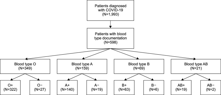
Among the 1,993 patients diagnosed with COVID-19, 1,397 patients were excluded due to insufficient ABO blood type documentation. Type and screen tests were not performed automatically on all patients admitted, limiting the final number of patients included in the final analysis. As a result, 596 patients were included in the final analysis. There was a total of 322 patients with blood type O+, and 27 with type O-, 140 with type A+, 19 with type A-, 63 with type B+, 6 with type B-, 19 with type AB+, and 2 with type AB-. The Fig. 1 consort diagram summaries the details. In the study, the mean age for blood types O, B, AB, and A were 51.48±19.5, 51.55±19.68, 48.05±20.65, and 53.48±20.20, respectively (P=0.578). Similarly, there was no statistical significance in mean age between rhesus factor positive and rhesus factor negative blood types. The differences in gender, body mass index, and ethnicity between groups were found to not be statistically significant as summarized in Table 1.
Fig. 1.
Consort diagram detailing the selection of patients with COVID-19 and their respective blood types.
Table 1.
Baseline patient characteristics based upon ABO blood type and Rh status separately with mortality data presented for each.
| Blood type O (N=349) |
Blood type B (N=69) |
Blood type AB (N=21) |
Blood type A (N=159) |
P | Rh positive (N=541) |
Rh negative (N=54) |
P | |
|---|---|---|---|---|---|---|---|---|
| Demographics | ||||||||
| Age | 51.48±19.5 | 51.55±19.68 | 48.05±20.65 | 53.48±20.20 | 0.578 | 48.81±19.14 | 52.21±19.87 | 0.221 |
| Body mass index | 30.65±7.8 | 28.7±5.6 | 27.5±7.09 | 29.35±7.73 | 0.047 | 30.20±6.35 | 29.94±7.74 | 0.785 |
| Gender | 0.642 | 0.126 | ||||||
| Female | 176 (51%) | 30 (43%) | 12 (57%) | 77 (48%) | 263 (49%) | 32 (61%) | ||
| Male | 173 (49%) | 39 (57%) | 9 (43%) | 82 (52%) | 281 (51%) | 22 (39%) | ||
| Ethnicity | 0.168 | 0.655 | ||||||
| Caucasian | 208 (60%) | 27 (39%) | 14 (67%) | 91 (57%) | 310 (57%) | 30 (56%) | ||
| African American | 21 (6%) | 6 (9%) | 0 | 8 (5%) | 34 (6%) | 1 (2%) | ||
| Asian | 5 (1.5%) | 3 (4%) | 0 | 4 (2.5%) | 11 (2%) | 1 (2%) | ||
| Hispanic | 112 (32%) | 33 (48%) | 7 (33%) | 53 (33%) | 184 (34%) | 21 (39%) | ||
| Other | 3 (0.5%) | 0 | 0 | 0 | 5 (1%) | 1 (1%) | ||
| Comorbid conditions | ||||||||
| Diabetes mellitus | 77 (22%) | 16 (23.2%) | 4 (19%) | 36 (22.6%) | 0.981 | 119 (22%) | 14 (25.9%) | 0.495 |
| Tobacco use | 7 (2%) | 1 (1.4%) | 0 (0%) | 2 (1.3%) | 0.854 | 9 (1.7%) | 1 (1.9%) | 0.914 |
| Cancer | 12 (3.4%) | 1 (1.4%) | 0 (0%) | 7 (4.4%) | 0.561 | 18 (3.3%) | 2 (3.7%) | 0.878 |
| Hypertension | 48 (13.8%) | 10 (14%) | 3 (14.3%) | 127 (79.9%) | 0.324 | 87 (16.1%) | 6 (11.1%) | 0.345 |
| Obesity | 56 (16%) | 8 (11.6%) | 2 (9.5%) | 21 (13.2%) | 0.621 | 76 (14%) | 11 (20.4%) | 0.203 |
| Chronic lung disease | 14 (4%) | 3 (4.3%) | 1 (4.8%) | 3 (1.9%) | 0.629 | 21 (3.9%) | 0 (0%) | 0.142 |
| Cirrhosis | 8 (2.3%) | 0 (0%) | 0 (0%) | 4 (2.5%) | 0.532 | 11 (6.9%) | 1 (1.9%) | 0.932 |
| Chronic kidney disease | 19 (5.4%) | 4 (5.8%) | 3 (14.3%) | 6 (3.8%) | 0.248 | 29 (5.4%) | 3 (5.6%) | 0.944 |
| Hospitalization mortality | 0.405 | <0.001 | ||||||
| Alive | 258 (73.9%) | 57 (82.6%) | 17 (81%) | 117 (73.6%) | 419 (77.4%) | 30 (55.6%) | ||
| Dead | 91 (26.1%) | 12 (17.4%) | 4 (19%) | 42 (26.4%) | 125 (23.1%) | 24 (44.4%) |
Patients with COVID-19 as grouped by blood type or rhesus factor did not have any statically significant medical comorbidities. The percentage of diabetes mellitus for blood type O, B, AB, A (22%, 23.2%, 19%, 22.6%, P=0.981), percentage of obesity (defined by body mass index greater than or equal to 30 kg/m2) for blood type O, B, AB, A (16%, 11.6%, 9.5%, 13.2%, P=0.621), and percentage of chronic lung disease for blood type O, B, AB, A (4%, 4.3%, 4.8%, 1.9%, P=0.629) were not statistically significant. Similarly, when the percentages of diabetes mellitus, obesity, and chronic lung disease [defined by the International Classification of Diseases, Tenth Revision (ICD-10)] were differentiated by rhesus factor, there was also no significant difference. The remainder of the medical comorbidities are summarized in Table 1. Of note, hospitalization mortality was assessed as well. While there was no significant difference in the blood type O, B, AB, A, there was a significant difference in mortality in the rhesus positive vs. rhesus negative group (23.1% vs. 44.4%, P<0.01). The remainder of the patient demographics are outlined in Table 1.
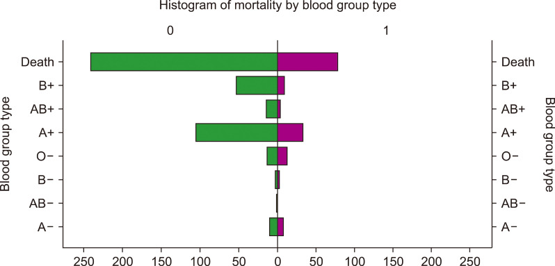
Patients requiring various modalities of respiratory treatment were assessed and compared against blood types O, B, AB, A, and rhesus factor status. Table 2 highlights the treatments. Of note, while blood type O, B, AB, A, and rhesus factor status approached significance, there was no statistically significant difference between ambient air, low-flow nasal cannula, high-flow nasal cannula, non-invasive mechanical ventilation, and invasive mechanical ventilation in any category. To assess for potential confounding, we compared individual groups of combined blood type O, B, AB, A, and rhesus factor status (O+, B+, AB+, A+, O-, B-, AB-, A-) in Table 3 and Table 4. As in Table 1, the only significant difference in patient characteristics was hospital mortality. Blood type B- and O- had the highest percentage of mortality at 50% and 48.1% (P=0.014). Fig. 2 additionally, highlights this finding graphically. Fig. 2 demonstrates a histogram that analyzes proportionality of death overall and based on each combined ABO blood group with Rh factor. This figure demonstrates that overall, the blood group types with Rh negative status had a higher proportion of patients who suffered from mortality (O-, B-, AB-, and A-). Table 4 highlights the highest percentage of intubated patients, who fell into the O- group at 29% (P=0.005).
Table 2.
Respiratory requirements of patients based on ABO blood type and Rh status separately.
| Blood type O (N=349) |
Blood type B (N=69) |
Blood type AB (N=21) |
Blood type A (N=159) |
P | Rh positive (N=541) |
Rh negative (N=54) |
P | |
|---|---|---|---|---|---|---|---|---|
| Respiratory treatment | 0.092 | 0.094 | ||||||
| Room air | 143 (41%) | 30 (43%) | 5 (24%) | 63 (40%) | 222 (41%) | 18 (33%) | ||
| Low flow oxygen | 82 (23%) | 26 (38%) | 7 (33%) | 42 (26%) | 147 (27%) | 10 (19%) | ||
| High flow oxygen | 35 (10%) | 7 (10%) | 3 (14%) | 10 (6%) | 50 (9%) | 5 (9%) | ||
| NIMV | 14 (4%) | 2 (3%) | 2 (9%) | 9 (7%) | 22 (4%) | 5 (9%) | ||
| Intubated | 75 (22%) | 4 (6%) | 4 (20%) | 35 (21%) | 102 (19%) | 16 (30%) |
Table 3.
Patient characteristics and mortality data of combined ABO and Rh blood types.
| Blood type O+ (N=322) |
Blood type B+ (N=63) |
Blood type AB+ (N=19) |
Blood type A+ (N=140) |
Blood type O- (N=27) |
Blood type B- (N=6) |
Blood type AB- (N=2) |
Blood type A- (N=19) |
P | |
|---|---|---|---|---|---|---|---|---|---|
| Demographics | |||||||||
| Age | 51.80±19.49 | 52.63±19.91 | 48.21±21.65 | 53.5±20.59 | 47.74±20.71 | 40.17±13.49 | 46.5±30.4 | 53.32±17.57 | 0.637 |
| Body mass index | 30.56±7.98 | 28.74±5.8 | 28.16±6.8 | 29.31±7.96 | 31.7±6.6 | 28.35±3.2 | 21.15±8.27 | 29.6±5.85 | 0.184 |
| Gender | 0.299 | ||||||||
| Female | 160 (49.6%) | 25 (39.6%) | 10 (52.6%) | 68 (48.6%) | 16 (59.3%) | 5 (83.3%) | 2 (100%) | 9 (47.4%) | |
| Male | 162 (50.4%) | 38 (60.4%) | 9 (47.4%) | 72 (51.4%) | 11 (40.7%) | 1 (16.7%) | 0 (0%) | 10 (52.6) | |
| Ethnicity | 0.597 | ||||||||
| Caucasian | 192 (59.6%) | 25 (39.7%) | 13 (68.4%) | 80 (57.1%) | 16 (59.3%) | 2 (33.3%) | 1 (50%) | 11 (57.8%) | |
| African American | 21 (6.5%) | 6 (9.5%) | 0 (0%) | 7 (5%) | 0 (0%) | 0 (0%) | 0 (0%) | 1 (5.3%) | |
| Asian | 5 (1.6%) | 3 (4.8%) | 0 (0%) | 3 (2.1%) | 0 (0%) | 0 (0%) | 0 (0%) | 1 (5.3%) | |
| Hispanic | 101 (31.4%) | 29 (46%) | 6 (31.6%) | 48 (34.3%) | 11 (40.7%) | 4 (66.7%) | 1 (50%) | 5 (26.3%) | |
| Other | 3 (0.9%) | 0 (0%) | 0 (0%) | 2 (1.5%) | 0 (0%) | 0 (0%) | 0 (0%) | 1 (5.3%) | |
| Comorbid conditions | |||||||||
| Diabetes mellitus | 70 (21.7%) | 15 (23.8%) | 3 (15.8%) | 31 (22.1%) | 7 (25.9%) | 1 (16.7%) | 1 (50%) | 5 (26.3%) | 0.961 |
| Tobacco use | 7 (2.2%) | 1 (1.6%) | 0 (0%) | 1 (0.7%) | 0 (0%) | 0 (0%) | 0 (0%) | 1 (5.3%) | 0.815 |
| Cancer | 11 (3.4%) | 1 (1.6%) | 0 (0%) | 6 (4.3%) | 1 (3.7%) | 0 (0%) | 0 (0%) | 1 (5.3%) | 0.951 |
| Hypertension | 46 (14.3%) | 10 (15.9%) | 3 (15.8%) | 28 (20%) | 2 (7.4%) | 0 (0%) | 0 (0%) | 4 (21.1%) | 0.565 |
| Obesity | 48 (14.9%) | 7 (11.1%) | 1 (5.3%) | 20 (14.3%) | 8 (29.6%) | 1 (16.7%) | 1 (50%) | 1 (5.3%) | 0.174 |
| Chronic lung disease | 14 (4.3%) | 3 (4.8%) | 1 (5.3%) | 3 (2.1%) | 0 (0%) | 0 (0%) | 0 (0%) | 0 (0%) | 0.795 |
| Cirrhosis | 7 (2.2%) | 0 (0%) | 0 (0%) | 4 (2.9%) | 1 (3.7%) | 0 (0%) | 0 (0%) | 0 (0%) | 0.867 |
| Chronic kidney disease | 17 (5.3%) | 4 (6.3%) | 3 (15.8%) | 5 (3.6%) | 2 (7.4%) | 0 (0%) | 0 (0%) | 1 (5.3%) | 0.567 |
| Hospitalization mortality | 0.014 | ||||||||
| Alive | 244 (75.8%) | 54 (85.7%) | 15 (78.9%) | 106 (75.7%) | 14 (51.9%) | 3 (50%) | 2 (100%) | 11 (57.9%) | |
| Dead | 78 (24.2%) | 9 (14.3%) | 4 (21.1%) | 34 (24.3%) | 13 (48.1%) | 3 (50%) | 0 (0%) | 8 (42.1%) |
Table 4.
Supplemental oxygen requirements of patients based on combined ABO and Rh blood types.
| Blood type O+ (N=322) |
Blood type B+ (N=63) |
Blood type AB+ (N=19) |
Blood type A+ (N=140) |
Blood type O- (N=27) |
Blood type B- (N=6) |
Blood type AB- (N=2) |
Blood type A- (N=19) |
P | |
|---|---|---|---|---|---|---|---|---|---|
| Respiratory treatment | 0.005 | ||||||||
| Room air | 134 (42%) | 27 (43%) | 4 (21%) | 58 (41%) | 9 (33%) | 3 (50%) | 1 (50%) | 5 (26%) | |
| Low flow oxygen | 78 (24%) | 26 (41%) | 6 (32%) | 37 (27%) | 4 (15%) | 0 | 1 (50%) | 5 (26%) | |
| High flow oxygen | 32 (10%) | 6 (10%) | 3 (16%) | 9 (7%) | 3 (11%) | 1 (17%) | 0 | 1 (6%) | |
| NIMV | 11 (3%) | 0 | 2 (11%) | 9 (6%) | 3 (11%) | 2 (33%) | 0 | 0 | |
| Intubated | 67 (21%) | 4 (6%) | 4 (21%) | 27 (19%) | 8 (30%) | 0 | 0 | 8 (42%) |
Fig. 2.
Histogram of patient mortality separated by combined ABO and Rh blood type. Green bars indicate the number of patients who were alive, which are separated by blood group types. Purple bars indicate the number of patients who died, which are separated by blood group types.
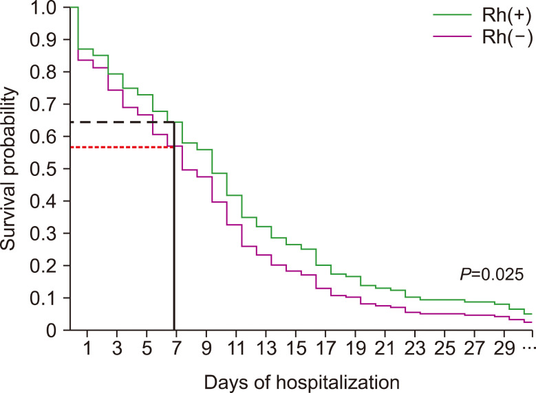
We utilized a Kaplan-Meier analysis to model event over time (measured in days) elapsed during the patient’s hospitalization. The Kaplan-Meier curve, Fig. 3, takes into consideration and calculates the 30-day survival probability differentiated by rhesus positive vs. rhesus negative status. Approximately by day 7, there is a 42% chance of mortality for rhesus negative patients and 37% chance of mortality for rhesus positive patients (P=0.025).
Fig. 3.
Kaplan-Meier survival probability based on RH status demonstrating higher risk of mortality for RH-patients where seven-day mortality risk for Rh-patients was 42% while seven-day mortality risk for Rh+ patients was 37% (P=0.025).
DISCUSSION
The ABO blood group system has been known to increase an individual’s risk for infection with viral and bacterial pathogens [1-4]. Many studies have cited relationships between ABO blood group types and SARS-CoV-2 infection susceptibility and severity [1-15]. Specifically, it has been demonstrated in SARS-CoV that the anti-A antibody produced by patients with blood group types O and B interfere with the binding of SARS-CoV to the angiotensin converting enzyme 2 receptor (ACE2R); thus, it would be reasonable to hypothesize that the same occurs with SARS-CoV-2, as this virus also binds to the same receptor [3, 4]. For this reason, various studies have demonstrated that blood type O patients are less susceptible to infection by SARS-CoV. This would then predict that patients infected by SARS-CoV-2 with blood types A and AB would have a higher propensity for worse outcomes, as they do not produce anti-A antibodies. However, our results in Table 1 do not indicate this, as blood types O and A proportionally had the highest rates of mortality when assessing each blood type individually, though the results are not statistically significant (P=0.405). This is also demonstrated in Fig. 2, which is a histogram graphically representing these observations. These results may be explained by the fact that blood type O is the most prevalent blood type in the United States, and has been previously demonstrated by other research groups [6, 9, 13].
Additionally, some studies have analyzed rhesus factor (Rh) associations with COVID-19 severity as well [10, 16-20]. Our study first demonstrates that ABO blood groups alone do not have an influence on COVID-19 mortality (P=0.405). This is consistent with the observations of several studies [5, 8, 12, 17, 21]. However, the results of many studies are inconsistent regarding ABO blood groups and their associations with COVID-19 severity, as some studies suggest a relationship between ABO blood type and mortality, while others do not. This disparity may be due to other risk factors that increase an individual’s risk for disease susceptibility and severity; consequently, the Rh factor is a point of interest that has been examined. We observed a statistically significant association between Rh and mortality (P<0.001). Of the 595 patients with an Rh factor documented, 90.8% were Rh positive, which may suggest that Rh positive patients have a higher susceptibility to COVID-19. This distribution of Rh status is consistent with the findings of Zietz et al. (2020) [18], Rana et al. (2021) [19], and Anderson et al. (2022) [20], and also consistent with blood type distributions around the world. Further, we note that COVID-19 patients with a Rh-negative status had a strong association with increasing COVID-19 mortality risk based on Kaplan-Meier analysis (Fig. 3). At seven days of hospitalization, patients in the Rh-negative group had a 5% increased risk of mortality (Fig. 3). This differs from the findings of Anderson et al. (2022) [20], who note that Rh positive patients with COVID-19 have worse outcomes. Though the mechanism of how Rh status affects COVID-19 pathophysiology remains unclear, it is known that Rh status mismatch between mothers and newborns is associated with hemolytic disease of the newborn. Additionally, it has been reported that Rh negative status has been associated with increased risk of West Nile Virus infection, and this is likely attributed to differential expression of glycosylated products on red blood cell membranes, facilitating viral entry into cells [22]. This facilitation may be due to glycan-glycan interactions of cells or lectin-glycan interactions, which has been reported. Though unclear, future studies may investigate this phenomenon by studying erythrocyte surface glycoproteins of individuals with COVID-19. As such, Rh factor may be an important factor in the assessment of immunological response to antigens.
Based on our observations, ABO blood groups alone do not have a significant association with COVID-19 severity and mortality. However, we found that combined ABO and Rh blood groups have a significant association with COVID-19 severity and mortality. Additionally, Rh status was associated with increasing mortality as demonstrated in our Kaplan-Meier analysis. A Kaplan-Meier analysis of ABO blood groups was not performed due to lack of statistically significant mortality differences amongst ABO blood groups. Due to our findings and the findings of other researchers, we do not recommend including ABO blood groups alone in risk stratification of COVID-19 disease severity; although, Rh status may be an important factor to consider in these types of risk calculators. We surmise that Rh status is an important marker that may be further studied for its role in COVID-19 pathogenesis in future analyses with larger sample sizes across multiple centers worldwide, as this may contribute to our understanding of COVID-19.
This study has two notable strengths. The first is that the data collected were obtained via manual chart review, rather than from an automated epidemiologic data set. Another strength is that our final analysis was conducted on approximately 600 patients, providing a large sample size for analysis enough to produce statistically significant values that represent our patient population in Southern California. This study’s primary limitation is that it was unable to derive a mechanism for the association between COVID-19 and Rh status, though this is a limitation of many observational studies. Another limitation is that our study was performed at a single center that serves a predominantly Hispanic population. This may confound our results, as ABO blood groups are known to be distributed based on ethnicity. Though this limitation exists, our findings provide valuable information for our region and the Hispanic population. Regardless of the limitations, we believe that they are minor and do not change the impact of our study.
In conclusion, our study found that ABO blood type did not have an association with COVID-19 severity and mortality. However, when combining ABO blood type and Rh status, there was a statistically significant association between each group and COVID-19 mortality and need for intubation. We also demonstrate that Rh status is significantly associated with COVID-19 severity and mortality, with Rh-negative patients having worse outcomes. Within each combined ABO and Rh blood group, O-, B-, and AB-had the highest rates of mortality; however, these relationships may not be attributed to the combined effect of ABO blood type and Rh status alone. Future multi-center studies may better assess these relationships and investigate the underlying role of Rh factor in the pathogenesis of COVID-19.
ACKNOWLEDGEMENTS
The authors would like to acknowledge the professional and exceptional care provided by healthcare providers, nursing, and ancillary staff at Arrowhead Regional Medical Center.
Footnotes
Authors’ Disclosures of Potential Conflicts of Interest
No potential conflicts of interest relevant to this article were reported.
REFERENCES
- 1.Fan Q, Zhang W, Li B, Li DJ, Zhang J, Zhao F. Association between ABO blood group system and COVID-19 susceptibility in Wuhan. Front Cell Infect Microbiol. 2020;10:404. doi: 10.3389/fcimb.2020.00404.a710bec4e5124e02be76c61b6ddaca1f [DOI] [PMC free article] [PubMed] [Google Scholar]
- 2.Wu BB, Gu DZ, Yu JN, Yang J, Shen WQ. Association between ABO blood groups and COVID-19 infection, severity and demise: a systematic review and meta-analysis. Infect Genet Evol. 2020;84:104485. doi: 10.1016/j.meegid.2020.104485. [DOI] [PMC free article] [PubMed] [Google Scholar]
- 3.Zaidi FZ, Zaidi ARZ, Abdullah SM, Zaidi SZA. COVID-19 and the ABO blood group connection. Transfus Apher Sci. 2020;59:102838. doi: 10.1016/j.transci.2020.102838. [DOI] [PMC free article] [PubMed] [Google Scholar]
- 4.Hoiland RL, Fergusson NA, Mitra AR, et al. The association of ABO blood group with indices of disease severity and multiorgan dysfunction in COVID-19. Blood Adv. 2020;4:4981–9. doi: 10.1182/bloodadvances.2020002623. [DOI] [PMC free article] [PubMed] [Google Scholar]
- 5.Gutiérrez-Valencia M, Leache L, Librero J, Jericó C, Enguita Germán M, García-Erce JA. ABO blood group and risk of COVID-19 infection and complications: a systematic review and meta-analysis. Transfusion. 2022;62:493–505. doi: 10.1111/trf.16748. [DOI] [PMC free article] [PubMed] [Google Scholar]
- 6.Jericó C, Zalba-Marcos S, Quintana-Díaz M, et al. Relationship between ABO blood group distribution and COVID-19 infection in patients admitted to the ICU: a multicenter observational Spanish study. J Clin Med. 2022;11:3042. doi: 10.3390/jcm11113042.6d9c6f6017a04f2f99b08ffcb72d41bc [DOI] [PMC free article] [PubMed] [Google Scholar]
- 7.Balaouras G, Eusebi P, Kostoulas P. Systematic review and meta-analysis of the effect of ABO blood group on the risk of SARS-CoV-2 infection. PLoS One. 2022;17:e0271451. doi: 10.1371/journal.pone.0271451.f56180babf894de7b61818620c884ec5 [DOI] [PMC free article] [PubMed] [Google Scholar]
- 8.Franchini M, Cruciani M, Mengoli C, et al. ABO blood group and COVID-19: an updated systematic literature review and meta-analysis. Blood Transfus. 2021;19:317–26. doi: 10.2450/2021.0049-21. [DOI] [PMC free article] [PubMed] [Google Scholar]
- 9.Kabrah SM, Kabrah AM, Flemban AF, Abuzerr S. Systematic review and meta-analysis of the susceptibility of ABO blood group to COVID-19 infection. Transfus Apher Sci. 2021;60:103169. doi: 10.1016/j.transci.2021.103169. [DOI] [PMC free article] [PubMed] [Google Scholar]
- 10.Liu N, Zhang T, Ma L, et al. The impact of ABO blood group on COVID-19 infection risk and mortality: a systematic review and meta-analysis. Blood Rev. 2021;48:100785. doi: 10.1016/j.blre.2020.100785. [DOI] [PMC free article] [PubMed] [Google Scholar]
- 11.Zhao J, Yang Y, Huang H, et al. Relationship between the ABO blood group and the coronavirus disease 2019 (COVID-19) susceptibility. Clin Infect Dis. 2021;73:328–31. doi: 10.1093/cid/ciaa1150. [DOI] [PMC free article] [PubMed] [Google Scholar]
- 12.Goel R, Bloch EM, Pirenne F, et al. ABO blood group and COVID-19: a review on behalf of the ISBT COVID-19 Working Group. Vox Sang. 2021;116:849–61. doi: 10.1111/vox.13076. [DOI] [PMC free article] [PubMed] [Google Scholar]
- 13.Pereira E, Felipe S, de Freitas R, et al. ABO blood group and link to COVID-19: a comprehensive review of the reported associations and their possible underlying mechanisms. Microb Pathog. 2022;169:105658. doi: 10.1016/j.micpath.2022.105658. [DOI] [PMC free article] [PubMed] [Google Scholar]
- 14.Soo KM, Chung KM, Mohd Azlan MAA, et al. The association of ABO and Rhesus blood type with the risks of developing SARS-CoV-2 infection: a meta-analysis. Trop Biomed. 2022;39:126–34. doi: 10.47665/tb.39.1.015. [DOI] [PubMed] [Google Scholar]
- 15.Ratiani L, Sanikidze TV, Ormotsadze G, Pachkoria E, Sordia G. Role of ABO blood groups in susceptibility and severity of COVID-19 in the Georgian population. Indian J Crit Care Med. 2022;26:487–90. doi: 10.5005/jp-journals-10071-24169. [DOI] [PMC free article] [PubMed] [Google Scholar]
- 16.Kerbage A, Haddad SF, Nasr L, et al. Impact of ABO and Rhesus blood groups on COVID-19 susceptibility and severity: a case-control study. J Med Virol. 2022;94:1162–6. doi: 10.1002/jmv.27444. [DOI] [PMC free article] [PubMed] [Google Scholar]
- 17.Hafez W, Ahmed S, Abbas N, et al. ABO blood group in relation to COVID-19 susceptibility and clinical outcomes: a retrospective observational study in the United Arab Emirates. Life (Basel) 2022;12:1157. doi: 10.3390/life12081157.bade0f8edd2c42638d63a2ede02f6dac [DOI] [PMC free article] [PubMed] [Google Scholar]
- 18.Zietz M, Zucker J, Tatonetti NP. Associations between blood type and COVID-19 infection, intubation, and death. Nat Commun. 2020;11:5761. doi: 10.1038/s41467-020-19623-x.fee6c7193bca4afdb6a44c97ae4c21a0 [DOI] [PMC free article] [PubMed] [Google Scholar]
- 19.Rana R, Ranjan V, Kumar N. Association of ABO and Rh blood group in susceptibility, severity, and mortality of coronavirus disease 2019: a hospital-based study from Delhi, India. Front Cell Infect Microbiol. 2021;11:767771. doi: 10.3389/fcimb.2021.767771.252b60b4f7984013b5559a0f9169b8e1 [DOI] [PMC free article] [PubMed] [Google Scholar]
- 20.Anderson JL, May HT, Knight S, Bair TL, Horne BD, Knowlton KU. Association of Rhesus factor blood type with risk of SARS-CoV-2 infection and COVID-19 severity. Br J Haematol. 2022;197:573–5. doi: 10.1111/bjh.18086. [DOI] [PubMed] [Google Scholar]
- 21.Bokhary DH, Bokhary NH, Seadawi LE, Moafa AM, Khairallah HH, Bakhsh AA. Variation in COVID-19 disease severity and clinical outcomes between different ABO blood groups. Cureus. 2022;14:e21838. doi: 10.7759/cureus.21838. [DOI] [PMC free article] [PubMed] [Google Scholar]
- 22.Arac E, Solmaz I, Akkoc H, et al. Association between the Rh blood group and the COVID-19 susceptibility. Int J Hematol Oncol. 2020;30:81–6. doi: 10.4999/uhod.204247. [DOI] [Google Scholar]


