

Key Clinical Message
Areca nut (AN) is an addictive substance widely used in different world regions. There are several side effects associated with the use of AN, which have already been reported. However, the reports on hepatotoxicity of AN are sporadic and non‐conclusive. In the present case report, we investigated the hepatotoxicity of AN in a four‐year‐old Iranian girl who was transferred to our medical center with abdominal pain, vomiting, diarrhea, fever, and other symptoms such as hematuria, decreased mental status, multiple seizure episodes. After a comprehensive evaluation, it was concluded that these signs and symptoms were all attributed to AN consumption, which was given by her mother to control diarrhea. Eventually, the patient medical conditions were managed successfully, and she survived by intense medical care. In conclusion, we suggest AN should be considered a potential hepatotoxic agent.
Keywords: an addictive substance, Areca nut, case report, hepatotoxic agent, hepatotoxicity
Is brought.

1. BACKGROUND
Areca nut (AN) is consumed by 600 million people worldwide, and betel quid, a preparation containing AN, is consumed by 10%–20% of the world's population. The world's largest consumers of AN are India and other Southeast Asian countries. 1 After nicotine, ethanol, and caffeine, AN is the most commonly used addictive substance globally. There is strong evidence that AN, whether with or without tobacco use, can cause cancer. 2 According to the IARC, the AN is carcinogenic in humankind. It has been linked to cancers in different body areas such as the mouth, pharynx, esophagus, liver, bile ducts, and the uterus. 3 This report describes a child's medical condition transferred to our medical center with altered mental status, seizure, elevated hepatic transaminases, hematuria, and hemolysis associated with AN toxicity.
2. CASE PRESENTATION
A four‐year‐old girl was referred to our hospital with hematuria and altered mental status, followed by frequent vomiting, diarrhea, and fever episodes. After talking to her mother, it was indicated that her general health status was excellent 2 days before her admission into the medical center. There was no remarkable health condition in her medical history. Her body weight was 12 kg. In the past 2 days, she developed a sudden abdominal pain, diarrhea, nausea, and three episodes of vomiting about 6 h after her meal. Before admission to the emergency department of Jahrom Hospital (Jahrom, Iran), she was given Ibuprofen syrup (3 mL) for her fever and AN (Figure 1) powder (two tablespoons) for her diarrhea by her mother. The patient's condition deteriorated immediately after AN ingestion. She developed a progressive paleness, and her extremities turned cold. She had blood in her urine (gross hematuria) and two episodes of generalized tonic–clonic seizure with an interval of 30 min. The duration of the first seizure episode was about 10 min, and the second episode lasted for 3 min. Eventually, she was brought to the ED of Jahrom hospital. During the initial evaluations in the ED, she was febrile, pale, and had tachycardia, gross hematuria, and non‐exudative conjunctivitis. Her heart rate was 140 beats/min, and her respiratory rate was 40/min. Oxygen saturation was 85%, blood pressure was 90/50 mmHg, and her body temperature was 39.4°C.
FIGURE 1.
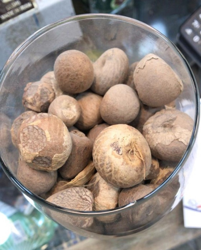
Areca nut.
Considering the patient's critical condition and lack of a PICU in Jahrom hospital, the medical team transferred her to Namazee Hospital, Shiraz, Iran. Due to high suspicion of a septic shock, intravenous hydration, and Vancomycin (15 mg/kg i.v. Q8H), and Ceftriaxone (100 mg/kg i.v. Q6h) were empirically prescribed. The patient was promptly transferred to PICU with GCS 13/15. At the PICU, rigorous intravenous hydration and the administration of previous antibiotics continued, and acyclovir (10 mg/kg i.v. Q8h) was started to treat possible herpetic encephalitis. Additionally, after pediatric neurology consultation, an anticonvulsant, Levetiracetam (10 mg i.v. Q12h), was administered.
Further evaluations for neurological symptoms were conducted, including biochemical and microbiological examination of CSF and brain computed tomographic scan. The results of these tests were unremarkable. Electroencephalography showed some paroxysmal sharp waves, contrary to a simple febrile seizure. Thus, intravenous acyclovir was stopped after neurology consultation.
Initial laboratory results showed normal electrolyte levels except for hypokalemia, high values of CRP (>150 mg/L), LDH (3570 U/L), and fibrinogen (611 mg/dL). Besides, the negative coombs' test and sufficient activity of G6PD all indicated non‐immune hemolysis. Venous blood gas assessment included pH: 7.43, PCO2: 14.3 kPa, and HCO3: 9.6 mEq/L. Echocardiography was normal. Renal function tests were within the normal range (BUN 14 mg/dL, Creatinine 0.42 mg/dL), which ruled out the hemolytic uremic syndrome followed by Escherichia coli gastroenteritis. Elevated hepatic transaminases AST: 2670, ALT: 970 U/L, and INR: 1.76 were observed. Total and direct bilirubin: 0.7, 0.3 g/dL, and alkaline phosphatase: 296 IU/L were reported within the normal range. Urine analysis showed +4 blood and +2 albumin, which could be due to hemolysis and fever. There was +3 occult blood and a high white blood cell count in the stool examination. Virology investigations were negative for HBV, HCV, HAV, SARS‐Cov‐2, and HSV 1 and 2 (Table 1).
TABLE 1.
Laboratory evaluation on the first and last days of admission (NC, not checked; NR, not reported).
| First day of admission | On the third day of admission | Last day of admission | First day of admission | On the third day of admission | Last day of admission | ||
|---|---|---|---|---|---|---|---|
| Hemoglobin (Hb) | 10.2 | 8.7 | 10.4 | Bilirubin Total | 0.7 | 0.2 | 0.3 |
| Hematocrit | 31.8 | 27.3 | 27.3 | Bilirubin Direct | 0.3 | 0.1 | 0.1 |
| MCHC | 32.1 | 31.9 | 30.0 | ALP | 296 | 320 | 363 |
| MCV | 82.2 | 87.3 | 87.8 | CPK | 173 | 3215 | 1250 |
| WBC count | 6.6 | 12.8 | 9.7 | LDH | 3570 | NC | 5510 |
| Neutrophil (Seg)% | 68.4 | NC | 47.1 | Ferritin | 209 | NC | NC |
| Lymphocyte (%) | 22.2 | NC | 43.4 | Lactate | 19 | NC | NC |
| Eosinophil | NR | NC | 3 | CRP | >150 | 13 | 4 |
| Platelet count | 243 | 301 | 526 | Coombs+ | Negative | NC | NC |
| RDW | 13.5 | 13.2 | 13.2 | Indirect Coombs | Negative | NC | NC |
| Retic count | 0.5 | NC | 1.5 | Urine analysis | |||
| RBC count | 3.87 | 3.28 | 3.11 | Specific Gravity | 1.014 | 1.004 | 1.009 |
| MCH | 26.4 | 26.5 | 26.4 | Glucose | Negative | Negative | Negative |
| PDW | 10.3 | 9.8 | 10.5 | Bilirubin | Negative | Negative | Negative |
| Mixed (%) | 9.4 | NC | 9.8 | Ketone | Trace | Negative | Negative |
| MPV | 8.5 | 9.1 | 8.8 | Blood | 4+ | Negative | Negative |
| P‐LCR | 14.4 | NC | 15.6 | Ascorbic acid | Negative | Negative | Negative |
| Monocyte % | 7 | NC | NC | Nitrite | Negative | Negative | Negative |
| ESR 1 | 36 | 67 | Alb | 3+ | Negative | Negative | |
| PT | 23.8 | 14.4 | 15.6 | RBC | 1–2 | 0–1 | 0–1 |
| INR | 1.76 | 1.07 | 1.16 | WBC | 0–1 | 2–3 | 6–8 |
| PTT | 41.8 | 30.3 | 31.1 | Bacteria | Few | Rare | Few |
| G6PD | Sufficient | NC | NC | Crystal | Many | NC | NC |
| Fibrinogen level | 611 | 311 | Appearance | Turbid | Clear | Clear | |
| BUN | 14 | 8 | 15 | pH | Acid | Alkaline | Alkaline |
| Cr | 0.42 | 0.43 | 0.44 | Color | Brown | Yellow | Yellow |
| K | 3.8 | 4.2 | 4.7 | Leukocyte esterase | Negative | Negative | Negative |
| Na | 136 | 140 | 141 | Stool Exam | |||
| Glucose (serum) | 126 | NC | 80 | Consistency | Mucoid | Soft | Soft |
| Calcium total | 0.42 | 9.5 | 10.1 | Stool OB | 3+ | Trace | Trace |
| Total protein | 8.6 | 4.5 | 5.8 | Stool WBC | Many | NR | |
| Albumin | 3.1 | 3.2 | 3.8 | Stool fat quantitative–acidified | Not seen | Not seen | Not seen |
| AST | 2670 | 920 | 62 | Blood culture | No growth | NC | No growth |
| ALT | 970 | 750 | 257 | Urine culture | No growth | NC | No growth |
| TG | 202 | NC | NC | CSF analysis | |||
| Chol | 162 | NC | NC | Total cell | 10 | NC | NC |
| HSV PCR | Not detected | NC | NC | RBC count | 10 | NC | NC |
| ABG assessment | CSF culture | Negative | NC | NC | |||
| pH | 7.43 | 7.53 | – | – | – | – | |
| PCO2 | 14.3 | 28.9 | – | – | – | – | |
| HCO3 | 9.6 | 24.5 | – | – | – | – | |
| BE | −11 | 1–9 | – | – | – | – | |
| PO2 | 152.5 | 77.8 | – | – | – | – |
On the second day of PICU admission, 120 mL packed cells were transfused due to the decreased hemoglobin level (initial value: 10.2 g/dL, follow‐up value: 9 g/dL). All the patient's signs and symptoms were alleviated gradually. On the sixth day of admission, the patient was transferred to the pediatric GI ward after stabilizing her vital signs, decreasing the levels of AST and ALT, and normal ABG and imaging. While she was admitted to the GI ward, antibiotic therapy, and Levetiracetam administration were continued. Zinc Sulfate (10 mg, p.o. daily), ursodeoxycholic acid (130 mg p.o. BID), and folic acid (5 mg daily) were added to her treatment. Eventually, The patient was discharged in a stable situation. The laboratory tests were repeated after 1 month, and all the results were within the normal range.
3. DISCUSSION AND CONCLUSION
AN is an addictive substance widely used in South and Southeast Asia and the Asia Pacific region, particularly among migrant communities in Africa, and North America. 4 If consumed in large quantities, AN can cause acute toxic symptoms such as dyspnea, tachypnea, tachycardia, palpitations, hypotension, chest tightness, nausea, vomiting, dizziness, abdominal colic, and even myocardial infarction and coma; however, in the majority of cases, the effects are transient, and patients recover quickly. 6
In the study reported by Sundqvist et al., the AN extract decreased cell survival and membrane integrity in a dose‐dependent manner. The ANs aqueous extract was highly cytotoxic and genotoxic and resulted in the formation of both DNA single‐strand breaks and DNA protein cross‐links. 5 Another study indicated that tumors could arise in the buccal mucosa after exposure to AN. 6 Furthermore, the AN showed cytotoxic effects on red blood cells, resulting in hemolysis, hematuria, and anemia. 7
In addition to AN being carcinogenic to the oral cavity, pharynx, esophagus, liver, and uterus, numerous adverse effects on the entire body organs have been reported. AN causes euphoria, tachycardia, hypertension, GABA inhibition, and neuronal damage while not affecting concentration or memory. It results in hyperlipidemia, vasospasm, and cardiac arrhythmias, increasing the risk of myocardial ischemia. Chronic consumption of AN results in hypothyroidism, prostate hyperplasia, infertility, and vitamin D deficiency. 2 Additionally, AN has been shown to increase the rate of preterm birth and stillbirth in tobacco smoker women. 8 Besides, in the review of Javed et al., obesity, increased waist circumference, elevated triacylglycerol level, metabolic syndrome, hyperglycemia, and Type 2 diabetes were reported as the side effects of AN. 9
In our current case report, hepatotoxicity was one of the clinical findings. Because of the patient's unstable hemodynamics, the hepatic shock was considered one of the differential diagnoses for elevated liver function. Various medical conditions cause hepatic shock, including hemorrhage, surgery, respiratory failure, infection, and chronic shock. Other causes include failure of microcirculation, a systemic inflammatory reaction, and adverse treatment effects in patients admitted into the intensive care unit. 10 Furthermore, Adediji et al. demonstrated that aqueous extract of AN changes the blood levels of ALT, ALP, and AST and has detrimental effects on the liver of adult Wistar rats. 11 In other studies, the AN was reported to be hepatotoxic, causing both cholestatic and hepatocellular hepatic injury and increased blood transaminases and alkaline phosphatase. 12 , 13 Considering the citation mentioned above, we propose that the observed elevation of liver enzymes in this case report is most likely due to both hepatic shock as a predisposing factor and the hepatotoxic effects of ANs.
Regarding the neurologic effects of the AN, it has been previously described that betel chewing causes widespread cortical desynchronization of the EEG, indicating a state of arousal, and betel nut‐induced extrapyramidal syndrome has also been well documented. 14 , 15 A betel nut‐induced seizure was reported in another case report in a young male. The patient had no other neuroradiologic or precipitating factors, and full recovery was observed without medication after abstinence. 16 Although these reports were in line with our observation of our patient, a recent study found that the social practice of chewing Areca catechu nuts was linked to fewer seizure episodes in people with epilepsy. This observation could be due to the nut's natural stimulant properties or other reasons. 17 This inconsistency should be investigated in future studies.
AN should be considered a cytotoxic and hepatotoxic agent. The patient's health conditions improved relatively rapidly after AN toxicity. In this case report, we concluded that the observed signs and symptoms, including decreased mental status, seizure, increased liver enzymes, and hemolysis, were due to AN's ingestion.
AUTHOR CONTRIBUTIONS
Seyede Maryam Mahdavi Mortazavi: Data curation; visualization; writing – original draft. Maryam Ataollahi: Conceptualization; project administration; supervision; validation. Amirali Mashhadiagha: Conceptualization; writing – original draft. Seyed Ali Moosavi: Visualization; writing – original draft. Reza Moshfeghinia: Conceptualization; writing – original draft; writing – review and editing. Marzieh Soheili: Conceptualization; data curation; writing – original draft; writing – review and editing.
FUNDING INFORMATION
The authors do not declare a specific grant for this research from any funding agency in the public, commercial or not‐for‐profit sectors.
CONFLICT OF INTEREST STATEMENT
The authors declare that they have no competing interests.
CONSENT STATEMENT
Written informed consent was obtained from the patient's legal guardian for publication of this case report. A copy of the written consent is available for review by the Editor‐in‐Chief of this journal.
ACKNOWLEDGMENTS
We would like to express our gratitude to Professor Mahmood Haghighat (Professor of Pediatric Gastroenterology, Department of Pediatrics, Gastroenterohepatology Research Center, Internal Medicine Research Institute, Namazi teaching Hospital, Shiraz University of Medical Sciences, Shiraz, Iran), Dr. Naser Honar (Associate Professor of Pediatric Gastroenterology, Department of Pediatrics, Namazi teaching Hospital, Abu Ali Sina center for Medicine & Organ transplant, Shiraz University of Medical Sciences, Shiraz, Iran), and Dr. Marzieh Davoodi (Pediatric department resident, Student Research Committee member, Shiraz University of Medical Sciences, Shiraz, Iran) from their constructive guidance, and valuable help in diagnosing and treating the patient.
Mahdavi Mortazavi SM, Ataollahi M, Mashhadiagha A, Moosavi SA, Moshfeghinia R, Soheili M. Acute toxic effects of areca nut on central nervous system and liver: A case report. Clin Case Rep. 2023;11:e7976. doi: 10.1002/ccr3.7976
Contributor Information
Reze Moshfeghinia, Email: rezamoshfeghinia@gmail.com.
Marzieh Soheili, Email: soheili_md@yahoo.com.
DATA AVAILABILITY STATEMENT
The data that support the findings of this study are from the corresponding author, Marzieh Soheili, upon reasonable request.
REFERENCES
- 1. Arora S, Squier C. Areca nut trade, globalization and its health impact: perspectives from India and South‐East Asia. Perspect Public Health. 2019;139(1):44‐48. [DOI] [PubMed] [Google Scholar]
- 2. Garg A, Chaturvedi P, Gupta PC. A review of the systemic adverse effects of areca nut or betel nut. Indian J Med Paediatr Oncol. 2014;35(1):3‐9. [DOI] [PMC free article] [PubMed] [Google Scholar]
- 3. Betel‐quid and areca‐nut chewing and some areca‐nut derived nitrosamines. IARC Monogr Eval Carcinog Risks Hum. 2004;85:1‐334. [PMC free article] [PubMed] [Google Scholar]
- 4. Gupta P, Ray CS. Epidemiology of betel quid usage. Ann Acad Med Singap. 2004;33:31‐36. [PubMed] [Google Scholar]
- 5. Sundqvist K, Liu Y, Nair J, Bartsch H, Arvidson K, Grafström RC. Cytotoxic and genotoxic effects of areca nut‐related compounds in cultured human buccal epithelial cells. Cancer res. 1989;49(19):5294‐5298. [PubMed] [Google Scholar]
- 6. Samuel K, Navani H, Logani KB. Epidemiology of oral carcinoma in eastern districts of Uttar Pradesh. J Indian Med Assoc. 1969;53(4):179. [PubMed] [Google Scholar]
- 7. Kolhe A, Patil N. Cytotoxicity of aqueous extract of areca nut and tobacco on human erythrocytes (red blood corpuscles). Int Res J. 2009;2:539‐540. [Google Scholar]
- 8. Kumar S. Tobacco and areca nut chewing—reproductive impairments: an overview. Reprod Toxicol. 2013;36:12‐17. [DOI] [PubMed] [Google Scholar]
- 9. Javed F, Al‐Hezaimi K, Warnakulasuriya S. Areca‐nut chewing habit is a significant risk factor for metabolic syndrome: a systematic review. J Nutr Health Aging. 2012;16(5):445‐448. [DOI] [PubMed] [Google Scholar]
- 10. Soleimanpour H, Safari S, Rahmani F, Nejabatian A, Alavian SM. Hepatic shock differential diagnosis and risk factors: a review article. Hepat Mon. 2015;15(10):e27063. [DOI] [PMC free article] [PubMed] [Google Scholar]
- 11. Adediji J, Adediji T, Eze GI. Effects of short term oral administration of aqueous extracts of areca nut (areca catechu) on liver profile of adult wistar rats. J Dent Med Sci. 2015;14(10):62‐67. [Google Scholar]
- 12. Chou WW, Guh JY, Tsai JF, et al. Arecoline‐induced growth arrest and p21WAF1 expression are dependent on p53 in rat hepatocytes. Toxicology. 2008;243(1–2):1‐10. [DOI] [PubMed] [Google Scholar]
- 13. Lin C‐F, Shiau T‐J, Ko Y‐C, Chen P‐H, Wang J‐D. Prevalence and determinants of biochemical dysfunction of the liver in Atayal aboriginal community of Taiwan: is betel nut chewing a risk factor? BMC Gastroenterol. 2008;8(1):13. [DOI] [PMC free article] [PubMed] [Google Scholar]
- 14. Chu NS. Effects of betel chewing on electroencephalographic activity: spectral analysis and topographic mapping. J Formos Med Assoc. 1994;93(2):167‐169. [PubMed] [Google Scholar]
- 15. Deahl M. Betel nut‐induced extrapyramidal syndrome: an unusual drug interaction. Mov Disord. 1989;4(4):330‐332. [DOI] [PubMed] [Google Scholar]
- 16. Huang Z, Xiao B, Wang X, Li Y, Deng H. Betel nut indulgence as a cause of epilepsy. Seizure. 2003;12(6):406‐408. [DOI] [PubMed] [Google Scholar]
- 17. Mateen FJ, Carone M, Tshering L, et al. Areca catechu (palm tree) nut chewing and seizures: an observational study. Epilepsy Behav. 2017;74:76‐80. [DOI] [PubMed] [Google Scholar]
Associated Data
This section collects any data citations, data availability statements, or supplementary materials included in this article.
Data Availability Statement
The data that support the findings of this study are from the corresponding author, Marzieh Soheili, upon reasonable request.