ABSTRACT
Xanthogranulomatous epithelial tumor (XGET) is an extremely rare and recently described mesenchymal neoplasm characterized by a distinctive histological appearance and clinical presentation. This case report describes a unique case of XGET in a 66-year-old female patient who presented with a 5 cm mass in the dorsal distal left thigh. The clinical, radiological, and pathological findings, as well as the management and follow-up, are discussed.
Keywords: soft tissue, emerging entities, xanthogranulomatous inflammation, cytokeratin, uncertain biologic potential
INTRODUCTION
Xanthogranulomatous epithelial tumor (XGET) is a recently described mesenchymal neoplasm characterized by xanthogranulomatous inflammation with foamy histiocytes, osteoclast-like giant cells and Touton giant cells as well as scattered keratin-expressing cells with distinctive eosinophilic cytoplasm. The lesion was first reported by Fritchie et al. in a series of 6 patients in 2020 [1]. The following year, Agaimy et al. reported 6 tumors similar to giant cell tumor of soft tissue but with a subpopulation of keratin-expressing cells and a novel HMGA2-NCOR2 fusion [2], which had only been described once previously in a tenosynovial giant cell tumor [3]. The authors proposed the term “keratin-positive giant cell-rich soft tissue tumor with HMGA2-NCOR2 fusion” (KPGCT) for this novel entity. In 2022, Dehner et al. described a series of 9 cases with the morphologic features of XGET, KPGCT, or intermediate between the two, 7 of which demonstrated the HMGA2-NCOR2 fusion previously described in KPGCT [4].
We here present the clinical, radiological, and pathological findings, as well as the management and follow-up, of a case with morphologic and immunohistochemical findings consistent with XGET but no HMGA2-NCOR2 fusion.
CASE PRESENTATION
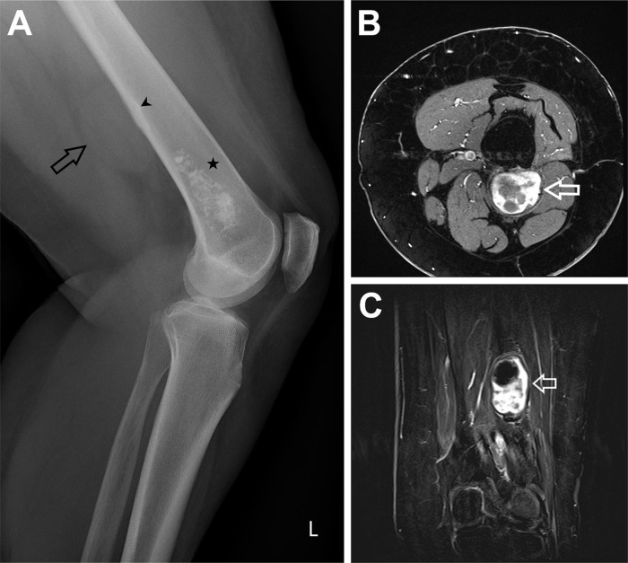
A 66-year-old woman presented to her general practitioner with a one-week history of bilateral knee pain. An X-ray showed a mass suspicious for enchondroma in the distal left femur as well as a more proximal non-calcified soft tissue lesion with discreet thickening of the endosteum and a discreet periosteal reaction (Figure 1A). A subsequent MRI of the left knee showed a well defined soft tissue lesion with suspected focal invasion of cortical bone (Figure 1B-C). An ultrasound-guided core biopsy was performed, and a diagnosis of XGET was made based on the core biopsy material.
Fig. 1.
(A) Radiograph Knee lateral view. Black star: chondroid lesion in the distal femur. Black arrow: Soft tissue lesion visible without any calcifications in the lesion. Black arrowhead: Smooth regular thickening of the endosteum. Discreet periosteal reaction, single layered. (B) MRI T1 TSE fat saturated after Gadolinium transversal view. White arrow: After Gadolinium heterogenous uptake of the contrast agent, the T2 hyperintense areas do not enhance, only the T2 hypointense areas show enhancement. (C) MRI T1 TSE subtraction after Gadolinium coronal view. White arrow: After Gadolinium the hypointense border shows discreet peripheral enhancement. Heterogenous enhancement of the lesion, the T2 hyperintense areas do not enhance.
The case was discussed within the interdisciplinary team of the Swiss Sarcoma Network. Due to the lack of data on this recently described lesion, a decision was made to resect the tumor completely without neoadjuvant therapy. A preoperative PET/CT with [18F]FDG detected no suspected metastases. The tumor was completely resected including the involved cortical bone. Macroscopically, the tumor was a smooth, reddish mass with a diameter of 5 cm. Due to the presence of an enchondroma in the distal femur, no osteosynthesis was performed. On the first postoperative day, the patient suffered a diaphyseal femur fracture when turning over in bed. A second operation was performed with osteosynthesis and curettage of the enchondroma. After the second operation, the postoperative course was uneventful, and the patient was discharged home 1 week later. A clinical and radiological follow-up after 6 months showed no further complications or signs of tumor manifestation.
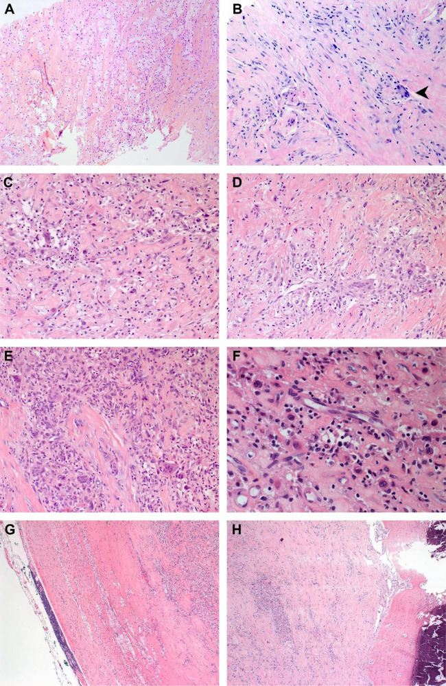
Microscopic examination of the core biopsy showed sheets of foamy histiocytes, osteoclast-like giant cells and Touton giant cells as well as scattered mononuclear cells with eosinophilic cytoplasm without cytological atypia and with no evident mitotic activity (Figure 2A-B). Subsequent examination of the resected mass further revealed a fibrous capsule containing lymphoid tissue as well as focal infiltration of the resected cortical bone (Figure 2C-H). There was no tumor necrosis, and the surgical margin was negative.
Fig. 2.
Microscopic examination showed sheets of foamy histiocytes (A) (HE, x100), Touton giant cells (B, arrowhead) (HE, x200), osteoclast-like giant cells (C-E) (HE, x200) and scattered bland mononuclear cells with eosinophilic cytoplasm (F) (HE, x400). The tumor was surrounded by a fibrous capsule containing lymphoid tissue (G) (HE x50) and focally infiltrated the resected cortical bone (H) (HE, x50).
The scattered mononuclear cells with eosinophilic cytoplasm stained positively for pancytokeratin and CK7 (Figure 3). There was also diffuse positivity for CD45 and CD99 and focal positivity for S100. The reactions for CD34, CD56, SATB2, GATA3, CK20, EMA, EpCAM, TTF1, PAX8, actin, and desmin were all negative. Focal nuclear positivity for MDM2 was noted, and a FISH analysis was performed to rule out an MDM2 gene amplification.
Fig. 3.
On immunohistochemistry, the scattered eosinophilic cells stained positively for pancytokeratin (A) (anti-CKAE1/AE3 Ab, x100) and CK7 (B) (anti-CK7 Ab, x100) and negatively for EpCAM (C) (anti-EpCAM Ab, x100).
No evidence of a fusion transcript was found using the NGS Archer™ FusionPlex™ (137 genes with fusion partner, including HMGA2) on the core biopsy material. No MDM2 gene amplification was found by FISH (ZytoLight SPEC MDM2/CEN 12 Dual Color Probe, Zytovision).
DISCUSSION
XGET is a novel and extremely rare neoplasm characterized by a unique histological appearance, including nests of epithelial cells with interspersed foamy macrophages. As the tumor was only recently described, most lesions in the published case series were diagnosed retrospectively. In this case, the diagnosis was made based on the core biopsy material, which was followed by a complete resection. The histological features were identical to those described in the previously published case series of XGET. However, the HMGA2-NCOR2 fusion found in some of the previously described cases of XGET and KPGCT could not be detected by NGS.
Due to the limited number of reported cases, the etiology and pathogenesis of XGET remain poorly understood, and its optimal management is yet to be established. However, the characteristic histological findings aid in its diagnosis. Surgical resection appears to be the primary treatment modality, and long-term follow-up is recommended to monitor for potential recurrence or metastasis, although the long-term outcomes are yet to be established. One patient described in the literature developed multiple suspected lung metastases, although a biopsy was not performed for confirmation [4]. XGET should therefore be regarded as a tumor of uncertain biologic potential. It is also not known whether XGET and KPGCT are distinct neoplasms or represent two different aspects of the same entity, as has been suggested by Dehner et al based on the finding of HMGA2-NCOR2 fusions in both tumors [4].
The nature of the keratin-expressing cells observed in XGET is not known. Keratin expression is seen in only a subset of the tumor cells and these cells have a distinct morphology. Whether or not these cells are themselves neoplastic is unknown. Keratin-expressing cells are not typically observed in other tumors similar to XGET and KPGCT such as juvenile xanthogranuloma and giant cell tumor of soft tissue or bone, which are far more common. Although keratin expression is characteristically present in some other mesenchymal tumors, such as synovial sarcoma, epithelioid sarcoma, adamantinoma, osteofibrous dysplasia and epithelioid angiosarcoma [5-8], these entities are morphologically distinct from XGET and KPGCT.
CONCLUSION
XGET is an exceedingly rare neoplasm with distinct histopathological features. This case report highlights the clinical presentation, diagnostic assessment, therapeutic intervention, and follow-up of a 66-year-old female patient diagnosed with XGET. The tumor was successfully resected, and the patient remained free of recurrence or metastasis during the 3-month follow-up period.
Further research and additional reported cases are necessary to enhance our understanding of XGET and develop standardized treatment guidelines. Long-term follow-up is crucial to monitor for any signs of recurrence or metastasis. Collaboration among clinicians, pathologists, and researchers is essential to advance our knowledge of this rare neoplasm and improve patient outcomes.
Conflict of interest
The authors declare that they have no competing interests.
Informed consent
Written informed consent was obtained from the patient for publication of this case report and accompanying images. A copy of the written consent is available for review by the Editor-in-Chief of this journal.
REFERENCES
- 1.Fritchie KJ, Torres-Mora J, Inwards C, et al. Xanthogranulomatous epithelial tumor: report of 6 cases of a novel, potentially deceptive lesion with a predilection for young women. Mod Pathol. 2020;33(10):1889–1895. doi: 10.1038/s41379-020-0562-8. [DOI] [PubMed] [Google Scholar]
- 2.Agaimy A, Michal M, Stoehr R, et al. Recurrent novel HMGA2-NCOR2 fusions characterize a subset of keratin-positive giant cell-rich soft tissue tumors. Mod Pathol. 2021;34(8):1507–1520. doi: 10.1038/s41379-021-00789-8. [DOI] [PMC free article] [PubMed] [Google Scholar]
- 3.Brahmi M, Alberti L, Tirode F, et al. Complete response to CSF1R inhibitor in a translocation variant of teno-synovial giant cell tumor without genomic alteration of the CSF1 gene. Ann Oncol. 2018;29(6):1488–1489. doi: 10.1093/annonc/mdy129. [DOI] [PubMed] [Google Scholar]
- 4.Dehner CA, Baker JC, Bell R, et al. Xanthogranulomatous epithelial tumors and keratin-positive giant cell-rich soft tissue tumors: two aspects of a single entity with frequent HMGA2-NCOR2 fusions. Mod Pathol. 2022;35(11):1656–1666. doi: 10.1038/s41379-022-01115-6. [DOI] [PubMed] [Google Scholar]
- 5.Miettinen M, Limon J, Niezabitowski A, et al. Patterns of keratin polypeptides in 110 biphasic, monophasic, and poorly differentiated synovial sarcomas. Virchows Arch. 2000;437(3):275–283. doi: 10.1007/s004280000238. [DOI] [PubMed] [Google Scholar]
- 6.Miettinen M, Fanburg-Smith JC, Virolainen M, et al. Epithelioid sarcoma: an immunohistochemical analysis of 112 classical and variant cases and a discussion of the differential diagnosis. Hum Pathol. 1999;30(8):934–942. doi: 10.1016/S0046-8177(99)90247-2. [DOI] [PubMed] [Google Scholar]
- 7.Benassi MS, Campanacci L, Gamberi G, et al. Cytokeratin expression and distribution in adamantinoma of the long bones and osteofibrous dysplasia of tibia and fibula. An immunohistochemical study correlated to histogenesis. Histopathology. 1994;25(1):71–76. doi: 10.1111/j.1365-2559.1994.tb00600.x. [DOI] [PubMed] [Google Scholar]
- 8.Al-Abbadi MA, Almasri NM, Al-Quran S, et al. Cytokeratin and epithelial membrane antigen expression in angiosarcomas: an immunohistochemical study of 33 cases. Arch Pathol Lab Med. 2007;131(2):288–292. doi: 10.5858/2007-131-288-CAEMAE. [DOI] [PubMed] [Google Scholar]


