Abstract
Background
The P-stalk is a conserved and vital structural element of ribosome. The eukaryotic P-stalk exists as a P0-(P1-P2)2 pentameric complex, in which P0 function as a base structure for incorporating the stalk onto 60S pre-ribosome. Prior studies have suggested that P0 genes are indispensable for survival in yeast and animals. However, the functions of P0 genes in plants remain elusive.
Results
In the present study, we show that rice has three P0 genes predicted to encode highly conserved proteins OsP0A, OsP0B and OsP0C. All of these P0 proteins were localized both in cytoplasm and nucleus, and all interacted with OsP1. Intriguingly, the transcripts of OsP0A presented more than 90% of the total P0 transcripts. Moreover, knockout of OsP0A led to embryo lethality, while single or double knockout of OsP0B and OsP0C did not show any visible defects in rice. The genomic DNA of OsP0A could well complement the lethal phenotypes of osp0a mutant. Finally, sequence and syntenic analyses revealed that OsP0C evolved from OsP0A, and that duplication of genomic fragment harboring OsP0C further gave birth to OsP0B, and both of these duplication events might happen prior to the differentiation of indica and japonica subspecies in rice ancestor.
Conclusion
These data suggested that OsP0A functions as the predominant P0 gene, playing an essential role in embryo development in rice. Our findings highlighted the importance of P0 genes in plant development.
Supplementary Information
The online version contains supplementary material available at 10.1186/s12870-023-04445-y.
Keywords: Ribosomal P0 protein, Embryo lethality, P stalk, Gene duplication, Oryza sativa
Background
Protein translation is a fundamental biological process, which is carried out by ribosome. A typical ribosome consists of two subunits, a large subunit and a small subunit, both of which are comprised of ribosomal RNAs and proteins in all organism [1, 2]. The large subunit has a conserved distinctive morphological feature, the stalk protuberance, that is responsible for recruiting translation factors and stimulating translation [3]. In eukaryotes, the stalk exists as a P0-(P1-P2)2 pentameric protein complex [4]. P0, P1 and P2 are all acidic ribosomal proteins, and all of them were identified as phosphorylated proteins in vivo, and for this reason, they were designated as P protein [5]. The P0 protein forms the base of the stalk. It contains two conserved domains including the RNA-interacting domain at the N-terminus and the P1/P2 interacting domain (P-domain) at the C-terminus [6–9]. The RNA-interacting domain is functionally conserved. It directly interacts with 28S rRNA and is responsible for anchoring the P0-(P1-P2)2 complex into the 60S subunit [8]. The ribosomal proteins from P1/P2 families form P1-P2 hetero-dimers, in which the N-terminus of P1/P2 proteins are responsible for the dimerization and interaction with the P-domain of P0 protein [9]. Yeast has only one P0 gene and four P1/P2 genes P1A, P1B, P2A and P2B. The P1A–P2B protein complex was proposed to be the key element in stalk formation, whereas the P1B–P2A protein complex was implicated in regulation of stalk function [7]. Loss of function of P0 gene resulted in a deficiency in active 60 S subunits, thus making the yeast cell nonviable [6]. The P1/P2 disrupted yeast strains were viable but grew with a doubling time threefold higher than wild-type cells [10]. However, ribosomes depleted of P1/P2 proteins were impaired in translation of some specific mRNAs, and translation fidelity at elongation and termination steps [10, 11]. These results suggested that P0 was essential for cell survival, while P1 and P2 were dispensable for cell viability [12]. However, a well-organized P0-(P1-P2)2 complex was required for optimal ribosomal activity and cell survival [12].
P0 family proteins are functionally conserved in eukaryotes. The yeast mutant lacking P0 gene could be complemented by the P0 genes from animal [13–15]. The first plant P0 gene was cloned as a light-induced gene from the callus of Chenopodium rubrum, and was proposed having a similar function as that of P0 family genes [16]. A comprehensive research on the maize 60S ribosomal stalk identified a P0 protein and four distinctive forms of P1/P2 family proteins, including P1, P2a, P2b and a plant specific P1/P2-like protein named as P3 [17]. The phosphorylation levels of P0 and P1/P2 proteins were upregulated by oxygen deprivation, therefore affecting ribosome assemble, indicating P proteins might be involved in stress response [17]. A cDNA of rice P0 gene was characterized, and deduced to encode a 34-KD protein [18]. Although plant P0 genes were cloned and reported more than two decades ago [16–18], the biological functions of plant P0 genes remain elusive.
Here, we characterized the three P0 (OsP0A, OsP0B and OsP0C) genes in rice, and revealed that OsP0A is essential for rice embryo development, while OsP0B and OsP0C were dispensable in plant development. Moreover, syntenic and sequences analyses revealed that the rice ancient P0 gene underwent two duplication events and OsP0A was proposed to be the ancestral P0 gene. In all, our results highlighted the critical function of OsP0A in rice development.
Results
Phylogenetic analysis of plant ribosomal protein P0s
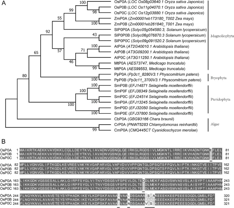
Homolog search of yeast P0 protein against the Nipponbare rice genome, identified three distinctive P0 proteins designated as P0A, P0B and P0C, which were encoded by LOC_Os08g03640, LOC_Os11g04070, and LOC_Os12g03880, respectively. To investigate the evolutionary patterns of plant P0 proteins, other 21 P0 proteins were identified from the nine representative species including unicell algae Cyanidioschyzon merolae and Chlamydomonas reinhardtii, charophyte Chara braunii, pteridophyte Selaginella moellendorffii, bryophyte Physcomitrium patens, and magnoliophyte Solanum lycopersicum, Arabidopsis thaliana, Medicago truncatula and Zea mays. Multiple sequences alignment and phylogenetic analysis showed that P0 proteins are highly conserved in plant kingdom (Fig. S1, Fig. 1A). Interestingly, each of the representative algae species has only one P0 gene, whereas each of the land plant species has at least two P0 genes (Fig. 1A), indicating that the plant P0 genes might be duplicated during the transition of plants from aquatic to terrestrial environments. The paralogues of rice P0 proteins are very similar in protein sequence. OsP0A shares 96% and 95% protein sequence identities with OsP0B and OsP0C respectively, while OsP0B and OsP0C exhibited 99% identity (Fig. 1B), indicating that OsP0B and OsP0C are more closely related during gene evolution.
Fig. 1.
Phylogenetic analysis of plant P0 proteins. P0 proteins were identified from various plant species, and the phylogenetic tree was built by MEGAX (A). The rice P0 protein sequences were aligned by Clustal X and the differences were highlighted with dark underline (B)
Subcellular localization of OsP0s proteins
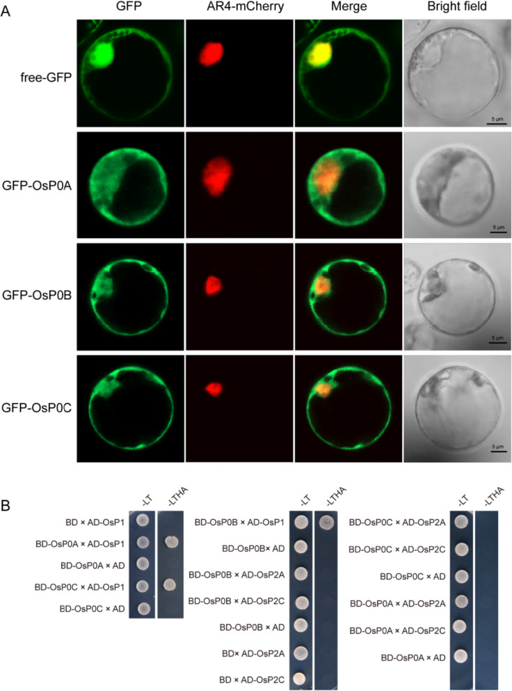
Proteins localized in appropriate cell compartments is vital for exerting their functions. Previous studies revealed that the yeast ScP0 protein was localized in the cytoplasm [19], and the C-terminus of ScP0 was critical for the protein localization [19]. OsP0A, OsP0B and OsP0C are highly similar in protein sequence, with variations mainly distributed in the C-termini. To test whether the C-terminal variations affect the P0 proteins localization in plant cells, a series of GFP tagged OsP0s were transiently expressed in rice protoplast. Examination of the fluorescence signals from the GFP-P0s showed that all of the GFP-OsP0s were localized in both the cytoplasm and nucleus (Fig. 2A), indicating that variations of the C-termini do not affect P0 protein distribution in rice cells.
Fig. 2.
Subcellular localization of OsP0s and the protein–protein interactions between OsP0s and OsP1/P2 proteins. (A) The localization of GFP-OsP0A, GFP-OsP0B and GFP-OsP0C in rice protoplast. Free GFP was used as a control. The Ar4-mCherry was used as the marker of nucleus. Scale bars = 5 μm. (B) Interaction analyses of P0 proteins with their partner proteins by the yeast two hybrid assay. -LT represents the synthetic medium lacking Leu and Trp, and -LTHA represents the synthetic medium lacking Leu, Trp, His and Ade
Protein–protein interactions between OsP0s and OsP1/P2 proteins
In eukaryotes, P1 directly interacted with P0, hence providing a ribosomal anchorage to P1-P2 heterodimers [20, 21]. The C-terminal fragment of P0 protein harbored the binding site for P1-P2 heterodimers [7, 9]. Homologue search of P1/P2 proteins in rice genomic database, identified a P1 gene named OsP1 and six P2 genes named OsP2A-F. Gene expression data from the public RNA-seq database showed that OsP1 and OsP2A-C were expressed in most of rice tissues, while the transcripts of OsP2D-F could not be detected in most of the tested tissues (Fig. S2B, C). OsP2A and OsP2C presented higher expression levels compared to the other OsP2 genes (Fig. S2C), suggesting they are probably the major OsP2 isoforms in rice. As we mentioned above, the rice P0 family proteins differ from each other mainly in the C-terminus that is important for interaction with partner proteins. Thereby, we examined the protein–protein interactions of OsP0s with OsP1, OsP2A and OsP2C by yeast two hybrid assay. The results showed that all the three OsP0s interacted with OsP1, but not OsP2A and OsP2C (Fig. 2B), indicating the interactions of P0 with the P1/P2 complex were evolutionarily conserved, and the variations of the C-termini of OsP0s did not affect the interactions.
Gene expression patterns of OsP0s
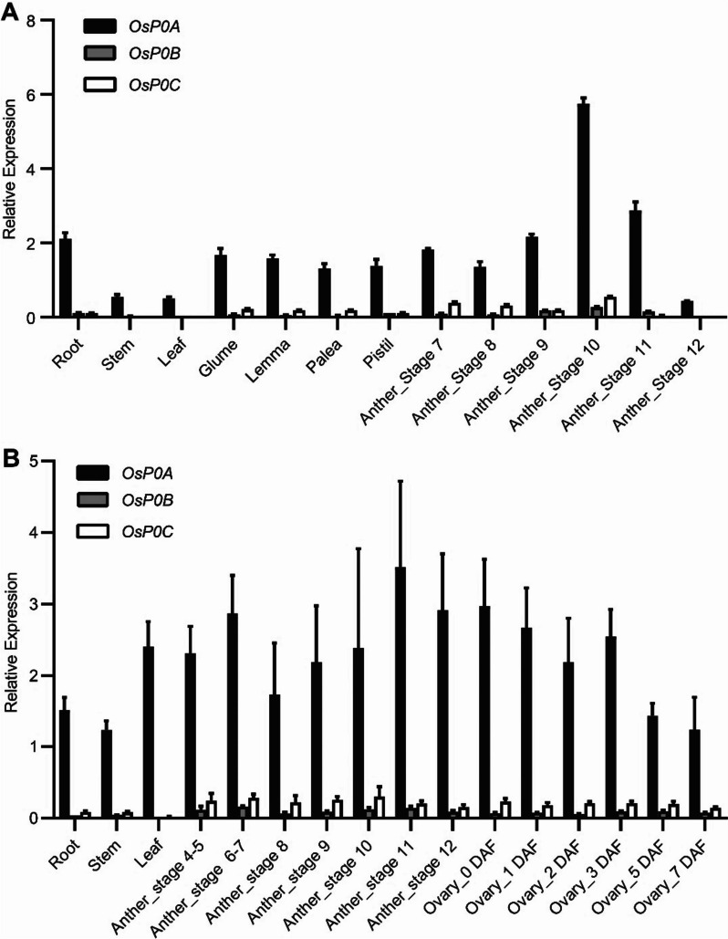
To understand the role of OsP0s in rice development, we examined the gene expression of OsP0s in the public rice RNA-seq database (http://expression.ic4r.org/) [22]. OsP0A was found to be expressed at a relative high level, while OsP0B and OsP0C were weakly expressed, or barely detectable in some rice tissues (Fig. S2A). We further analyzed the expression of OsP0s in various tissues of HHZ (an indica variety) by qRT-PCR. The transcripts of OsP0A, OsP0B and OsP0C were detected in most of the tested tissues. The OsP0A transcripts was more abundant, presenting ≥ 90% of the total P0 transcripts (Fig. 3A). The same experiment was also conducted in WYG (a japonica variety), in which OsP0s displayed the similar expression patterns (Fig. 3B). Ribosomal protein-encoding genes are always transcribed in a well-coordinated manner [23]. Gene co-expression analysis revealed that the co-expressed genes of OsP0A were enriched in the group of structural constituents of ribosome (Fig. S3). These results suggested OsP0A is the predominant P0 gene in rice. There are three P0 genes in Arabidopsis, and two P0 genes in maize. It was surprising that in both Arabidopsis and maize, a predominant P0 gene contributes to the majority of the P0 transcripts (Figs. S4, S5), which was similar to the P0 family genes in rice. These results suggested convergent evolution of P0 genes among various plant species.
Fig. 3.
Expression of OsP0A, OsP0B and OsP0C. Vegetative tissues were collected from HHZ (A) and WYG (B) at heading stage. Anthers were collected from stage 4–5 to stage 12. Ovaries were collected from WYG before pollination and 1, 2, 3, 5, and 7 d after pollination. The levels of OsP0A, OsP0B, and OsP0C transcripts were determined by qRT-PCR using OsUbq5 as an internal control. Data are shown as means ± SD (n = 3)
Phenotypes of the null mutants of the rice P0 genes
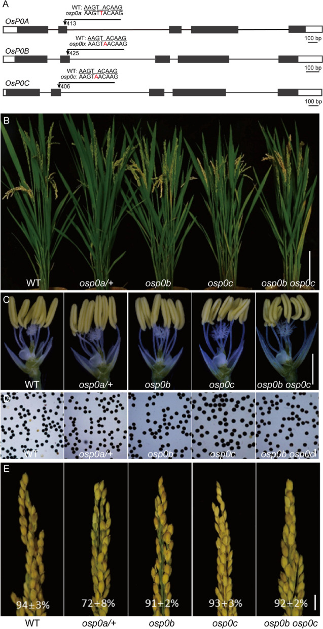
As a way to investigate the functions of OsP0s in plant development, we deployed the CRISPR/Cas9 technology to knock out OsP0s in rice. A guiding sequence was designed to target the conserved region of the three OsP0s in exon 2 (Fig. 4A). No homozygous mutant was obtained for OsP0A, but a heterozygous mutant was obtained with a single nucleotide insertion (T413) in exon 2 that disrupted the open reading frame (Fig. 4A). This plant (osp0a/ +) looked normal in vegetative growth and anther development (Fig. 4B, C). The pollen grains from osp0a/ + plants were round, and could be stained darkly with I2-KI solution, appearing similar to that of the WT plants (Fig. 4D). However, the seed-setting rate of osp0a/ + plants (72 ± 8%) was significantly decreased compared with that of the WT plants (94 ± 3%) (Fig. 4E). A homozygous mutant of osp0b with a single nucleotide insertion in exon 2 of OsP0B and a homozygous mutant of osp0c with a single nucleotide insertion in exon 2 of OsP0C were identified. Both of these mutations disrupted the corresponding genes, but the homozygous mutants displayed normal vegetative growth, anther development, pollen appearance, and normal seed setting (Fig. 4E). We then crossed the two mutant genes together and allowed self-pollination of the F1 plant. Double mutant of OsP0B and OsP0C genes were isolated from the F2 progeny by High Resolution Melting (HRM) analysis of the mutation sites. The osp0b osp0c double mutant also showed similar phenotypes to the WT plants (Fig. 4B-E), suggesting that OsP0B and OsP0C did not play a significant role in the plant growth and development.
Fig. 4.
Phenotypes of osp0a/ + , osp0b, osp0c, and osp0b osp0c mutants. (A) Structure of OsP0A, OsP0B, and OsP0C genes and mutation sites. (B-E) Comparison of phenotypes of WT, osp0a/ + , osp0b, osp0c, and osp0b osp0c mutant plants in plant morphology (B), anther (C), pollen grains stained with I2–KI (D), and panicles with seed-setting rates (E). Scale bar = 10 cm (B), 2 mm (C), 100 μm (D) and 1 cm (E)
Embryo development of osp0a/ + in rice
Failure in male and female gametophyte development and embryo abortion could all lead to the absence of homozygous mutant progeny from self-pollination. To differentiate these possibilities, we first examined the seeds derived from self-pollination of the heterozygous osp0a/ + plants. Genotyping of the progenies of osp0a/ + plants obtained 57 WT plants, 126 heterozygous osp0a/ + plants and 0 homozygous osp0a plants, suggesting the homozygous osp0a seeds were aborted during development. The ratio of WT plants and heterozygous osp0a/ + plants was consistent with the expected value 1:2 of a single gene (Table 1). We further used the heterozygous osp0a/ + plant as pollen donor to cross-pollinate the male sterile mutant osnp1-2 that carried a homozygous mutation of the OsNP1 gene [24]. 52 WT plants and 43 heterozygous osp0a/ + plants were obtained from the F1 generation. The χ2 value of the F1 population fall well within the range of that expected for the ratio 1:1 (Table 1). We finally used the heterozygous osp0a/ + plants as female parent to cross with WT WYG (Table 1). Again, the segregation data also fit the hypothesis that gametophyte development was normal in osp0a/ + plants (Table 1). Together, these results suggested that osp0a homozygous mutation may cause embryo lethality.
Table 1.
Progeny genotypes from self-pollination and reciprocal pollination of the osp0a/ + plant
| Pollination types | Progeny genotype (W:H:M) | χ2 (χ20.95 = 3.841) |
|---|---|---|
| osp0a/ + self-pollination | 57:126:0 (expect, 1:2:0) | 0.393 |
| osnp1-2(♀)xosp0a/ + (♂) | 52:43:0 (expect, 1:1:0) | 0.853 |
| osp0a/ + (♀)xWT(♂) | 51:37:0 (expect, 1:1:0) | 2.227 |
W, H, and M represent progenies with the wild-type, osp0a/ + , and osp0a homozygous genotypes
To determine when the abnormal embryo development occurred, we observed the ovule development following self-pollination of the heterozygous osp0a/ + mutant. In the WT plant, the ovules gradually enlarged from 1 to 7 day after pollination (DAP) (Fig. 5). The ovules of the heterozygous osp0a/ + plant looked similar to the WT ovules at 1 DAP, but at 3 DAP, enlargement was obviously delayed or even stopped in a portion of ovules (Fig. 5). The embryo development inside the ovule was also examined. It was clear that, embryo development stopped at the globular stage inside the abnormal small ovules but was largely normal inside the large normal ovules at 5 and 7 DAP (Fig. 6).
Fig. 5.
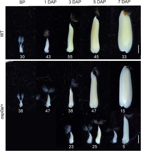
Ovule development in WT and osp0a/ + heterozygous plant after self-pollination. WT and osp0a/ + ovules before pollination (BP) and 1–7 day after pollination (DAP). The numbers under the ovules are numbers of samples. In osp0a/ + , two ovules at 5 and 7 DAP were shown to represent the smallest and largest ovules in the abnormal group of samples. Scale bar = 1 mm
Fig. 6.
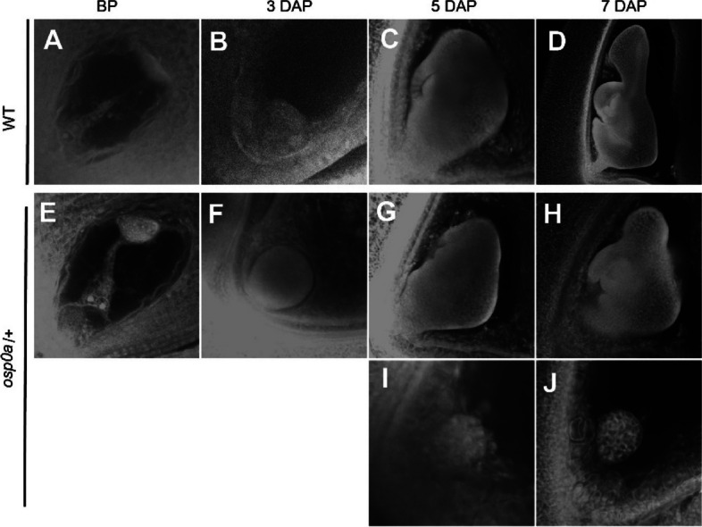
Embryo development in WT and osp0a/ + heterozygous plant after self-pollination. Ovules were collected from the WT (A-D) and osp0a/ + (E-J) before pollination (BP) and 3–7 day after pollination (DAP) and were treated with whole-mount staining to make the embryos visible. I and J show embryos inside the smallest ovules collected at 5 and 7 DAP
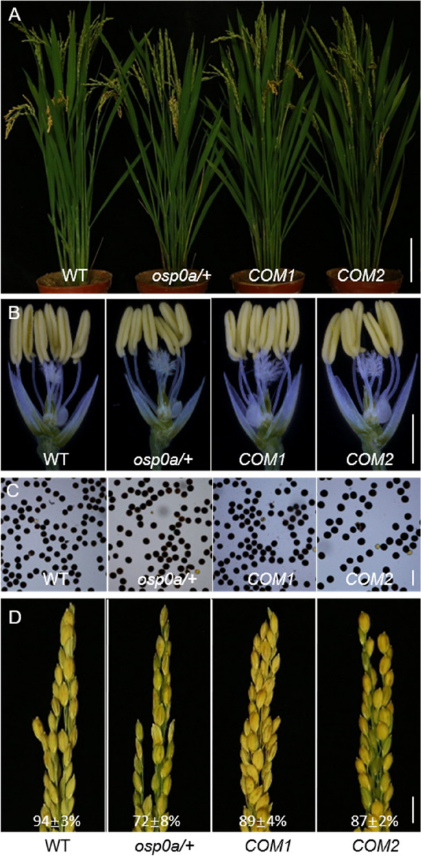
To further confirm that the osp0a seed abortion was caused by the disruption of OsP0A, a genomic fragment of OsP0A was introduced to the osp0a/ + plants, and homozygous osp0a plants containing complementary genomic fragment were identified. The homozygous complementary plants (COM1 and COM2) showed normal plant vegetative growth, anther development and pollen appearance (Fig. 7A-C). However, the seed-setting rates of the complementary plants (89 ± 4% and 87 ± 2%) were lower than that of the WT plants (94 ± 3%) (Fig. 7D), indicating a more appropriate genomic fragment of OsP0A is required for fulfilling the function of OsP0A. Taken together, these data suggested OsP0A is essential for embryo development in rice.
Fig. 7.
Transgenic complementation of the osp0a mutant. Comparison of phenotypes of WT, osp0a/ + heterozygote, and two independent complementary lines for osp0a with genomic DNA. (A) plant morphology, (B) anther, (C) pollen grains stained with I2–KI, and (D) panicles with seed-setting rates. Scale bar = 10 cm (A), 2 mm (B), 100 μm (C) and 1 cm (D)
Evolution of OsP0s in rice genomes
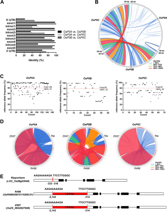
Genome annotation indicated that all of OsP0A, OsP0B and OsP0C had five exons and four introns (Fig. 8A). However, comparison of the genomic DNA sequences of OsP0A, OsP0B, and OsP0C indicated that OsP0B and OsP0C are more similar in sequence, and the sequence identity between OsP0A and OsP0C is higher than that between OsP0A and OsP0B (Fig. 8A). Syntenic analysis of 50 kb genomic sequences flanking the three genes indicated that OsP0B and OsP0C are located in large duplicated genomic regions that have little synteny with the genomic region harboring OsP0A (Fig. 8B). Sequence variation search indicated more variants in OsP0A than in OsP0B and OsP0C among the 4,726 rice varieties downloaded from the RiceVarMap2 database [25], especially the numbers of variations with reference allele frequency < 80% (Fig. 8C). These results suggested that OsP0A was probably evolved earlier before the duplication of genomic regions harboring OsP0B and OsP0C.
Fig. 8.
Characteristics of OsP0A and its family members in rice genome. (A) Gene structures OsP0A, OsP0B and OsP0C and their sequence identity. Black boxes, exon; white boxes, UTR; black lines, intron. The sequences of OsP0A, OsP0B and OsP0C were aligned with ClustalW for calculating identities for each exon, intron, 5’-UTR, and 3’-UTR. (B) Synteny analysis of the genomic regions harboring OsP0A, OsP0B, and OsP0C. The 50 kb genomic fragments flanking OsP0A, OsP0B and OsP0C were extracted from the Nipponbare genome and aligned to each other. The results were visualized with Circoletto. The position of OsP0A, OsP0B and OsP0C were marked with red boxes. Red lines, 99 ~ 100% identity; orange lines, 75 ~ 99% identity; green, 50 ~ 75% identity; blue lines, < 50% identity. (C) Variations in OsP0A, OsP0B, and OsP0C. The variations in the OsP0A, OsP0B and OsP0C genic regions and 2 kb upstream and 1 kb downstream of each gene were downloaded from the RiceVarMap2. Red lines represent the threshold of 80% reference allele frequency. (D) Synteny analysis of OsP0A, OsP0B and OsP0C in Nipponbare, R498 and ZS97 genomes. The alignments of flanking 50 kb sequences among the three rice genomes were visualized with Circoletto. Red lines, 99 ~ 100% identity; orange lines, 75 ~ 99% identity; green, 50 ~ 75% identity; blue lines, < 50% identity. (E) Characteristics of OsP0A in Nipponbare, R498 and ZS97 genomes. The red boxes represent the 1,819 bp transposon insertion in the promoters of OsP0A in R498 and ZS97. The sequences flanking the transposon insertion were shown for each rice genome
Other than the Nipponbare (NIP) genome, there are two assembled indica rice genomes (R498 and ZS97) available [26]. Alignment of the 50 kb genomic sequences flanking the three genes among the indica and japonica genomes indicated high synteny for all the three loci (Fig. 8D), indicating the presence of the three genomic fragments in Oryza sativa ancestry plant before differentiation of indica and japonica subspecies. Sequence alignment identified an 1819 bp mutator transposon sequence in the promoter region of OsP0A in R498 and ZS97 genomes but absent in NIP genome (Fig. 8E). We also PCR-amplified the OsP0A genomic DNA from HHZ, an indica variety and WYG, a japonica variety that were used as experimental materials in this study. Sequence analysis indicated that the OsP0A genomic DNA in HHZ, WYG, and R498 were identical, and all of them have the transposon sequence in the promoter. The transposon sequence was likely inserted into the OsP0A promoter after the splicing of indica and japonica subspecies, and crossbreeding between indica and japonica materials moved the gene across subspecies. To determine if the transposon insertion has an impact on the gene expression, the OsP0A transcript levels were examined in different tissues collected from the three varieties. The OsP0A mRNA levels were slightly higher in NIP than in HHZ and WYG (Fig. S6), suggesting that the transposon insertion does not have a big impact on the gene expression.
Discussion
OsP0A is essential for rice embryo development
Protein biosynthesis conducted by ribosome is a basic biological event in all living cells. Ribosome is a well-organized apparatus composed of rRNAs and a large number of ribosomal proteins [27]. Losses of ribosomal proteins cause phenotypes with various severity, which are largely dependent on the degree of damage on the ribosome activity [28]. One typical feature of the mutants of the conserved cytoplasmic ribosomal protein-encoding genes is embryo lethal. For instance, the mutants of each gene of RPS5A, RPS11A, RPL3A, RPL8A, RPL10A, RPL19A, RPL23C and RPL40B presented severe embryo-lethal phenotypes in Arabidopsis [29–31]. In these mutants, embryos appeared normal before globular stage, but could not advance any further [29–31]. P0 protein together with P1/P2 proteins form the stalk of ribosome in eukaryote, which is indispensable for ribosomal function in yeast and animal [3]. In the absence of P0 protein, a deficient 60S ribosome was assembled, which was impaired in protein synthesis resulting in cell death in yeast [17]. Gene silencing analysis demonstrated that the ribosomal protein P0 is required for the viability of Haemaphysalis longicornis [32]. In this study, three P0 family genes named OsP0A, OsP0B and OsP0C were identified in rice. Among the three P0 genes, OsP0A was the predominant gene expressed in all the tested tissues, whereas OsP0B and OsP0C were expressed at very low levels (Fig. S2A, Fig. 3). Consistently, knockouts of OsP0B or OsP0C or both of them had no visible impact on the plant growth and development, while knockout of OsP0A resulted in embryo-lethality (Figs. 4, 5). Cytological analysis showed that the osp0a mutant embryos were arrested at globular stage (Fig. 6), which was consistent with the typical phenotypes of the essential ribosomal protein mutants [27]. The embryo-lethal phenotype is expected because P0 protein is required for ribosome function, and OsP0A as the predominant P0 gene in rice, when knocked out, the mutation would certainly impair the ribosome function, and in turn, abolish the protein synthesis.
The evolution of P0 family genes in rice
Gene duplication provides resources for further evolution of the family members with inherited function, specialized function or loss of function, and/or evolved new functions [28, 33]. In plants, each cytoplasmic ribosomal protein is encoded by a gene family comprised of two or more homologous members. For example, 213 and 235 cytoplasmic ribosomal protein encoding genes were identified in Arabidopsis and rice respectively, and these genes were categorized into 81 gene families in both species [1]. This enormous increase in members of each cytoplasmic ribosomal protein family makes the ribosomes highly heterogeneous, and provides freedom for gene evolution [33]. For instance, RPL10 is an essential integral component of the large subunit of eukaryotic ribosomes. There are three RPL10 paralogs in Arabidopsis, AtRPL10A, AtRPL10B and AtRPL10C. All of them could complement a yeast mutant in RPL10, indicating that they are functional in translation [34]. However, AtRPL10A and AtRPL10B appearing to be more important in plant development and AtRPL10A appeared to play a major role in the regulation of protein synthesis under UV-B stress [30]. Arabidopsis has three RPL9 genes, AtRPL9B, AtRPL9C, and AtRPL9D. Although these genes are ubiquitously expressed, the transcript of AtRPL9C is more abundant, and is twofold and threefold higher than that of AtRPL9D and AtRPL9B respectively [35]. The knockdown mutant of atrpl9c/pgy displayed pointed leaves and more prominent marginal serrations [35], while the null mutant atrpl9d showed no visible differences from the wild type [36]. However, the double mutant atrpl9c atrpl9d was embryo lethal [36], suggesting a dosage-dependent requirement of RPL9 for ribosome activity. The ribosomal P0 proteins are very conserved in eukaryotic kingdom. Heteroexpression of P0 genes from worm, mammals, or protozoa could complement the genetic defect of P0 gene in yeast [13–15]. In the present study, we identified three P0 genes in rice. Syntenic analysis revealed that the presence of the three P0 genes in rice ancestor before differentiation of indica and japonica subspecies, and OsP0B and OsP0C are located in large duplicated genomic regions (Fig. 8B). Moreover, OsP0A presented more sequence variants than OsP0B and OsP0C in a larger collection of rice varieties (Fig. 8C), indicating the duplication event of OsP0B and OsP0C happened after the emergence of OsP0A. Additionally, OsP0A was more similar to OsP0C than to OsP0B in DNA sequences. These data suggest P0 gene has undergone two gene duplication events in rice. OsP0A may be the parent copy, and the first duplication event gave rise to OsP0C, and OsP0B emerged from a duplication of OsP0C. A transposon was found in the promoter of OsP0A in indica varieties, but not in the japonica variety NIP (Fig. 8D), indicating the transposon was likely inserted into the OsP0A promoter after the splicing of indica and japonica subspecies. The presence of the transposon in the promoter of OsP0A in WYG might come from the crossbreeding between indica and japonica materials.
The OsP0A, OsP0B, and OsP0C share high identities in amino acid sequences. Consistent with the relationship of P0 and P1 in other eukaryotes, OsP0s directly interacts with OsP1 (Fig. 2B). The yeast P0 protein was reported to be localized in cytoplasm and proposed to be incorporated into the 60 s ribosomal subunit in the late cytoplasmic stage of ribosome assembly [19]. However, all the OsP0s appeared to be located in both cytoplasm and nucleus (Fig. 2A), which was the same as the HsP0 distribution in human cells [37], providing the possibilities that OsP0s and HsP0 may be assembled onto pre-ribosomes in nucleus. The transcripts of OsP0A presented ≥ 90% of the total P0 transcripts in most of the tested rice tissues (Fig. 3). The null mutant of OsP0A was embryo lethal (Fig. 6), while knockout of OsP0B and OsP0C simultaneously did not show any visible phenotype in rice (Fig. 4). These results suggested that OsP0A is the predominant P0 gene in rice.
In addition to the fundamental functions in protein translation, lots of ribosomal proteins evolved extraribosomal functions in plants, such as RPL10A, RPL24B, RPS6, and the organelle ribosomal protein uL18-L1, uL18-L8, OsPRPL18 and so on [38–42]. The eukaryotic P0 family were also documented to be functional in ribosome-independent manners. The virus protein VPg (viral protein genome-linked) protects the RNA of Potato virus A from degradation and facilitats its translation [43]. The tobacco NbP0 was essential for the activity of VPg and, together with VPg and eIF(iso)4E synergistically enhanced viral translation [44]. Biochemical analysis confirmed that the Drosophila ribosomal protein P0 contained apurinic/apyrimidinic endonuclease activity for both single- and double-stranded DNA, and might act as a DNA repair protein [45]. NONO is an essential component of paraspeckle, playing a pivotal role in the repair of DNA double-strand breaks (DSB) [46]. The human HsP0 directly bind to the RRM1 and RRM2 domains of NONO, therefore enhancing non-homologous end joining-mediated DSB repair [47]. OsSec18 is a conserved ATPase required for vesicle membrane fusion [48]. OsSec18 interacts with Os60sP0/OsP0A and together with other proteins constitutes a 290-kDa complex in rice endosperm cells. However, the function of OsP0A in the complex remains unclear [48]. As P0 proteins are highly conserved, and OsP0s localizes both in cytoplasm and nucleus, we speculate that OsP0s probably also have evolved extraribosomal functions as its homologs did. However, more studies are required to support this notion.
In summary, we here demonstrate the essential role of P0 family genes in plant survival and provide the evidences that OsP0A is the predominant P0 gene in rice. Also, an evolutionary model is proposed that OsP0C is duplicated from OsP0A, and OsP0B is duplicated from OsP0C. Together, our data highlight the functions of P0 family genes in plants.
Materials and methods
Plant materials and growth conditions
The japonica variety Wuyungeng 7 (WYG) was kindly provided by Dr. Jianmin Wan (Nanjing Agricultural University). The indica variety Huanghuazhan (HHZ) was obtained form the Rice Research Institute of Guangdong Academy of Agricultural Sciences. WYG and HHZ were used as the wild-type in this study. Rice seeds were sterilized by 0.3% hydrogen peroxide, and then germinated in pans. The seedlings at the 4–5 leaf stage were transplanted into the paddy field with regular care.
Phylogenetic analysis
P0 family proteins from Cyanidioschyzon merolae, reinhardtii, Chara braunii, Selaginella moellendorffii, Physcomitrium patens, Solanum lycopersicum, Arabidopsis thaliana, Medicago truncatula, Zea mays and oryza sativa were obtained from the Ensembl Plants Database (http://plants.ensembl.org/index.html) with the yeast P0 protein sequence as a query. Protein multiple sequence alignment was performed using ClustalW, and the neighbor-joining tree was constructed using MEGA11 with default parameters [49].
Protein subcellular localization analysis
Plasmids comprising the expression cassettes GFP-OsP0A, GFP-OsP0B, and GFP-OsP0C driven by 35S promoter were constructed respectively. The resultant plasmids together with the nuclear marker ARF19IV-mCherry were introduced into rice protoplast by the polyethylene glycol (PEG)–calcium mediated method as previous described [50]. After 12 h incubation, the fluorescence signal in the transformed protoplasts were examined by a laser confocal scanning microscope (LSM-800; Carl Zeiss).
Yeast two hybrid assay
The CDSs of OsP0A, OsP0B, and OsP0C were individually cloned into pGBKT7 (containing a GAL4 DNA-binding domain, BD construct), and the CDSs of OsP1 OsP2A and OsP2C were cloned into pGADT7 (containing a GAL4 activating domain, AD construct) respectively. Each of the resultant BD constructs in combination with the various AD constructs were introduced into the yeast strain AH109. The transformed yeast cells were further plated on the -LT (SD-Leu/-Trp) medium. The positive clones were transferred on the -LTHA (SD-Leu/-Trp/-His/-Ade) medium for testing the protein–protein interactions.
RNA extraction and qRT-PCR assay
For gene expression analyses, qRT-PCR assay was performed as previously described [51]. Briefly, total RNA was extracted from various rice tissues and used for cDNA synthesis. qPCR was performed using SYBR Premix Ex Taq II (TaKaRa, Dalian, China) with gene specific primers (Table S1). The OsUbq5 gene was employed as a normalizing reference gene.
Generation of p0 mutants and complementation of p0a mutant
The plasmid and method described by Liu et al. [52] were deployed for construction of the CRISPR/Cas9 gene knockout plasmid to create mutations in OsP0A, OsP0B, and OsP0C genes with the target site sequence AGTACAAGGTATAACTGGCA.
To generate the Com-OsP0A vector for p0a mutant complementation, the OsP0A genomic fragment, including 4 kb upstream promoter and 1.4 kb downstream region was PCR-amplified using Com-OsP0A-F/R primers. The PCR product was cloned into binary vector pCAMBIA1300 using In-Fusion HD Cloning Kit (Takara, Dalian, China).
All the constructs were sequence-confirmed before introduced into the Agrobacterium tumefaciens AG10 strain for rice transformation. The transgenic plants were examined for mutations in OsP0A, OsP0B, and OsP0C genes by High Resolution Melt (HRM) method [53] with primer sets osp0a-HRM-F/R, osp0b-HRM-F/R, and osp0c-HRM-F/R, respectively. To identify the background genotype of osp0a complementary transgenic plants, primer sets Com-OsP0A-1-BJ-F/R and Com-OsP0A-2-BJ-F/R were used to amply the specific genomic fragment covering the mutation site in osp0a/ + mutant. The PCR product was diluted 1000–2000 times for HRM assay using the primers osp0a-HRM-F/R. All the primers were listed in Table S1.
Synteny analysis
The sequences for OsP0A, OsP0B, and OsP0C gene were retrieved from the Nipponbare genome (Release 7 of the MSU Rice Genome Annotation Project, http://rice.plantbiology.msu.edu/) and aligned with ClustalW for calculating identities for each exon, intron, 5’-UTR, and 3’-UTR. The promoter sequences upstream of OsP0A were extracted from the Nipponbare, ZS97 and R498 genomes [54, 55] for characterization of the transposon insertion. The 50 kb genomic fragments flanking OsP0A, OsP0B and OsP0C were extracted from the Nipponbare, ZS97 and R498 genomes. Alignments of the flanking 50 kb sequences were performed and visualized with Circoletto [56]. The variations in the OsP0A, OsP0B and OsP0C genic regions and 2 kb upstream and 1 kb downstream of each gene were downloaded from the RiceVarMap2 (http://ricevarmap.ncpgr.cn/) [25].
Supplementary Information
Additional file 1: Fig. S1. Multiple sequence alignment of plant P0 proteins. Fig. S2. The gene expression patterns of OsP0s, OsP1, and OsP2s in rice. The expression data were retrieved from the public RNA-seq database (http://expression.ic4r.org/). Fig. S3. GO enrichment of the co-expression genes of OsP0A. The co-expression genes of OsP0A were predicted by riceFREND (http://ricefrend.dna.affrc.go.jp/), and the top 100 ranking co-expression genes were selected for GO analysis. Fig. S4. The gene expression patterns of AtP0s in Arabidopsis. The expression data were retrieved from the public gene expression database (https://bar.utoronto.ca/efp_arabidopsis/cgi-bin/efpWeb.cgi).Fig. S5. The gene expression patterns of ZmP0s in maize. The expression data were retrieved from the public gene expression database (https://maizemine.rnet.missouri.edu/maizemine/begin.do).Fig S6. Comparison of the OsP0A gene expression in different rice varieties. Root, shoot and leaf tissues were collected from the 5-week-old plants of WYG, NIP and HHZ. The levels of OsP0A transcripts were determined by qRT-PCR using OsUbq5 as internal control. Data are shown as means ± SD (n=3). Table S1. Primers used in this study.
Acknowledgements
We thank Dr. Yaoguang Liu (South China Agricultural University) and Dr. Jianmin Wan (Nanjing Agricultural University) for providing the CRISPR-Cas9 vectors and the seeds of Wuyungeng 7, respectively.
Authors’ contributions
Z.Chang, X.W., X.P., W.W., Y.Z., D.W., S.Y., D.L and C.X. performed the experiments. W.Y. and Z.L. did structural and evolutionary analyses of OsP0 genes. J.W. and Z. C analyzed the data. X.T., J.W., and Z.C designed the experiments. J.W. and X.T. wrote the paper. All authors read and approved the manuscript.
Funding
This work was supported by the Laboratory of Lingnan Modern Agriculture Project (NZ2021003), Major Program of Guangdong Basic and Applied Research (2019B030302006), National Natural Science Foundation of China (U1901203), and Natural Science Foundation of Guangdong Province (2021A1515010507, 2022A1515012353).
Availability of data and materials
The datasets supporting the conclusions of this article are included within the article and its additional files.
Declarations
Ethics approval and consent to participate
Not applicable.
Consent for publication
Not applicable.
Competing interests
The authors declare that they have no competing interests.
Footnotes
Publisher's Note
Springer Nature remains neutral with regard to jurisdictional claims in published maps and institutional affiliations.
Zhenyi Chang and Xia Wang contributed equally to this work.
Contributor Information
Zi Sheng Chen, Email: czs6868@126.com.
Xiaoyan Tang, Email: txy@frontier-ag.com.
Jianxin Wu, Email: wjxin@m.scnu.edu.cn.
References
- 1.Lan T, Xiong W, Chen X, Mo B, Tang G. Plant cytoplasmic ribosomal proteins: an update on classification, nomenclature, evolution and resources. Plant J. 2022;110(1):292–318. doi: 10.1111/tpj.15667. [DOI] [PubMed] [Google Scholar]
- 2.Wilson DN, Doudna Cate JH. The structure and function of the eukaryotic ribosome. Cold Spring Harb Perspect Biol. 2012; 4(5). [DOI] [PMC free article] [PubMed]
- 3.Liljas A, Sanyal S. The enigmatic ribosomal stalk. Q Rev Biophys. 2018;51:e12. doi: 10.1017/S0033583518000100. [DOI] [PubMed] [Google Scholar]
- 4.Grela P, Krokowski D, Gordiyenko Y, Krowarsch D, Robinson CV, Otlewski J, Grankowski N, Tchorzewski M. Biophysical properties of the eukaryotic ribosomal stalk. Biochemistry. 2010;49(5):924–933. doi: 10.1021/bi901811s. [DOI] [PubMed] [Google Scholar]
- 5.Zinker S, Warner JR. The ribosomal proteins of Saccharomyces cerevisiae. Phosphorylated and exchangeable proteins. J Biol Chem. 1976; 251(6):1799–1807. [PubMed]
- 6.Santos C, Ballesta JP. The highly conserved protein P0 carboxyl end is essential for ribosome activity only in the absence of proteins P1 and P2. J Biol Chem. 1995;270(35):20608–20614. doi: 10.1074/jbc.270.35.20608. [DOI] [PubMed] [Google Scholar]
- 7.Krokowski D, Tchorzewski M, Boguszewska A, Grankowski N. Acquisition of a stable structure by yeast ribosomal P0 protein requires binding of P1A–P2B complex: in vitro formation of the stalk structure. Biochim Biophys Acta. 2005;1724(1–2):59–70. doi: 10.1016/j.bbagen.2005.03.009. [DOI] [PubMed] [Google Scholar]
- 8.Santos C, Ballesta JP. Characterization of the 26S rRNA-binding domain in Saccharomyces cerevisiae ribosomal stalk phosphoprotein P0. Mol Microbiol. 2005;58(1):217–226. doi: 10.1111/j.1365-2958.2005.04816.x. [DOI] [PubMed] [Google Scholar]
- 9.Krokowski D, Boguszewska A, Abramczyk D, Liljas A, Tchorzewski M, Grankowski N. Yeast ribosomal P0 protein has two separate binding sites for P1/P2 proteins. Mol Microbiol. 2006;60(2):386–400. doi: 10.1111/j.1365-2958.2006.05117.x. [DOI] [PubMed] [Google Scholar]
- 10.Remacha M, Jimenez-Diaz A, Santos C, Briones E, Zambrano R, Rodriguez Gabriel MA, Guarinos E, Ballesta JP. Proteins P1, P2, and P0, components of the eukaryotic ribosome stalk. New structural and functional aspects. Biochem Cell Biol. 1995; 73(11–12):959–968. [DOI] [PubMed]
- 11.Wawiorka L, Molestak E, Szajwaj M, Michalec-Wawiorka B, Molon M, Borkiewicz L, Grela P, Boguszewska A, Tchorzewski M. Multiplication of ribosomal p-stalk proteins contributes to the fidelity of translation. Mol Cell Biol. 2017; 37(17). [DOI] [PMC free article] [PubMed]
- 12.Santos C, Ballesta JP. Ribosomal protein P0, contrary to phosphoproteins P1 and P2, is required for ribosome activity and Saccharomyces cerevisiae viability. J Biol Chem. 1994;269(22):15689–15696. doi: 10.1016/S0021-9258(17)40736-8. [DOI] [PubMed] [Google Scholar]
- 13.Kouyanou S, Santos C, Koliaraki V, Ballesta JP. Protein BmP0 from the silkworm Bombyx mori can be assembled and is functional in the Saccharomyces cerevisiae ribosomal stalk in the absence of the acidic P1 and P2 proteins. Gene. 2003;314:173–179. doi: 10.1016/S0378-1119(03)00731-5. [DOI] [PubMed] [Google Scholar]
- 14.Aruna K, Chakraborty T, Rao PN, Santos C, Ballesta JP, Sharma S. Functional complementation of yeast ribosomal P0 protein with Plasmodium falciparum P0. Gene. 2005;357(1):9–17. doi: 10.1016/j.gene.2005.04.007. [DOI] [PubMed] [Google Scholar]
- 15.Rodriguez-Gabriel MA, Remacha M, Ballesta JP. The RNA interacting domain but not the protein interacting domain is highly conserved in ribosomal protein P0. J Biol Chem. 2000;275(3):2130–2136. doi: 10.1074/jbc.275.3.2130. [DOI] [PubMed] [Google Scholar]
- 16.Kaldenhoff R, Richter G. Light induction of genes preceding chloroplast differentiation in cultured plant cells. Planta. 1990;181(2):220–228. doi: 10.1007/BF02411542. [DOI] [PubMed] [Google Scholar]
- 17.Bailey-Serres J, Vangala S, Szick K, Lee CH. Acidic phosphoprotein complex of the 60S ribosomal subunit of maize seedling roots. Components and changes in response to flooding. Plant Physiol. 1997; 114(4):1293–1305. [DOI] [PMC free article] [PubMed]
- 18.Hihara Y, Umeda M, Hara C, Toriyama K, Uchimiya H. Nucleotide sequence of a rice acidic ribosomal phosphoprotein P0 cDNA. Plant Physiol. 1994;105(2):753–754. doi: 10.1104/pp.105.2.753. [DOI] [PMC free article] [PubMed] [Google Scholar]
- 19.Francisco-Velilla R, Remacha M, Ballesta JP. Carboxy terminal modifications of the P0 protein reveal alternative mechanisms of nuclear ribosomal stalk assembly. Nucleic Acids Res. 2013;41(18):8628–8636. doi: 10.1093/nar/gkt637. [DOI] [PMC free article] [PubMed] [Google Scholar]
- 20.Zurdo J, Parada P, van den Berg A, Nusspaumer G, Jimenez-Diaz A, Remacha M, Ballesta JP. Assembly of Saccharomyces cerevisiae ribosomal stalk: binding of P1 proteins is required for the interaction of P2 proteins. Biochemistry. 2000;39(30):8929–8934. doi: 10.1021/bi000362j. [DOI] [PubMed] [Google Scholar]
- 21.Gonzalo P, Lavergne JP, Reboud JP. Pivotal role of the P1 N-terminal domain in the assembly of the mammalian ribosomal stalk and in the proteosynthetic activity. J Biol Chem. 2001;276(23):19762–19769. doi: 10.1074/jbc.M101398200. [DOI] [PubMed] [Google Scholar]
- 22.Xia L, Zou D, Sang J, Xu X, Yin H, Li M, Wu S, Hu S, Hao L, Zhang Z. Rice Expression Database (RED): An integrated RNA-Seq-derived gene expression database for rice. J Genet Genomics. 2017;44(5):235–241. doi: 10.1016/j.jgg.2017.05.003. [DOI] [PubMed] [Google Scholar]
- 23.Petibon C, Malik Ghulam M, Catala M, Abou Elela S. Regulation of ribosomal protein genes: An ordered anarchy. Wiley Interdiscip Rev RNA. 2021;12(3):e1632. doi: 10.1002/wrna.1632. [DOI] [PMC free article] [PubMed] [Google Scholar]
- 24.Chang Z, Chen Z, Wang N, Xie G, Lu J, Yan W, Zhou J, Tang X, Deng XW. Construction of a male sterility system for hybrid rice breeding and seed production using a nuclear male sterility gene. Proc Natl Acad Sci U S A. 2016;113(49):14145–14150. doi: 10.1073/pnas.1613792113. [DOI] [PMC free article] [PubMed] [Google Scholar]
- 25.Zhao H, Yao W, Ouyang Y, Yang W, Wang G, Lian X, Xing Y, Chen L, Xie W. RiceVarMap: a comprehensive database of rice genomic variations. Nucleic Acids Res. 2015, 43(Database issue):D1018–1022. [DOI] [PMC free article] [PubMed]
- 26.Song JM, Lei Y, Shu CC, Ding Y, Xing F, Liu H, Wang J, Xie W, Zhang J, Chen LL. Rice information gateway: a comprehensive bioinformatics platform for indica rice genomes. Mol Plant. 2018;11(3):505–507. doi: 10.1016/j.molp.2017.10.003. [DOI] [PubMed] [Google Scholar]
- 27.Carroll AJ. The arabidopsis cytosolic ribosomal proteome: from form to function. Front Plant Sci. 2013;4:32. doi: 10.3389/fpls.2013.00032. [DOI] [PMC free article] [PubMed] [Google Scholar]
- 28.Martinez-Seidel F, Beine-Golovchuk O, Hsieh YC, Kopka J. Systematic review of plant ribosome heterogeneity and specialization. Front Plant Sci. 2020;11:948. doi: 10.3389/fpls.2020.00948. [DOI] [PMC free article] [PubMed] [Google Scholar]
- 29.Tzafrir I, Pena-Muralla R, Dickerman A, Berg M, Rogers R, Hutchens S, Sweeney TC, McElver J, Aux G, Patton D, et al. Identification of genes required for embryo development in Arabidopsis. Plant Physiol. 2004;135(3):1206–1220. doi: 10.1104/pp.104.045179. [DOI] [PMC free article] [PubMed] [Google Scholar]
- 30.Falcone Ferreyra ML, Pezza A, Biarc J, Burlingame AL, Casati P. Plant L10 ribosomal proteins have different roles during development and translation under ultraviolet-B stress. Plant Physiol. 2010;153(4):1878–1894. doi: 10.1104/pp.110.157057. [DOI] [PMC free article] [PubMed] [Google Scholar]
- 31.Weijers D, Franke-van Dijk M, Vencken RJ, Quint A, Hooykaas P, Offringa R. An arabidopsis minute-like phenotype caused by a semi-dominant mutation in a RIBOSOMAL PROTEIN S5 gene. Development. 2001;128(21):4289–4299. doi: 10.1242/dev.128.21.4289. [DOI] [PubMed] [Google Scholar]
- 32.Gong H, Liao M, Zhou J, Hatta T, Huang P, Zhang G, Kanuka H, Nishikawa Y, Xuan X, Fujisaki K. Gene silencing of ribosomal protein P0 is lethal to the tick Haemaphysalis longicornis. Vet Parasitol. 2008;151(2–4):268–278. doi: 10.1016/j.vetpar.2007.11.015. [DOI] [PubMed] [Google Scholar]
- 33.Norris K, Hopes T, Aspden JL. Ribosome heterogeneity and specialization in development. Wiley Interdiscip Rev RNA. 2021;12(4):e1644. doi: 10.1002/wrna.1644. [DOI] [PMC free article] [PubMed] [Google Scholar]
- 34.Falcone Ferreyra ML, Casadevall R, Luciani MD, Pezza A, Casati P. New evidence for differential roles of l10 ribosomal proteins from Arabidopsis. Plant Physiol. 2013;163(1):378–391. doi: 10.1104/pp.113.223222. [DOI] [PMC free article] [PubMed] [Google Scholar]
- 35.Pinon V, Etchells JP, Rossignol P, Collier SA, Arroyo JM, Martienssen RA, Byrne ME. Three PIGGYBACK genes that specifically influence leaf patterning encode ribosomal proteins. Development. 2008;135(7):1315–1324. doi: 10.1242/dev.016469. [DOI] [PubMed] [Google Scholar]
- 36.Devis D, Firth SM, Liang Z, Byrne ME. Dosage sensitivity of RPL9 and concerted evolution of ribosomal protein genes in plants. Front Plant Sci. 2015;6:1102. doi: 10.3389/fpls.2015.01102. [DOI] [PMC free article] [PubMed] [Google Scholar]
- 37.Tchorzewski M, Krokowski D, Rzeski W, Issinger OG, Grankowski N. The subcellular distribution of the human ribosomal "stalk" components: P1, P2 and P0 proteins. Int J Biochem Cell Biol. 2003;35(2):203–211. doi: 10.1016/S1357-2725(02)00133-4. [DOI] [PubMed] [Google Scholar]
- 38.Wang C, Fourdin R, Quadrado M, Dargel-Graffin C, Tolleter D, Macherel D, Mireau H. Rerouting of ribosomal proteins into splicing in plant organelles. Proc Natl Acad Sci U S A. 2020;117(47):29979–29987. doi: 10.1073/pnas.2004075117. [DOI] [PMC free article] [PubMed] [Google Scholar]
- 39.Carvalho CM, Santos AA, Pires SR, Rocha CS, Saraiva DI, Machado JP, Mattos EC, Fietto LG, Fontes EP. Regulated nuclear trafficking of rpL10A mediated by NIK1 represents a defense strategy of plant cells against virus. PLoS Pathog. 2008;4(12):e1000247. doi: 10.1371/journal.ppat.1000247. [DOI] [PMC free article] [PubMed] [Google Scholar]
- 40.Li S, Liu K, Zhang S, Wang X, Rogers K, Ren G, Zhang C, Yu B. STV1, a ribosomal protein, binds primary microRNA transcripts to promote their interaction with the processing complex in Arabidopsis. Proc Natl Acad Sci U S A. 2017;114(6):1424–1429. doi: 10.1073/pnas.1613069114. [DOI] [PMC free article] [PubMed] [Google Scholar]
- 41.Kim YK, Kim S, Shin YJ, Hur YS, Kim WY, Lee MS, Cheon CI, Verma DP. Ribosomal protein S6, a target of rapamycin, is involved in the regulation of rRNA genes by possible epigenetic changes in Arabidopsis. J Biol Chem. 2014;289(7):3901–3912. doi: 10.1074/jbc.M113.515015. [DOI] [PMC free article] [PubMed] [Google Scholar]
- 42.Xiong W, Lan T, Mo B. Extraribosomal functions of cytosolic ribosomal proteins in plants. Front Plant Sci. 2021;12:607157. doi: 10.3389/fpls.2021.607157. [DOI] [PMC free article] [PubMed] [Google Scholar]
- 43.Eskelin K, Hafren A, Rantalainen KI, Makinen K. Potyviral VPg enhances viral RNA Translation and inhibits reporter mRNA translation in planta. J Virol. 2011;85(17):9210–9221. doi: 10.1128/JVI.00052-11. [DOI] [PMC free article] [PubMed] [Google Scholar]
- 44.Hafren A, Eskelin K, Makinen K. Ribosomal protein P0 promotes potato virus A infection and functions in viral translation together with VPg and eIF(iso)4E. J Virol. 2013;87(8):4302–4312. doi: 10.1128/JVI.03198-12. [DOI] [PMC free article] [PubMed] [Google Scholar]
- 45.Yacoub A, Kelley MR, Deutsch WA. Drosophila ribosomal protein PO contains apurinic/apyrimidinic endonuclease activity. Nucleic Acids Res. 1996;24(21):4298–4303. doi: 10.1093/nar/24.21.4298. [DOI] [PMC free article] [PubMed] [Google Scholar]
- 46.Krietsch J, Caron MC, Gagne JP, Ethier C, Vignard J, Vincent M, Rouleau M, Hendzel MJ, Poirier GG, Masson JY. PARP activation regulates the RNA-binding protein NONO in the DNA damage response to DNA double-strand breaks. Nucleic Acids Res. 2012;40(20):10287–10301. doi: 10.1093/nar/gks798. [DOI] [PMC free article] [PubMed] [Google Scholar]
- 47.Wang YL, Zhao WW, Bai SM, Ma Y, Yin XK, Feng LL, Zeng GD, Wang F, Feng WX, Zheng J, et al. DNA damage-induced paraspeckle formation enhances DNA repair and tumor radioresistance by recruiting ribosomal protein P0. Cell Death Dis. 2022;13(8):709. doi: 10.1038/s41419-022-05092-1. [DOI] [PMC free article] [PubMed] [Google Scholar]
- 48.Sun Y, Ning T, Liu Z, Pang J, Jiang D, Guo Z, Song G, Yang D. The OsSec18 complex interacts with P0(P1–P2)2 to regulate vacuolar morphology in rice endosperm cell. BMC Plant Biol. 2015;15:55. doi: 10.1186/s12870-014-0324-1. [DOI] [PMC free article] [PubMed] [Google Scholar]
- 49.Tamura K, Stecher G, Kumar S. MEGA11: Molecular evolutionary genetics analysis Version 11. Mol Biol Evol. 2021;38(7):3022–3027. doi: 10.1093/molbev/msab120. [DOI] [PMC free article] [PubMed] [Google Scholar]
- 50.Chang Z, Xu C, Huang X, Yan W, Qiu S, Yuan S, Ni H, Chen S, Xie G, Chen Z, et al. The plant-specific ABERRANT GAMETOGENESIS 1 gene is essential for meiosis in rice. J Exp Bot. 2020;71(1):204–218. doi: 10.1093/jxb/erz441. [DOI] [PubMed] [Google Scholar]
- 51.Peng X, Wang M, Li Y, Yan W, Chang Z, Chen Z, Xu C, Yang C, Deng XW, Wu J, et al. Lectin receptor kinase OsLecRK-S.7 is required for pollen development and male fertility. J Integr Plant Biol. 2020; 62(8):1227–1245. [DOI] [PubMed]
- 52.Liu W, Xie X, Ma X, Li J, Chen J, Liu YG. DSDecode: a web-based tool for decoding of sequencing chromatograms for genotyping of targeted mutations. Mol Plant. 2015;8(9):1431–1433. doi: 10.1016/j.molp.2015.05.009. [DOI] [PubMed] [Google Scholar]
- 53.Lochlainn SO, Amoah S, Graham NS, Alamer K, Rios JJ, Kurup S, Stoute A, Hammond JP, Ostergaard L, King GJ, et al. High Resolution Melt (HRM) analysis is an efficient tool to genotype EMS mutants in complex crop genomes. Plant Methods. 2011;7(1):43. doi: 10.1186/1746-4811-7-43. [DOI] [PMC free article] [PubMed] [Google Scholar]
- 54.Zhang J, Chen LL, Sun S, Kudrna D, Copetti D, Li W, Mu T, Jiao WB, Xing F, Lee S, et al. Building two indica rice reference genomes with PacBio long-read and Illumina paired-end sequencing data. Sci Data. 2016;3:160076. doi: 10.1038/sdata.2016.76. [DOI] [PMC free article] [PubMed] [Google Scholar]
- 55.Du H, Yu Y, Ma Y, Gao Q, Cao Y, Chen Z, Ma B, Qi M, Li Y, Zhao X, et al. Sequencing and de novo assembly of a near complete indica rice genome. Nat Commun. 2017;8:15324. doi: 10.1038/ncomms15324. [DOI] [PMC free article] [PubMed] [Google Scholar]
- 56.Darzentas N. Circoletto: visualizing sequence similarity with Circos. Bioinformatics. 2010;26(20):2620–2621. doi: 10.1093/bioinformatics/btq484. [DOI] [PubMed] [Google Scholar]
Associated Data
This section collects any data citations, data availability statements, or supplementary materials included in this article.
Supplementary Materials
Additional file 1: Fig. S1. Multiple sequence alignment of plant P0 proteins. Fig. S2. The gene expression patterns of OsP0s, OsP1, and OsP2s in rice. The expression data were retrieved from the public RNA-seq database (http://expression.ic4r.org/). Fig. S3. GO enrichment of the co-expression genes of OsP0A. The co-expression genes of OsP0A were predicted by riceFREND (http://ricefrend.dna.affrc.go.jp/), and the top 100 ranking co-expression genes were selected for GO analysis. Fig. S4. The gene expression patterns of AtP0s in Arabidopsis. The expression data were retrieved from the public gene expression database (https://bar.utoronto.ca/efp_arabidopsis/cgi-bin/efpWeb.cgi).Fig. S5. The gene expression patterns of ZmP0s in maize. The expression data were retrieved from the public gene expression database (https://maizemine.rnet.missouri.edu/maizemine/begin.do).Fig S6. Comparison of the OsP0A gene expression in different rice varieties. Root, shoot and leaf tissues were collected from the 5-week-old plants of WYG, NIP and HHZ. The levels of OsP0A transcripts were determined by qRT-PCR using OsUbq5 as internal control. Data are shown as means ± SD (n=3). Table S1. Primers used in this study.
Data Availability Statement
The datasets supporting the conclusions of this article are included within the article and its additional files.


