

Summary
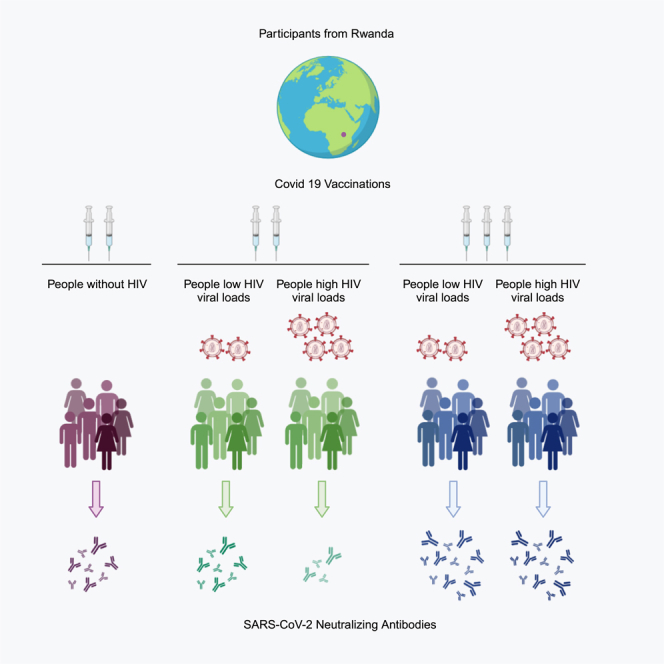
SARS-CoV-2 (severe acute respiratory syndrome coronavirus 2) causing COVID-19 (coronavirus disease 2019) poses a greater health risk to immunocompromized individuals including people living with HIV (PLWH). However, most studies on PLWH have been conducted in higher-income countries. We investigated the post-vaccination antibody responses of PLWH in Rwanda by collecting peripheral blood from participants after receiving a second or third COVID-19 vaccine. Virus-binding antibodies as well as antibody neutralization ability against all major SARS-CoV-2 variants of concern were analyzed. We found that people with high HIV viral loads and two COVID-19 vaccine doses had lower levels of binding antibodies that were less virus neutralizing and less cross-reactive compared to control groups. A third vaccination increased neutralizing antibody titers. Our data suggest that people with high HIV viral loads require a third dose of vaccine to neutralize SARS-CoV-2 virus and new variants as they emerge.
Subject areas: Virology, Public health
Graphical abstract
Highlights
-
•
SARS-CoV-2 poses a greater health risk to immunocompromized people living with HIV
-
•
COVID-19 vaccine outcomes in low- to middle-income countries are poorly understood
-
•
2 vaccines produced lower neutralizing antibody levels in people with high HIV
-
•
3 vaccines produced similar antibody levels in people with high or low HIV levels
Virology; Public health
Introduction
The COVID-19 (coronavirus disease 2019) pandemic caused by SARS-CoV-2 (severe acute respiratory syndrome coronavirus 2) is the largest public health crisis in modern history. Risk factors for severe COVID-19 include age as well as immunodeficiencies such as HIV.1 To initiate infection, coronaviruses use a spike (S) protein to gain entry into host cells,2 and following infection, the host elicits antibodies to the S protein for virus neutralization and subsequent protection. The S protein is therefore a common target for vaccine development. Among people living with HIV (PLWH), routine vaccination is associated with suboptimal neutralizing antibody responses.3 Following the rapid deployment of novel COVID-19 vaccines, there has been limited immunogenicity data specific to PLWH. While some studies have suggested reduced antibody levels and durability with more PLWH experiencing a hyporesponse to COVID-19 vaccination,4,5,6 other studies report normal responses.7 Third doses of COVID-19 vaccines have shown to be beneficial to immunocompromized groups such as transplantation recipients and also to PLWH hyporesponders.8,9,10,11
The COVID-19 pandemic experiences and responses per country have not been uniform worldwide. Each jurisdiction had region-specific case fatality rates, differing public health restrictions, as well as non-universal access to COVID-19-related health care, diagnostics, and vaccines.12 Within African nations, as elsewhere if not more so, it is suspected that asymptomatic/mild cases of COVID-19 have been significantly under-reported.12 Rwanda had a more proactive response, as a total lockdown was imposed on 22 March 2020.13 However, the COVID-19 testing rate in Rwanda was still below western countries, such as the US, with 1 per 100,000 versus 15.4 per 100,000 people tested daily, respectively, during peak pandemic periods.12 Despite the COVAX (COVID-19 Vaccine Global Access) program, COVID-19 vaccines were also deployed later and slower in continental Africa.14,15 It was estimated in October 2022 that over 9 million people in Rwanda had at least one dose of a COVID-19 vaccine. At this time we know very little about the COVID-19 vaccine responses in PLWH in continental Africa since most studies have been conducted in higher-income countries.4,8,10,16,17 Additional health challenges, such as malnutrition and parasitic infections, affecting people in LMICs (low- and middle-income countries) are known to significantly influence vaccine responses.12,18,19 Therefore, COVID-19 vaccine studies conducted in North America and Europe may not represent the global needs of PLWH.
To address this research priority, we established a cohort of people in Rwanda living with high and low HIV viral loads (VLs) to evaluate plasma antibody levels and neutralization to variants of concern (VOCs) including original SARS-CoV-2 lineage B (ancestral), B.1.1.7 (Alpha), B.1.351 (Beta), B.1.617 (Delta), and BA.1.18 (or BA.1) (Omicron) following a second or third dose of mRNA or adenoviral vectored COVID-19 vaccine. Due to the organization with the health care system and vaccine rollout program, Rwanda was an ideal choice for conducting a vaccine study.
Research in context
Evidence before this study
Understanding the COVID-19 situation in African countries has been less straightforward due to the lack of COVID-19 tests, organized case reporting, and variant characterization. The effectiveness of third COVID-19 vaccine doses has been demonstrated in healthy adults, older individuals, and some immunocompromized populations; however, in PLWH, especially those in LMICs, responses to a third vaccine dose remain poorly understood. Our PubMed search of COVID-19 vaccination and HIV-positive people identified studies performed in higher-income countries such as Canada, Germany, Italy, and the Netherlands, with only one such study from Africa. The published studies, in general, undertook minimal investigation of the virus neutralization capacity of COVID-19 vaccine-induced antibodies among PLWH and none used live SARS-CoV-2 virus or VOCs in their assessments.
Added value of this study
Most PLWH globally reside in sub-Saharan Africa where there are unique health challenges such as access to medical care, malnourishment, and co-infections with parasites that do not pose risks in other areas. Here we analyzed the antibody responses elicited after mRNA and/or viral vectored COVID-19 vaccination in 2021–2022 among Rwandans with high and low HIV VL) during 2021–2022. We found that PLWH with high VL had significantly lower neutralizing antibody titers after a primary series of COVID-19 vaccinations compared to those with low VL that received two doses. We also found that these antibodies were not as broadly neutralizing against VOCs suggesting these people may be less protected against new variants. Our data are more representative of the immunity status of PLWH in Rwanda than studies performed in higher-income nations.
Implications of all the available evidence
Our findings demonstrate that after two COVID-19 vaccine doses, PLWH with high VL have decreased virus neutralizing antibody titers against both the ancestral SARS-CoV-2 virus as well as VOCs. These data suggest reduced protection against COVID-19 and emerging variants among PLWH with high versus low or no detectable HIV VL. Neutralizing antibody titers were significantly increased with a third dose, reinforcing that PLWH with high VL require at least three COVID-19 vaccine doses and should be prioritized during booster dose campaigns. Better reflecting the community of PLWH in low-income nations and within sub-Saharan Africa, our findings have significant global implications for health equity, public health policy, and vaccine prioritization.
Results
Participant profiles
In total, 91 participants were vaccinated either 2 times (2x) or 3 times (3x) (Table 1; Figure 1). Volunteers were categorized as HIV negative, high HIV VL (>1,000 copies/mL), or low HIV VL (≤1,000 copies/mL). First and second vaccine doses were either mRNA (Pfizer-BioNTech Comirnaty or Moderna Spikevax) or viral vector (AstraZeneca Vaxzevria), or mixed. All third doses were mRNA vaccines. More than half of the participants were female, and the average age per group was between 35 and 47. Participants reported low percentages of comorbidities. Differences were detected between the median interval between doses and the median time from the last dose to blood collection among some groups.
Table 1.
Characteristics of study participants
| All participants (N = 90) | HIV-negative (N = 17) |
HIV-positive, overall (N = 73) |
HIVHigh Viral Load (N = 34) |
HIVLow Viral Load (N = 39) |
||||
|---|---|---|---|---|---|---|---|---|
| Two-dose (N = 17) | Two-dose (N = 42) | Three-dose (N = 31) | Two-dose (N = 20) | Three-dose (N = 14) | Two-dose (N = 22) | Three-dose (N = 17) | ||
| Female Sex, n (%) | 62 (69) | 12 (71) | 32 (76) | 18 (58) | 18 (90) | 7 (50) | 14 (64) | 11 (65) |
| Age (years) | ||||||||
| 19-35, n (%) | 38 (42) | 12 (71) | 22 (52) | 4 (13) | 12 (60) | 2 (14) | 10 (45) | 2 (12) |
| 36-49, n (%) | 32 (36) | 4 (24) | 16 (38) | 12 (39) | 5 (25) | 7 (50) | 11 (50) | 5 (29) |
| 50-64, n (%) | 20 (22) | 1 (6) | 4 (10) | 15 (48) | 3 (15) | 5 (36) | 1 (5) | 10 (59) |
| Median (IQR) | 39 (30–47) | 29 (23–37) | 35 (30–42) | 49 (41–53) | 34 (29–39) | 44 (40–52) | 39 (30–42) | 50 (42–53) |
| Range | 24–64 | 20–53 | 23–59 | 24–64 | 23–59 | 34–61 | 24–54 | 24–64 |
| Education status | ||||||||
| No formal education, n (%) | 11 (12) | 2 (71) | 5 (12) | 4 (13) | 2 (10) | 2 (14) | 3 (14) | 2 (12) |
| Primary school, n (%) | 47 (52) | 8 (47) | 26 (62) | 13 (42) | 12 (60) | 7 (50) | 14 (63) | 6 (35) |
| Secondary school, n (%) | 23 (26) | 6 (35) | 9 (21) | 8 (26) | 6 (30) | 2 (14) | 3 (14) | 6 (35) |
| Post-secondary, n (%) | 9 (10) | 1 (6) | 2 (5) | 6 (19) | 0 (0) | 3 (21) | 2 (9) | 3 (18) |
| Doses one and two, COVID-19 vaccination | ||||||||
| mRNA, n (%) | 64 (71) | 16 (94) | 38 (90) | 10 (32) | 17 (85) | 5 (36) | 21 (95) | 5 (29) |
| AstraZeneca, n (%) | 22 (25) | 0 (0) | 2 (5) | 20 (65) | 2 (10) | 9 (64) | 0 (0) | 11 (65) |
| Mixed mRNA/AstraZeneca, n (%) | 4 (4) | 1 (6) | 2 (5) | 1 (3) | 1 (5) | 0 (0) | 1 (5) | 1 (6) |
| Interval doses one and two, Median (IQR) (days) | 28 (22–75) | 23 (21–68) | 25 (22–36) | 75 (28–77) | 31 (23–67) | 74 (23–76) | 22 (21–27) | 76 (29–78) |
| Interval doses one and two, Range (days) | 16–280 | 18–87 | 16–243 | 21–280 | 22–243 | 21–93 | 16–71 | 21–280 |
| Interval doses two and three, COVID-19 vaccination (all third doses mRNA) | ||||||||
| Median (IQR) (days) | 189 (169–203) | NA | NA | 189 (169–204) | NA | 197 (146–204) | NA | 185 (183–203) |
| Range (days) | 12–244 | NA | NA | 12–244 | NA | 79–236 | NA | 12–244 |
| Time from last vaccine dose to blood draw | ||||||||
| Median (IQR) (days) | 53 (37–105) | 101 (54–105) | 102 (53–110) | 26 (16–38) | 75 (50–105) | 16 (14–26) | 106 (79–111) | 38 (25–38) |
| Range (days) | 11–222 | 21–112 | 17–222 | 11–67 | 17–222 | 13–34 | 37–125 | 11–67 |
| Duration of HIV infection (years) | ||||||||
| <10, n (%) | NA | NA | 25 (60) | 11 (35) | 12 (60) | 7 (50) | 13 (59) | 4 (24) |
| 10-19, n (%) | NA | NA | 15 (36) | 17 (55) | 6 (30) | 7 (50) | 9 (41) | 10 (59) |
| 20+, n (%) | NA | NA | 2 (5) | 3 (10) | 2 (10) | 0 (0) | 0 (0) | 3 (18) |
| Antiviral (ART) regimen | ||||||||
| First-line, n (%) | NA | NA | 39 (93) | 24 (77) | 17 (85) | 10 (71) | 22 (100) | 14 (82) |
| Second-line, n (%) | NA | NA | 3 (7) | 6 (19) | 3 (15) | 4 (29) | 0 (0) | 2 (12) |
| Third-line, n (%) | NA | NA | 0 (0) | 1 (3) | 0 (0) | 0 (0) | 0 (0) | 1 (6) |
| Duration of ART regimen (years) | ||||||||
| <10, n (%) | NA | NA | 27 (64) | 13 (42) | 13 (65) | 8 (57) | 14 (64) | 5 (29) |
| 10-19, n (%) | NA | NA | 14 (33) | 17 (55) | 6 (30) | 6 (43) | 8 (36) | 11 (65) |
| 20+, n (%) | NA | NA | 1 (2) | 1 (3) | 1 (5) | 0 (0) | 0 (0) | 1 (6) |
| Most recent CD4 T cell count among HIV-positive participants with available counts | ||||||||
| <200 copies/uL, n/N (%) | NA | NA | 7/40 (17) | 2/28 (7) | 6/18 (33) | 2/11 (18) | 1/22 (5) | 0/17 (0) |
| ≥200 copies/uL, n/N (%) | NA | NA | 33/40 (83) | 26/28 (93) | 12/18 (67) | 9/11 (82) | 21/22 (95) | 17/17 (100) |
Figure 1.

Study participant profile
91 participants were recruited for sample collection including 17 HIV− and 73 PLWH. PLWH were further defined as having high or low HIV viral loads at >1,000 copies/mL or ≤1000 copies/mL, respectively. HIV− individuals received 2 doses of a COVID-19 while PLWH received either 2 or 3 doses.
People living with high HIV VL and two doses of COVID-19 vaccines had lower SARS-CoV-2 binding antibodies
Immunoglobulin G (IgG) antibody levels were assessed and the geometric mean titer (GMT) was determined in the plasma of participants against the S1 subunit, the receptor binding domain (RBD), and the N protein of the SARS-CoV-2 virus using ELISA (Figures 2A–2C). The coronavirus spike (S) protein is composed of two subunits, the S1 and S2, and is responsible for virus entry into the cell.20 Within the S1 domain, the RBD is where binding directly occurs between the virus and the host cell receptor angiotensin-converting enzyme 2 (ACE2). Antibodies that specifically bind RBD are often correlated with virus neutralization while antibodies that bind other domains within the S1 such as the NTD (N Terminal Domain) are less likely to block virus entry.20 Therefore, we assessed both the cumulative antibodies elicited toward the entire S1 molecule and then those specific to the RBD in an effort to determine if any group had a skewed response where antibodies were directed away from the RBD. The control group (HIV− 2x) had a GMT of 17,021 for S1 binding antibodies, while the HIVHigh 2x group had lower S1 binding antibodies (GMT of 7,672) although not statistically significant. The HIVLow 2x group had statistically higher S1 titers at 23,502 than the HIVHigh 2x at 7,672 (Figure 2A). Both of the HIVHigh 3x and HIV low HIVLow 3x had significantly higher S1 antibody titers than their respective groups that had only 2 doses. Similar trends were observed for RBD antibody titers (Figure 2B). Antibodies binding the N protein were assessed to determine previous exposure to SARS-CoV-2, and minimal variation was seen among the groups with only statistical differences between HIVHigh 2x and HIVLow 2x groups (Figure 2C).
Figure 2.

Participants with high HIV viral loads and two doses of COVID-19 vaccine have lower antibodies to the receptor binding domain (RBD) of the S protein
Plasma IgG antibody levels were determined against the SARS-CoV-2 S1 domain of the S protein (A), the RBD of the S protein (B), and the N protein (C) by ELISA. Blood samples were collected from 4 groups that had received COVID-19 vaccines: HIV− 2x (control), HIVHigh 2x, HIVHigh 3x, HIVLow 2x, and HIVLow 3x. Plasma was isolated and used for direct ELISA against proteins or domains of SARS-CoV-2. High HIV viral load is equivalent to >1,000 copies/mL and Low viral load is equivalent to ≤1,000 copies/mL. LOD = Limit of Detection. The calculated geometric mean titer (GMT) for each group is indicated above and by the thick line in each column. Statistical differences between groups were determined by Mann-Whitney U test. ns represents p > 0.05; ∗ represents p ≤ 0.05; ∗∗ represents p ≤ 0.01; ∗∗∗ represents p ≤ 0.001; ∗∗∗∗ represents p ≤ 0.0001.
Antibody virus neutralization and cross-neutralization were improved in PLWH with a third vaccine dose
The ability of antibodies to neutralize the SARS-CoV-2 virus has been directly linked to protection from COVID-19; however, only a few studies of HIV-positive vaccinated individuals have directly evaluated the ability of COVID-19 vaccine-elicited antibodies in PLWH to neutralize SARS-CoV-2.21 We performed neutralization assays against ancestral SARS-CoV-2 as well as the VOCs Alpha, Beta, Delta, and Omicron. Titers per group were graphed, and the GMT of each group was noted. Moreover, each group was graphed per variant across groups or each group across the variants in Figure 3, (see also Figure S1) and Figure 4, respectively. When comparing the neutralization titers across the groups for the ancestral virus (Figure 3A), the triple vaccinated groups had the highest virus neutralization titers. The GMTs were 320 for the HIV− 2x group and 807 and 1,002 for HIVhigh 3x and HIVlow 3x, respectively (Figure 3A). The titers of the groups that received 3 vaccine doses were also statistically increased compared to the groups that only received 2 doses. Moreover, the titers for HIVHigh 2x and HIVLow 2x groups were close or below those of the control group. Similar trends were found when analyzing across the groups for the other variants, where PLWH receiving 3 doses of COVID-19 vaccines had titers statistically above the groups of PLWH that only received 2 doses. The HIVLow 2x vaccinated group had levels of neutralizing titers similar to the control group for variants Alpha, Beta, Delta, and Omicron with titers of 113, 193, 141, and 84, respectively. In comparison, the HIVHigh 2x group had very low GMTs of 78, 50, 85, and 8 for the Alpha, Beta, Delta, and Omicron, respectively. We also found more low responders (participants with titers at 1:80 or below) in the HIVHigh 2x group compared to other groups (Table 2). These results suggest that the control of HIV VL is important for antibody responses to COVID-19 vaccination and that a third vaccine dose can improve neutralizing antibody titers regardless of HIV VL levels.
Figure 3.

Participants with high HIV viral loads and 3 doses of COVID-19 vaccines have similar neutralizing antibody levels to those with controlled HIV and 3 doses
(A–E) Neutralizing antibodies to SARS-CoV-2 and variants of concern (original B-lineage, Alpha, Beta, Delta, and Omicron) were determined by microneutralization assay. The data from each group were analyzed against the other groups per variant original B-lineage (A), Alpha (B), Beta (C), Delta (D), and Omicron (E) to determine statistical differences between the study groups. The violin graphs show the median and quartiles. High HIV viral load is equivalent to >1,000 copies/mL and Low viral load is equivalent to ≤1,000 copies/mL. Statistical differences between groups were determined by Mann-Whitney U test. ns represents p > 0.05; ∗ represents p ≤ 0.05; ∗∗ represents p ≤ 0.01; ∗∗∗ represents p ≤ 0.001; ∗∗∗∗ represents p ≤ 0.0001 (See also Figure S1).
Figure 4.

Participants with high HIV viral loads and only two doses of COVID-19 vaccine have low cross-neutralizing antibodies
(A–E) Neutralizing antibodies to SARS-CoV-2 original B-lineage virus and variants of concern Alpha, Beta, Delta, and Omicron were determined by microneutralization assay. Data were then analyzed by each experimental study group across the viruses: HIV− 2x (control) (A), HIVHigh 2x (B), HIVLow 2x (C), HIVHigh 3x (D), and HIVLow 3x (E). The violin graphs show the median and quartiles. Scatterplots joining samples across assays are shown. The GMT for each assay for each group is indicated numerically and on the graph. High HIV viral load is equivalent to >1,000 copies/mL and Low viral load is equivalent to ≤1,000 copies/mL. Statistical differences between virus assays for each group were determined by Mann-Whitney U test. ns represents p > 0.05; ∗ represents p ≤ 0.05; ∗∗ represents p ≤ 0.01; ∗∗∗ represents p ≤ 0.001; ∗∗∗∗ represents p ≤ 0.0001.
Table 2.
Study participants classified as low vaccine responders
| Study group | Wuhan |
Alpha |
Beta |
Delta |
Omicron |
|---|---|---|---|---|---|
| Participant number, n (row %) with neutralizing GMT ≤1:80 | |||||
| HIV− 2x (N = 17) | 1 (6) | 4 (24) | 5 (29) | 3 (18) | 11 (65) |
| HIVHigh 2x (N = 20) | 6 (30) | 11 (55) | 13 (65) | 8 (40) | 20 (100) |
| HIVLow 2x (N = 22) | 2 (9) | 11 (50) | 7 (32) | 10 (45) | 10 (45) |
| HIVHigh 3x (N = 14) | 2 (14) | 3 (21) | 3 (21) | 2 (14) | 6 (43) |
| HIVLow 3x (N = 17) | 0 (0) | 2 (12) | 0 (0) | 1 (6) | 5 (29) |
Next, the neutralization titers of each group were graphed to compare across variants to visualize how cross-reactive antibodies were per group (Figure 4). Trend lines marked the same sample across the variant neutralization evaluations (Figure 4A [HIV− 2x], B [HIVHigh 2x], C [HIVLow 2x], D [HIVHigh 3x], and E [HIVLow 3x]). All groups showed statistical decreases of the neutralizing ability when comparing neutralization of the ancestral virus to the VOCs with antibody titers against Omicron being the lowest. Statistical differences between titers for the ancestral and Omicron virus were noted for each group. Some differences were also detected for variants such as neutralization of the Beta variant which was statistically lower than that of the ancestral virus for the HIV− 2x and HIVHigh 2x groups. Taken together, this analysis suggests that 3 doses of COVID-19 vaccines increase neutralizing antibody titers for VOCs in people with high HIV VL.
COVID-19 vaccination type and antibody responses in PLWH
It has been noted that virus neutralization ability of antibodies after COVID-19 vaccination may be dependent on the vaccine platform used for immunization;22 however, the comparative responses of PLWH have not been thoroughly investigated. We compared the virus neutralization antibody titers against the ancestral virus per participant group per vaccine platform (Figure 5). All participants in the HIV− 2x, HIVHigh 2x, and HIVLow 2x groups had two doses of COVID-19 vaccines with the mRNA platform with the exception of one individual in each group who received an mRNA and a viral vector dose (mixed) and two people who had two viral vector doses in the HIVHigh 2x group. All third doses were with an mRNA vaccine. GMTs for 2 doses of mRNA vaccines were 293, 194, and 408, for the HIV−, HIVHigh 2x, and HIVLow 2x groups, respectively. The participant with mixed vaccine regimes in the HIV− group had a robust titer of 1,280, while the participants in the HIV+ 2x vaccinated groups had much lower neutralization titers of 80 (HIVHigh 2x) and 20 (HIVLow 2x). In the HIVHigh 3x group, the subgroup that received an mRNA primary series had markedly lower neutralizations titers than the participants that received a viral vector primary series. Conversely, all participants in the HIVlow 3x group had similar GMTs regardless of initial vaccination series. While the differences observed between vaccine regimes in the 3x groups were not significant, this analysis indicated that participants with high HIV VL who received a viral vector COVID-19 primary vaccine series followed by an mRNA boost had higher levels of neutralizing antibodies.
Figure 5.

Neutralizing antibody titers to SARS-CoV-2 original B-lineage for each participant study group were analyzed by vaccine platform
Microneutralization assays were performed as previously described against the original B-lineage virus. Data were plotted per study group and further stratified by vaccine regime: 2 doses of mRNA, 2 doses of AstraZeneca, or mixed vaccine doses. All participants that were vaccinated 3 times received an mRNA vaccine as the third dose. GMT for each vaccine regime within each group is indicated. The number of participants per vaccine regime per group is also indicated.
Discussion
Here we investigated the humoral responses to COVID-19 vaccination in response in people living with high and low HIV VL living in Rwanda. We found significant difference in binding antibodies as well as virus neutralizing antibodies after COVID-19 vaccination among our groups. Importantly, PLWH with high VL that received a third COVID-19 vaccine dose had significantly higher virus neutralizing antibody levels compared to those that only received two doses. Moreover, those with high HIV VL and only two doses of COVID-19 vaccines had extremely low cross-neutralizing antibodies toward the Omicron variant suggesting these participants are unprotected from antigenically distinct SARS-CoV-2 variants. Our study is the first study of which we are aware to investigate the virus neutralizing capacity of COVID-19 vaccine-elicited antibodies in PLWH from sub-Saharan Africa using live virus and all the major VOCs. Considering the essential role T cells play in both coordinating the humoral response to vaccination as well as in cell-mediated immunity for viral clearance,23 we found a trend where people with higher VL had higher CD4 counts, but follow-up studies should investigate further the T cell responses in PLWH in sub-Saharan Africa among more participants.
Few studies of COVID-19 vaccine responses in PLWH have been conducted with most studies focused on people living in European and North American countries.4,6,7,8,10,16,17,24,25,26,27 We found only one report, a clinical trial conducted in South Africa, from continental Africa.17 Moreover, the majority of published studies were designed to compare antibody responses following COVID-19 vaccination in people who had not previously had a COVID-19 experience thus allowing a straightforward and uncomplicated analysis. In our study, we found that participants had evidence of a previous COVID-19 experience due to the positive anti-N ELISA results for each person. These findings could possibly be due to participants being unaware they had a SARS-CoV-2 infection as COVID-19 testing was infrequent. Additionally, since the samples were collected in 2022, there was ample time for the individuals to be infected prior to study enrollment throughout 2020 and 2021. Similarly, a 2020 ChAdOx1 nCoV-19 (AZD1222) vaccine clinical trial conducted early in the pandemic (2020) in South Africa found 31% of participants were previously infected with SARS-CoV-2.17 Although controlling the participant groups for previous exposure to SARS-CoV-2 would improve the stratification of immune responses, our findings are representative of the actual immunity in the community in Rwanda which is an important comparison to data acquired from North American and European studies.
COVID-19 primary series vaccine studies conducted in COVID-19-naive PLWH have identified more non-responders and more hyporesponsive individuals compared to control groups with respect to antibody titers.16 Further investigation of initial hyporesponders has shown increases in antibody titers after a third vaccine dose was given.8 We found more hyporesponders in our group of people living with high HIV VL after 2 vaccine doses, although the sample size is small to draw definitive conclusions. However, since we have evidence that our participants were previously exposed to the virus due to detectable N antibody titers, it was surprising that the virus neutralization antibody levels were not higher since hybrid immunity has been shown to induce more robust antibody responses in healthy adults.28 Hybrid immunity is also associated with antibodies that have increased cross-neutralizing capacity,29 but we found most participants in the HIVHigh 2x group to have undetectable neutralization titers against Omicron. Titers were restored in those that had received a third dose. These results were similar to finding of adults living with and without HIV in European and North American countries.4,8,11,16 Interestingly, a study from Italy investigating the effect of a bivalent (BA.4/5 and ancestral) booster dose in PLWH who had previously been infected with SARS-CoV-2 found increased virus cross-neutralizing antibody titers suggesting that the decreased antibody breadth which we found even in those with hybrid immunity can be overcome with a bivalent vaccine dose.27 Time since COVID-19 experience is most likely important for understanding the effects of hybrid immunity of antibody titers. In our study we do not know how long prior infection may have occurred, and the sera were drawn from among our two-dose participants (HIV-negative or HIV-positive) much longer after their last dose (∼100 days) than among our three-dose participants (∼30 days). Therefore, the time since the COVID-19 experience may have influenced our results, which is a limitation of our study.
Vulnerable populations have been prioritized for COVID-19 vaccination, both in primary series and for follow-up booster vaccinations, as they have historically had suboptimal responses following vaccination for diseases such as influenza.3,30,31,32 These groups include those of older age, people living with immunosuppressive conditions including HIV, and those on immunosuppressant therapies for conditions such as rheumatoid arthritis (RA), solid organ transplantation recipients, and cancer patients. Although, these groups are at greater risk for severe disease following viral infection and therefore vaccination is critically important, the responses to COVID-19 vaccination have been understudied across all groups and not just in those living with HIV. Recently published studies have found similar trends as those emerging for vaccine responses in PLWH. Specifically, booster or third doses of COVID-19 vaccines in people living with multiple sclerosis (MS) and people with RA increased antibody levels to levels more similar to healthy controls, similar to what we have reported in our study as well as in other studies in PLWH.33,34 In MS patients, the elicitation of antibodies was dependent on specific MS immunosuppressive treatment where individuals who received ocrelizumab did not have a similar increase in antibodies.33 Interestingly, in the MS and RA studies, T cell responses were investigated and indicated that these vulnerable groups had reduced T cell activation profiles after S protein stimulation characterized by impaired interleukin-2 (IL-2) responses. Another study investigating vaccine responses in liver transplantation recipients found that recipients were capable of eliciting both humoral and cell-mediated immunity after a two-dose vaccine regime but that responses were also dictated by immunosuppressive therapy as well as time since transplantation.35 Specifically, those on mycophenolate mofetil had reduced responses which have been suggested in other vaccine studies to be due to lower IL-4+CD4+ T cell numbers and reduced B cell activation.36 In our study, we did not assess the T cell arm of the adaptive immune response; therefore we cannot make direct comparisons to these investigations. As impaired IL-2 responses were common for people with RA and MS, future work with PLWH from Rwanda should determine if IL-2 responses are different following COVID-19 vaccination in PLWH who have high and low VLs. Overall, understanding how each vulnerable group and any associated treatments impact COVID-19 vaccine responses will be valuable in implementing public health vaccine policy as well as determining vaccine mechanisms associated with protection.
Limitations of our study include the absence of PLWH who have not been infected with SARS-CoV-2 but have been vaccinated. Although this limits the interpretation of our study, we feel the data are more representative of the immunity within the HIV community in Rwanda. Also, in this study we focused on the humoral responses and did not investigate the cellular phenotypes or the CD4+ or CD8+ T cell compartments as done in some other studies.6 We used convenience sampling which led to variation in the timing of blood collection and time since a COVID-19 experience which may influence our immunogenicity observations; however, knowing stability of antibody levels after boost to 4 months post-vaccination supports our study design. It is important to note the importance of T cell responses that are mounted after vaccination which aid in protection.23 Robust T cell responses have been associated with mild COVID-19 disease as CD4+ T cells are essential for effective antibody responses and CD8+ T cell response helps clear infected cells.23 Following SARS-CoV-2 infection, some studies have indicated that T cell responses are more stable in healthy individuals than humoral immunity and antibody levels. Although the importance of T cell immunity is understood during viral infection, it is understudied in vaccine research and little is known about how T cell immunity is regulated in vulnerable populations after vaccination.23 As we were focused on the antibody responses which were also acquired through easier sampling methods in Rwanda due to resource limitations, our study did not address cell-mediated immunity. Although we analyzed CD4 counts in our participants, we did not investigate T cell activation, T cell subsets, and SARs-CoV-2-specific responses. Follow-up studies should address this knowledge gap for PLWH in LMICs such as Rwanda as insights may aid in the development of more effective vaccines or vaccination strategies. The vaccinations received by participants in this study were comprised of a primary series of mRNA, viral vector, or mixed doses of the two. Our study was limited in its ability to perform a robust statistical analysis of the different vaccine regimes by the low number of participants within each regime received. Participants receiving only 2x vaccines predominantly received two doses of mRNA vaccines which did not allow for any statistical analysis to be performed. Within the cohort of participants that received 3x vaccines, a non-parametric Wilcoxon Mann-Whitney test determined that there was no significant difference between participants that received the mRNA or viral vector primary series of vaccines in either the HIVhigh or HIVlow groups. Although the low number of participants in this study limited the statistical analysis, the outcome of different vaccine types on the immune status of PLWH is an important question that should be addressed in future studies. Finally, our study employed a cross-sectional design and the participants who had two doses were not followed for their third dose, thereby limiting our understanding of the direct effects of a third dose. Further evaluation with larger sample size to stratify the data further would be an important future objective; however, considering the progression of the COVID-19 pandemic, circulating variants, and additional vaccine doses, it may not be possible to recruit more participants.
Taken together, our data give geographically specific information on the COVID-19 vaccine responses of people living with high and low HIV VL during an on-going pandemic in Rwanda. We have specifically shown that two doses of COVID-19 vaccines would be insufficient to induce high levels of virus neutralizing and cross-neutralizing antibodies, with these improved in all groups who received a third dose. Additionally, immunocompromized individuals, including those with HIV, have an increased tendency to harbor SARS-CoV-2 new variants during a SARS-CoV-2 infection.37,38,39 It has been postulated that new variants such as Omicron may have emerged from those who are immunocompromized due to the continued viral replication leading to mutations within the virus which can occur when the immune response is not robust.39 Therefore, our data suggest that a third or additional vaccine dose/s may be important for controlling SARS-CoV-2 infection, viral mutations, and the emergence of new SARS-CoV-2 variants. Taken together, the results from our study are important for maintaining public health as well as implementing public health guidance and policies.
Limitations of the study
Limitations of our study include the absence of PLWH who have not been infected with SARS-CoV-2 but have been vaccinated. Therefore, in our population the robustness of the vaccine responses in naive PLWH is unclear which would have been useful to compare to those who have also had COVID-19. Also, our study did not address cell-mediated immunity as we were focused on the antibody responses. In addition, we were unable to perform a robust statistical analysis of the different vaccine regimes due to the low number of participants within each regime received. This would be important to follow up as understanding what vaccine platform is the most effective in PLWH would be an essential step forward in designing optimal vaccines for this population. Finally, our study employed a cross-sectional design and the participants who had two doses were not followed for their third dose, thereby limiting our understanding of the direct effects of a third dose.
STAR★Methods
Key resources table
| REAGENT or RESOURCE | SOURCE | IDENTIFIER |
|---|---|---|
| Antibodies | ||
| Anti-human IgG-HRP | Jackson ImmunoResearch Laboratories Inc. | 109-035-088; RRID: AB_2337584 |
| Bacterial and virus strains | ||
| SARS-CoV-2 isolate/Canada/ON/VIDO-01/2020 (ancestral) | VIDO20, Saskatoon, SK | GISAID–EPI_ISL_425177 |
| SARS-CoV-2 Alpha variant | Alberta Health | GenBank no. OQ781006 |
| SARS-CoV-2 Beta variant | Roy Romanow Provincial Laboratory | Bioproject No. SAMN24618764 |
| SARS-CoV-2 Delta variant | National Microbiology Laboratory | NCBI Bioproject No. SAMN24618763 |
| SARS-CoV-2 Omicron variant (lineage BA.1.19) | BC CDC | GISAID Accession: EPI_ISL_7370259 |
| Biological samples | ||
| Plasma isolated from peripheral blood | National Reference Laboratory, Kigali, Rwanda | N/A |
| Chemicals, peptides, and recombinant proteins | ||
| SARS-CoV-2 S1 subunit peptide | Sino Biological | 40591-V08H |
| SARS-CoV-2 RBD peptide | Sino Biological | 40592-V08H |
| SARS-CoV-2 N peptide | Abeomics | 21–1003 |
| Deposited data | ||
| Raw and analyzed data | This paper | N/A |
| Experimental models: Cell lines | ||
| African green monkey: Vero cell line | ATCC | CCL-81; RRID:CVCL_0059 |
| Software and algorithms | ||
| GraphPad Prism8 | Dotmatics, San Diego, USA | RRID:SCR_002798; https://www.graphpad.com/features |
| BioRender | 2023 BioRender | www.biorender.com |
| Other | ||
| 96 well microtiter plates, Immulon 2HB | ThermoFisher Scientific | Part# 3655 |
Resource availability
Lead contact
Further information and requests for resources and reagents should be directed to and will be fulfilled by the lead contact, Alyson Kelvin (Alyson.kelvin@usask.ca). Sharing of reagents may require MTA agreements.
Materials availability
This study did not generate any unique reagents.
Experimental model and study participant details
Study participant Recruitment
In total, 91 participants living in Rwanda between the ages of 18 and 65 were recruited between December 31, 2021 and April 4, 2022. Participants that were either HIV negative or positive and had had either two or three Covid19 vaccinations were recruited. All study categories contained both female and male participants from diverse educational backgrounds with the majority of the participants being female (62%) and having completed primary school education (52%). One participant was excluded from the study due to insufficient information on vaccination and HIV status being provided by the participant. Detailed characteristics of the study participants can be found in Table 1, in the Participant Categories section in method details and under the Participant Profiles heading in the Results section.
Ethics Statement
Ethics approval was obtained from the Rwanda National Ethics Committee (RNEC) (Reference number. No.832/RNEC/2021), authorized from the Ministry of Health, and registered as a study in the Rwanda national health research registry. All participants provided informed consent, were assigned a research code, and data was confidential. Secondary use of samples was conducted in accordance with Biomedical Research Ethics Board (Bio-REB) at the University of Saskatchewan under the protocol ID#2387.
Method details
Participant Categories
Volunteers were categorized as HIV negative, high HIV VL (>1000 copies/mL), or low HIV VL (≤1000 copies/mL). The following study groups were defined by HIV and COVID-19 vaccine status, respectively: (a) high VL, twice-vaccinated (HIVHigh 2x); (b) high VL, thrice-vaccinated (HIVHigh 3x); (c) low VL, twice-vaccinated (HIVLow 2x); (d) low VL, thrice-vaccinated (HIVLow 3x) and (e) HIV-negative, twice-vaccinated (HIV- 2x) (Figure 1; Table 1). Participants completed a questionnaire to provide anonymized information and samples were collected 2 weeks to 4 months following the participant’s last vaccination event. Efforts were made to recruit participants without a history of COVID-19; however, many participants were suspected to have a history of COVID-19 due to positive SARS-CoV-2 S or N ELISAs.
SARS-CoV-2 viruses and variants
SARS-CoV-2 isolate /Canada/ON/VIDO-01/2020 was used as a representative for the original Wuhan virus B-lineage40 (GISAID–EPI_ISL_425177) (ancestral). SARS-CoV-2 variants Alpha (Genbank no. OQ781006) and Beta (Bioproject No. SAMN24618764) were obtained from the Alberta Health and Roy Romanow Provincial Laboratory, respectively. Delta was obtained from the National Microbiology Laboratory (NCBI Bioproject No. SAMN24618763). VOC Omicron (lineage BA.1.19, Pango v4.2, PANGO-v.1.18) was acquired from the BC CDC (GISAID Accession: EPI_ISL_7370259). All SARS-CoV-2 variants were cultured in vDMEM (DMEM (Dulbecco’s Modified Eagle Medium) (Wisent Bioproducts (Cat # 319-005-CL)), 2% fetal calf serum (Wisent Bioproducts (Cat # 090–150)), 5 mL 100x penicillin (10,000 U/mL)/streptomycin (10,000 μg/mL), and 2 μg/mL TPCK-trypsin in Vero cells. Virus work was performed in the Vaccine and Infectious Disease Organization’s (VIDO) Containment Level 3 (CL3) facility (InterVac) in Saskatoon, Canada.
Blood sampling
Peripheral blood was collected from participants in K2EDTA spray coated tubes (BD cat# 367844), transported to the National Reference Laboratory in Kigali, Rwanda, and centrifuged for 10 m at 200 x g. Plasma was isolated and stored in conical vials (Fisher 1495949B) at −80°C. Samples were shipped on dry ice to VIDO (Saskatoon, Canada) for analysis.
Enzyme-linked Immunosorbent assay (ELISA)
IgG antibody levels were assessed in the plasma of participants against the S1 subunit of the spike protein, the RBD (receptor binding domain of the spike protein), and to the N (nucleocapsid protein) of ancestral SARS-CoV-2 virus using ELISA assays, according to standard protocol. Briefly, microtiter plates (96-well, Immulon® 2HB) were coated with 0.5 μg/mL SARS-CoV-2 S1 subunit (Sino Biological 40591-V08H), RBD (Sino Biological 40592-V08H), and SARS-CoV-2 N protein (Abeomics 21-1003). Plates were washed with 1x TBS with 0.05% Tween-20 (Sigma P1379) (TBS-T) then blocked for 1 h. 100 μl of human plasma samples were serially diluted in 1% skim milk TBS-T then incubated at room temperature followed by washing and secondary antibody (anti-human IgG-HRP 1:20,000 (Cedarlane 109-035-088)) incubations. After washing, OPD substrate (Thermo Fisher 34062) and stop solution were added and the plates were read at 490 nm on a BioTek 800TS reader. Samples were considered positive if average optical density (OD) was greater than 0.1 and greater than the mean OD in SARS-CoV-2 unexposed samples plus 3 standard deviations at the same dilution. Negative samples are denoted as “1” for display on a logarithmic scale.
Virus neutralization assay
Virus neutralization assays were conducted as previous described.41 Briefly, Vero cells were seeded in 96-well tissue plates (20,000 cells/well) and cultured overnight in DMEM (Dulbecco’s Modified Eagle Medium) (Wisent Bioproducts (Cat # 319-005-CL)), containing 2% fetal calf serum (Wisent Bioproducts (Cat # 090–150)), and 5 mL 100x penicillin (10,000 U/mL)/streptomycin (10,000 μg/mL). Plasma samples were heat-inactivated at 56°C and serially diluted 1:2 in vDMEM then added to virus (50 TCID50) at a 1:1 ratio and incubated at 37°C for 1 h in a 5% CO2 chamber. The plasma-virus mixture was added to cultured cells in 96-well plates for absorption for 1 hour, media was replaced, and cells were incubated for 5 days. Endpoint neutralization titer was based on inhibition of CPE (cytopathic effect) observed on day 5. Antibody titer was calculated as the inverse of the most diluted sample without CPE was detected. Negative samples are denoted as “1”.
Quantification and statistical analysis
For each analysis, the number of participants in each group is indicated as “n” in each figure.
Results were analysed using GraphPad Prism8. Non-parametric Wilcoxon Mann-Whitney tests were conducted using GraphPad Prism8 (San Diego, USA) to determine statistical differences between viral titres. A p value of ≤ 0.05 was considered statistically significant.
The use of lines indicating medians, geometric mean titres, quartiles, assay limits of detection and symbols indicating levels of significant differences are described in the figure legends for each figure. For calculation of geometric mean titres, samples that had non-detectable results were given a value of “1”.
Acknowledgments
We thank Dr. Danuta Skowronski for her critical review of this manuscript and the BC CDC for supplying the Omicron variant. This study was supported by a Canadian Institutes of Health Research (CIHR) Operating Grant: Emerging COVID-19 Research Gaps and Priorities. VIDO receives operational funding from the Canada Foundation for Innovation through the Major Science Initiatives Fund and from the Government of Saskatchewan through Innovation Saskatchewan. This article is published with the permission of the Director of VIDO-InterVac with article number 1025. The authors thank the participants of the study. All authors approved the manuscript and agreed with the decision to submit the manuscript for publication.
Author contributions
AAK and LB conceived and co-designed the study and analyses. VD, PN, SS, BT, JL, and AF acquired ethics approvals, generated study protocols, and processed clinical samples. CLS and RB performed antibody quantification and neutralizations assays, performed statistical analyses, and generated figures. AAK, LB, VD, MEF, and PN verified all the data. TY and CS performed or analyzed viral sequencing to verify variants. VG, DF, and DJK provided reagents, viruses, or tools. All authors contributed to data interpretation and manuscript writing. All authors have read and approved the final manuscript. The corresponding authors had access to all raw and analyzed data. They made the decision to submit for publication. AAK acquired the funding.
Declaration of interests
The authors have no competing interests to declare.
Inclusion and diversity
We support inclusive, diverse, and equitable conduct of research.
Published: September 19, 2023
Footnotes
Supplemental information can be found online at https://doi.org/10.1016/j.isci.2023.107959.
Supplemental information
Data and code availability
-
•
All study data are available from the lead contact (Alyson.kelvin@usask.ca) upon reasonable request.
-
•
This paper does not report original code.
-
•
Any additional information required to reanalyze the data reported in this paper is available from the lead contact upon request.
References
- 1.Centers for Disease Control and Prevention Underlying Medical Conditions Associated with Higher Risk for Severe COVID-19: Information for Healthcare Professionals. 2023. https://www.cdc.gov/coronavirus/2019-ncov/hcp/clinical-care/underlyingconditions.html [PubMed]
- 2.Walls A.C., Park Y.J., Tortorici M.A., Wall A., McGuire A.T., Veesler D. Structure, Function, and Antigenicity of the SARS-CoV-2 Spike Glycoprotein. Cell. 2020;181:281–292.e6. doi: 10.1016/j.cell.2020.02.058. [DOI] [PMC free article] [PubMed] [Google Scholar]
- 3.Crum-Cianflone N.F., Sullivan E. Vaccinations for the HIV-Infected Adult: A Review of the Current Recommendations, Part I. Infect. Dis. Ther. 2017;6:303–331. doi: 10.1007/s40121-017-0166-x. [DOI] [PMC free article] [PubMed] [Google Scholar]
- 4.Costiniuk C.T., Singer J., Lee T., Langlois M.A., Arnold C., Galipeau Y., Needham J., Kulic I., Jenabian M.A., Burchell A.N., et al. COVID-19 vaccine immunogenicity in people with HIV. AIDS. 2023;37:F1–F10. doi: 10.1097/QAD.0000000000003429. [DOI] [PMC free article] [PubMed] [Google Scholar]
- 5.Hassold N., Brichler S., Ouedraogo E., Leclerc D., Carroue S., Gater Y., Alloui C., Carbonnelle E., Bouchaud O., Mechai F., et al. Impaired antibody response to COVID-19 vaccination in advanced HIV infection. AIDS. 2022;36:F1–F5. doi: 10.1097/QAD.0000000000003166. [DOI] [PubMed] [Google Scholar]
- 6.Bessen C., Plaza-Sirvent C., Simsek A., Bhat J., Marheinecke C., Urlaub D., Bonowitz P., Busse S., Schumann S., Blanco E.V., et al. Impact of SARS-CoV-2 vaccination on systemic immune responses in people living with HIV. Front. Immunol. 2022;13:1049070. doi: 10.3389/fimmu.2022.1049070. [DOI] [PMC free article] [PubMed] [Google Scholar]
- 7.Chambers C., Samji H., Cooper C.L., Costiniuk C.T., Janjua N.Z., Kroch A.E., Arbess G., Benoit A.C., Buchan S.A., Chung H., et al. Coronavirus disease 2019 vaccine effectiveness among a population-based cohort of people living with HIV. AIDS. 2022;36:F17–F26. doi: 10.1097/QAD.0000000000003405. [DOI] [PMC free article] [PubMed] [Google Scholar]
- 8.Jongkees M.J., Geers D., Hensley K.S., Huisman W., GeurtsvanKessel C.H., Bogers S., Gommers L., Papageorgiou G., Jochems S.P., den Hollander J.G., et al. Immunogenicity of an Additional mRNA-1273 SARS-CoV-2 Vaccination in People With HIV With Hyporesponse After Primary Vaccination. J. Infect. Dis. 2023;227:651–662. doi: 10.1093/infdis/jiac451. [DOI] [PMC free article] [PubMed] [Google Scholar]
- 9.Hassold N., Brichler S., Gater Y., Leclerc D., Gordien E., Bouchaud O., Carbonnelle E., Mechai F., Cordel H., Delagreverie H. Mixed outcomes following a third SARS-CoV-2 vaccine dose in previously unresponsive people with HIV. J. Clin. Virol. 2023;158:105347. doi: 10.1016/j.jcv.2022.105347. [DOI] [PMC free article] [PubMed] [Google Scholar]
- 10.Lamacchia G., Salvati L., Kiros S.T., Mazzoni A., Vanni A., Capone M., Carnasciali A., Farahvachi P., Lagi F., Di Lauria N., et al. Fourth Dose of mRNA COVID-19 Vaccine Transiently Reactivates Spike-Specific Immunological Memory in People Living with HIV (PLWH) Biomedicines. 2022;10:3261. doi: 10.3390/biomedicines10123261. [DOI] [PMC free article] [PubMed] [Google Scholar]
- 11.Kamar N., Abravanel F., Marion O., Couat C., Izopet J., Del Bello A. Three Doses of an mRNA Covid-19 Vaccine in Solid-Organ Transplant Recipients. N. Engl. J. Med. 2021;385:661–662. doi: 10.1056/NEJMc2108861. [DOI] [PMC free article] [PubMed] [Google Scholar]
- 12.Margolin E., Burgers W.A., Sturrock E.D., Mendelson M., Chapman R., Douglass N., Williamson A.L., Rybicki E.P. Prospects for SARS-CoV-2 diagnostics, therapeutics and vaccines in Africa. Nat. Rev. Microbiol. 2020;18:690–704. doi: 10.1038/s41579-020-00441-3. [DOI] [PMC free article] [PubMed] [Google Scholar]
- 13.Semakula M., Niragire F., Umutoni A., Nsanzimana S., Ndahindwa V., Rwagasore E., Nyatanyi T., Remera E., Faes C. The secondary transmission pattern of COVID-19 based on contact tracing in Rwanda. BMJ Glob. Health. 2021;6:e004885. doi: 10.1136/bmjgh-2020-004885. [DOI] [PMC free article] [PubMed] [Google Scholar]
- 14.Nkengasong J.N., Ndembi N., Tshangela A., Raji T. COVID-19 vaccines: how to ensure Africa has access. Nature. 2020;586:197–199. doi: 10.1038/d41586-020-02774-8. [DOI] [PubMed] [Google Scholar]
- 15.Baptista S., Naidoo S., Suliman S., Nepolo E., Kanoi B.N., Gitaka J., Blessing O.M., Enany S. COVID-19 vaccinology landscape in Africa. Front. Immunol. 2022;13:955168. doi: 10.3389/fimmu.2022.955168. [DOI] [PMC free article] [PubMed] [Google Scholar]
- 16.Hensley K.S., Jongkees M.J., Geers D., GeurtsvanKessel C.H., Mueller Y.M., Dalm V.A.S.H., Papageorgiou G., Steggink H., Gorska A., Bogers S., et al. Immunogenicity and reactogenicity of SARS-CoV-2 vaccines in people living with HIV in the Netherlands: A nationwide prospective cohort study. PLoS Med. 2022;19:e1003979. doi: 10.1371/journal.pmed.1003979. [DOI] [PMC free article] [PubMed] [Google Scholar]
- 17.Madhi S.A., Koen A.L., Izu A., Fairlie L., Cutland C.L., Baillie V., Padayachee S.D., Dheda K., Barnabas S.L., Bhorat Q.E., et al. Safety and immunogenicity of the ChAdOx1 nCoV-19 (AZD1222) vaccine against SARS-CoV-2 in people living with and without HIV in South Africa: an interim analysis of a randomised, double-blind, placebo-controlled, phase 1B/2A trial. Lancet. HIV. 2021;8:e568–e580. doi: 10.1016/S2352-3018(21)00157-0. [DOI] [PMC free article] [PubMed] [Google Scholar]
- 18.Prendergast A.J. Malnutrition and vaccination in developing countries. Philos. Trans. R. Soc. Lond. B Biol. Sci. 2015;370:20140141. doi: 10.1098/rstb.2014.0141. [DOI] [PMC free article] [PubMed] [Google Scholar]
- 19.Wait L.F., Dobson A.P., Graham A.L. Do parasite infections interfere with immunisation? A review and meta-analysis. Vaccine. 2020;38:5582–5590. doi: 10.1016/j.vaccine.2020.06.064. [DOI] [PubMed] [Google Scholar]
- 20.Huang Y., Yang C., Xu X.F., Xu W., Liu S.W. Structural and functional properties of SARS-CoV-2 spike protein: potential antivirus drug development for COVID-19. Acta Pharmacol. Sin. 2020;41:1141–1149. doi: 10.1038/s41401-020-0485-4. [DOI] [PMC free article] [PubMed] [Google Scholar]
- 21.Boyarsky B.J., Werbel W.A., Avery R.K., Tobian A.A.R., Massie A.B., Segev D.L., Garonzik-Wang J.M. Immunogenicity of a Single Dose of SARS-CoV-2 Messenger RNA Vaccine in Solid Organ Transplant Recipients. JAMA. 2021;325:1784–1786. doi: 10.1001/jama.2021.4385. [DOI] [PMC free article] [PubMed] [Google Scholar]
- 22.Hvidt A.K., Baerends E.A.M., Søgaard O.S., Stærke N.B., Raben D., Reekie J., Nielsen H., Johansen I.S., Wiese L., Benfield T.L., et al. Comparison of vaccine-induced antibody neutralization against SARS-CoV-2 variants of concern following primary and booster doses of COVID-19 vaccines. Front. Med. 2022;9:994160. doi: 10.3389/fmed.2022.994160. [DOI] [PMC free article] [PubMed] [Google Scholar]
- 23.Petrone L., Sette A., de Vries R.D., Goletti D. The Importance of Measuring SARS-CoV-2-Specific T-Cell Responses in an Ongoing Pandemic. Pathogens. 2023;12:862. doi: 10.3390/pathogens12070862. [DOI] [PMC free article] [PubMed] [Google Scholar]
- 24.Liu W.D., Pang M.W., Wang J.T., Sun H.Y., Huang Y.S., Lin K.Y., Wu U.I., Li G.C., Liu W.C., Su Y.C., et al. Evolution of anti-SARS-CoV-2 spike protein titers after two-dose of COVID-19 vaccination among people living with HIV. J. Virus Erad. 2022;8:100308. doi: 10.1016/j.jve.2022.100308. [DOI] [PMC free article] [PubMed] [Google Scholar]
- 25.Maponga T.G., Jeffries M., Tegally H., Sutherland A., Wilkinson E., Lessells R.J., Msomi N., van Zyl G., de Oliveira T., Preiser W. Persistent Severe Acute Respiratory Syndrome Coronavirus 2 Infection With accumulation of mutations in a patient with poorly controlled Human Immunodeficiency Virus infection. Clin. Infect. Dis. 2023;76:e522–e525. doi: 10.1093/cid/ciac548. [DOI] [PMC free article] [PubMed] [Google Scholar]
- 26.Lombardi A., Butta G.M., Donnici L., Bozzi G., Oggioni M., Bono P., Matera M., Consonni D., Ludovisi S., Muscatello A., et al. Anti-spike antibodies and neutralising antibody activity in people living with HIV vaccinated with COVID-19 mRNA-1273 vaccine: a prospective single-centre cohort study. Lancet Reg. Health. Eur. 2022;13:100287. doi: 10.1016/j.lanepe.2021.100287. [DOI] [PMC free article] [PubMed] [Google Scholar]
- 27.Vergori A., Matusali G., Lepri A.C., Cimini E., Fusto M., Colavita F., Gagliardini R., Notari S., Mazzotta V., Mariotti D., et al. Neutralizing activity and T-cell response after bivalent fifth dose of messenger RNA vaccine in people living with HIV. Int. J. Infect. Dis. 2023;134:195–199. doi: 10.1016/j.ijid.2023.06.010. [DOI] [PMC free article] [PubMed] [Google Scholar]
- 28.Bates T.A., McBride S.K., Leier H.C., Guzman G., Lyski Z.L., Schoen D., Winders B., Lee J.Y., Lee D.X., Messer W.B., et al. Vaccination before or after SARS-CoV-2 infection leads to robust humoral response and antibodies that effectively neutralize variants. Sci. Immunol. 2022;7:eabn8014. doi: 10.1126/sciimmunol.abn8014. [DOI] [PMC free article] [PubMed] [Google Scholar]
- 29.Andreano E., Paciello I., Piccini G., Manganaro N., Pileri P., Hyseni I., Leonardi M., Pantano E., Abbiento V., Benincasa L., et al. Hybrid immunity improves B cells and antibodies against SARS-CoV-2 variants. Nature. 2021;600:530–535. doi: 10.1038/s41586-021-04117-7. [DOI] [PMC free article] [PubMed] [Google Scholar]
- 30.Mombelli M., Rettby N., Perreau M., Pascual M., Pantaleo G., Manuel O. Immunogenicity and safety of double versus standard dose of the seasonal influenza vaccine in solid-organ transplant recipients: A randomized controlled trial. Vaccine. 2018;36:6163–6169. doi: 10.1016/j.vaccine.2018.08.057. [DOI] [PubMed] [Google Scholar]
- 31.Oren S., Mandelboim M., Braun-Moscovici Y., Paran D., Ablin J., Litinsky I., Comaneshter D., Levartovsky D., Mendelson E., Azar R., et al. Vaccination against influenza in patients with rheumatoid arthritis: the effect of rituximab on the humoral response. Ann. Rheum. Dis. 2008;67:937–941. doi: 10.1136/ard.2007.077461. [DOI] [PubMed] [Google Scholar]
- 32.Koff W.C., Schenkelberg T., Williams T., Baric R.S., McDermott A., Cameron C.M., Cameron M.J., Friemann M.B., Neumann G., Kawaoka Y., et al. Development and deployment of COVID-19 vaccines for those most vulnerable. Sci. Transl. Med. 2021;13:eabd1525. doi: 10.1126/scitranslmed.abd1525. [DOI] [PubMed] [Google Scholar]
- 33.Aiello A., Coppola A., Ruggieri S., Farroni C., Altera A.M.G., Salmi A., Vanini V., Cuzzi G., Petrone L., Meschi S., et al. Longitudinal characterisation of B and T-cell immune responses after the booster dose of COVID-19 mRNA-vaccine in people with multiple sclerosis using different disease-modifying therapies. J. Neurol. Neurosurg. Psychiatry. 2023;94:290–299. doi: 10.1136/jnnp-2022-330175. [DOI] [PMC free article] [PubMed] [Google Scholar]
- 34.Farroni C., Aiello A., Picchianti-Diamanti A., Laganà B., Petruccioli E., Agrati C., Garbuglia A.R., Meschi S., Lapa D., Cuzzi G., et al. Booster dose of SARS-CoV-2 messenger RNA vaccines strengthens the specific immune response of patients with rheumatoid arthritis: A prospective multicenter longitudinal study. Int. J. Infect. Dis. 2022;125:195–208. doi: 10.1016/j.ijid.2022.10.035. [DOI] [PMC free article] [PubMed] [Google Scholar]
- 35.D'Offizi G., Agrati C., Visco-Comandini U., Castilletti C., Puro V., Piccolo P., Montalbano M., Meschi S., Tartaglia E., Sorace C., et al. Coordinated cellular and humoral immune responses after two-dose SARS-CoV2 mRNA vaccination in liver transplant recipients. Liver Int. 2022;42:180–186. doi: 10.1111/liv.15089. [DOI] [PMC free article] [PubMed] [Google Scholar]
- 36.Egli A., Humar A., Widmer L.A., Lisboa L.F., Santer D.M., Mueller T., Stelling J., Baluch A., O'Shea D., Houghton M., Kumar D. Effect of Immunosuppression on T-Helper 2 and B-Cell Responses to Influenza Vaccination. J. Infect. Dis. 2015;212:137–146. doi: 10.1093/infdis/jiv015. [DOI] [PubMed] [Google Scholar]
- 37.Baang J.H., Smith C., Mirabelli C., Valesano A.L., Manthei D.M., Bachman M.A., Wobus C.E., Adams M., Washer L., Martin E.T., Lauring A.S. Prolonged Severe Acute Respiratory Syndrome Coronavirus 2 Replication in an Immunocompromised Patient. J. Infect. Dis. 2021;223:23–27. doi: 10.1093/infdis/jiaa666. [DOI] [PMC free article] [PubMed] [Google Scholar]
- 38.Avanzato V.A., Matson M.J., Seifert S.N., Pryce R., Williamson B.N., Anzick S.L., Barbian K., Judson S.D., Fischer E.R., Martens C., et al. Case Study: Prolonged Infectious SARS-CoV-2 Shedding from an Asymptomatic Immunocompromised Individual with Cancer. Cell. 2020;183:1901–1912.e9. doi: 10.1016/j.cell.2020.10.049. [DOI] [PMC free article] [PubMed] [Google Scholar]
- 39.Kemp S.A., Collier D.A., Datir R.P., Ferreira I.A.T.M., Gayed S., Jahun A., Hosmillo M., Rees-Spear C., Mlcochova P., Lumb I.U., et al. SARS-CoV-2 evolution during treatment of chronic infection. Nature. 2021;592:277–282. doi: 10.1038/s41586-021-03291-y. [DOI] [PMC free article] [PubMed] [Google Scholar]
- 40.Marchand-Senécal X., Kozak R., Mubareka S., Salt N., Gubbay J.B., Eshaghi A., Allen V., Li Y., Bastien N., Gilmour M., et al. Diagnosis and Management of First Case of COVID-19 in Canada: Lessons Applied From SARS-CoV-1. Clin. Infect. Dis. 2020;71:2207–2210. doi: 10.1093/cid/ciaa227. [DOI] [PMC free article] [PubMed] [Google Scholar]
- 41.Foley M.K., Searle S.D., Toloue A., Booth R., Falkenham A., Falzarano D., Rubino S., Francis M.E., McNeil M., Richardson C., et al. Centenarians and extremely old people living with frailty can elicit durable SARS-CoV-2 spike specific IgG antibodies with virus neutralization functions following virus infection as determined by serological study. EClinicalMedicine. 2021;37:100975. doi: 10.1016/j.eclinm.2021.100975. [DOI] [PMC free article] [PubMed] [Google Scholar]
Associated Data
This section collects any data citations, data availability statements, or supplementary materials included in this article.
Supplementary Materials
Data Availability Statement
-
•
All study data are available from the lead contact (Alyson.kelvin@usask.ca) upon reasonable request.
-
•
This paper does not report original code.
-
•
Any additional information required to reanalyze the data reported in this paper is available from the lead contact upon request.