Abstract
Lingonberry are underutilised due to the lack of evaluating active compounds in different parts. In this study, the phytochemical profiles, antioxidant and antiproliferative activities of lingonberry's fruits, leaves and stems from different regions of China were compared. Ninety-five bioactive compounds were rapidly identified using a molecular network based on UPLC-Q-Exactive Orbitrap mass spectrometry. The UPLC-QqQ-MS/MS method combined with principal component analysis (PCA) quantified 18 bioactive components in 6 classes. The highest content of arbutin (15 mg/100 g DW) was found in leaves of Huzhong (P6). Ursolic acid and cyanidin-3-O-galactoside were highest in fruits of Tahe (P4) (4.5 mg/100 g DW and 3.2 mg/100 g DW, respectively). Antioxidant activities determined by DPPH, ABTS+ and FRAP methods were significantly correlated with total phenolic content (TPC), total flavonoid content (TFC) and total anthocyanin content (TAC). The results indicate that the strongest antioxidant activity and antiproliferative efficacy are observed in the fruits of Tahe (P4) and leaves of Huzhong (P6), respectively. Our results provide valuable insights into lingonberry's comprehensive development and utilization.
Comparative comprehensive chemical profiles of major metabolites of lingonberries from different origins.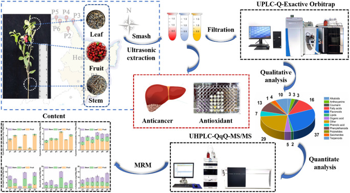
Introduction
Plant-driven natural antioxidant-sourced foods with proven human health benefits are receiving more attention.1 Lingonberry (Vaccinium vitis-idaea L.) is a wild evergreen dwarf shrub known as a “superfood” owing to its high antioxidant content.2 It is widespread in northern and central Europe, Canada and Asia, including alpine areas such as the Greater Khingan Range and Lesser Hinggan Mountains in North-eastern China.3 The fruits and aerial parts of lingonberries are rich in nutrients, including vitamins C, A, E, fibre and minerals.4 In addition to nutrients, lingonberries are abundant in functional compounds such as polyphenols, flavonoids, anthocyanins and triterpenoids. These compounds have biological activities such as antioxidant, anti-inflammatory, antimicrobial, antitumour and vasoprotective effects.5–8 Given its nutritional and functional properties, lingonberry has high potential as a functional food.
Plants have inherited genes that regulate their biological clocks and are able to respond to changes in the external environment by regulating the synthesis of biologically active compounds, thus adapting to different growing conditions.9 However, external conditions strongly influence the phytochemical content, quality and safety of plant foods. Plants exposed to changes in the external environment during the growing season. The effects of external abiotic stresses on the transport, accumulation and storage of plant secondary phytometabolites need to be elucidated. They may differ according to plant species, organ parts and metabolite types.10 Therefore, it is important to examine the relationship between the external environment and secondary metabolites from the perspective of quality.11
It has been shown that different parts of the lingonberry (fruit, leaves, and stems) are used for other purposes based on varying chemical contents.12 Lingonberry leaves are used as an important raw material for making tea, as they contain high levels of polyphenols and possess strong antimicrobial and antioxidant properties.13 Compared to other parts of the plant, the fruit of the lingonberry has a higher accumulation of triterpenoid constituents. Plants triterpenoids exist in free and bound forms with different polarities and solubilities. In particular, the low-polarity compounds are mainly present in the edible peel. The epidermis was usually removed during processing, resulting in a loss of active compounds. For this reason, lingonberries with edible peel are of particular interest when consumed fresh or frozen, and can also be processed into juices, jams, jellies and pastries. They can be considered as a dietary source rich in naturally occurring bioactives.12 Therefore, it is imperative to investigate the types of functional agents present in different parts of lingonberry. Analyzing the content and efficacy of these components is crucial for both application and assessment of biosafety in functional foods. In view of this, an effective analytical strategy must be in place for a comprehensive evaluation of the functional constituents of lingonberry in different geographical regions.14
However, current studies mainly focus on individual parts of lingonberries such as fruits, leaves or are reported for a separate group of components,3,11 polyphenols, flavonoids and triterpenes.8,12 In addition, metabolites in plants are influenced by a number of factors such as genotype, environmental conditions and geographical location.
To our knowledge, there have been no reports on the varying metabolic profile of lingonberry in different alpine regions in China. The aim of this study was to carry out the first comparative investigation of the bioactive constituents and their corresponding bioactivities in lingonberries from different geographical regions in China. The extracts of lingonberry were identified based on a comprehensive strategy of UPLC-Q-Exactive Orbitrap MS, Global Natural Products Society (GNPS) molecular network and chemical standards. The target compounds were rapidly quantified by UPLC-QqQ-MS/MS. In vitro antioxidant and antiproliferative activities of lingonberry extracts were also investigated. Overall, a comparative study of the functional components and in vitro activities of lingonberries from different geographical regions provides new insights into their comprehensive utilisation.
Experimental
Material and methods
Plant materials
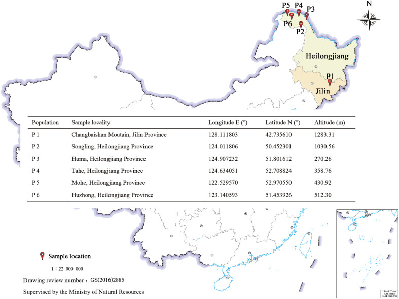
Lingonberry was collected from six major distribution fields, including P1 (Changbaishan Moutain, Jilin, Province), P2 (Songling, Heilongjiang, Province), P3 (Huma, Heilongjiang, Province), P4 (Tahe, Heilongjiang, Province), P5 (Mohe, Heilongjiang, Province) and P6 (Huzhong, Heilongjiang, Province) (Fig. 1). The fruits, leaves, and stems were lyophilized under vacuum freeze and ground into a powder of approximately 60 mesh size. All samples were stocked at −20 °C for subsequent experiments.
Fig. 1. Geographic conditions of original habitat of lingonberries.
Chemicals and reagents
Standard compounds of cyanidin-3-O-glucoside, cyanidin-3-O-galactoside, cyanidin-3-O-arabinoside, astragalin, quercetin, (+)-catechin, (−)-epicatechin, oleanolic acid, ursolic acid, chlorogenic acid, p-coumaric, benzoic acid, quinic acid, succinic acid, citric acid, malic acid, ascorbic acid and arbutin were purchased from Nakeli Biological Technology Co., Ltd (Chengdu, China). All reference compounds were identified with a purity higher than 98%. The structures of all compounds were displayed in Fig. S4.† UPLC-MS grade acetonitrile, methanol and formic acid were purchased from Fisher Scientific (Geel, Belglum). The pure water for the analysis of UPLC-QqQ-MS/MS was obtained from Wahaha Group Corporation (Hangzhou, China). All samples were filtered through a 0.22 μm poly tetra fluoroethylene (PTFE) membrane (Millipore, MA, USA).
Sample preparation
The sample powders were weighed precisely 3 g, dissolved in 90 mL of ice ethanol water (30 : 70, v/v), sonicated for 25 min at 4 °C and 300 W, and centrifuged at 10 000 rpm for 10 min. After centrifugation for 2 times, the supernatant was collected. Subsequently, the extracts were rotary dried under vacuum at 45 °C, redissolved in 10 mL of methanol solution. Then, 100 μL were piped to a constant volume of 10 mL, and filtered through a 0.22 μm PTFE membrane, followed by injection into a UPLC-QqQ-MS/MS and UPLC-Q-Exactive Orbitrap MS system for subsequent analysis. Water : ethanol (30 : 70, v/v) and quality control (QC) samples were also included for identification of untargeted metabolites. QC samples were prepared by mixing all samples tested. The blank and QC samples were placed at the beginning and the end of the sample sequence of the assay at 10 sample intervals.
Determination of total phenol content (TPC)
TPC was determined according to the method of Gan et al. with modifications as follows.15 In short, 40 μL of the sample solution was accurately diluted with 1.8 mL of 20-fold diluted Folin–Ciocalteu reagent and protected from light for 5 min. A solution of 1.2 mL of 7.5% (w/v) Na2CO3 was added and reacted in the dark at 25 °C for 2 h to determine an absorption wavelength of 760 nm. The blank was used as the control. Gallic acid was used as a standard (0.03125–1.0 mg mL−1, R2 = 0.993) and the results were expressed as mg of gallic acid equivalent (GAE) per hundred-gram dry weight (mg GAE/100 g DW) of lingonberry.
Determination of total flavonoid content (TFC)
TFC was calculated by referring to Bai et al. with slight modifications.16 Briefly, 1 mL of diluted sample solution was added to 3.5 mL of aqueous solution, then 0.3 mL of 5% NaNO2 solution was injected to react for 6 min, and 0.3 mL of 10% AlCl3 solution was inserted. Finally, 1 mL of 4% NaOH solution was added and mixed uniformly, and then the aqueous solution was supplemented to a total volume of 10 mL, followed by recording the absorbance of the blank sample at 510 nm (UV-1800, MAPADA). Catechin (CE) was used as a standard (0.0625–2.0 mg mL−1, R2 = 0.993). The TFC content results were expressed as CE equivalents (mg CE/100 g DW).
Determination of total anthocyanin content (TAC)
TAC in the samples was determined by pH difference method with slight modification according to reported.17 Briefly, the buffer solutions were prepared as follows: KCl and CH3COONa solution were adjusted to pH 1.0 and pH 4.5 with HCl solution, respectively. Then 200 μL of the sample solution was added to 5 mL of pH 1 KCl solution and pH 4.5 CH3COONa solution, mixed well and protected from light for 15 minutes. Finally, the absorbance A was evaluated at 510 nm and 700 nm, respectively, and measured according to the following eqn (1) and (2). The TAC content results were expressed as cyanidin-3-O-glucoside (C3G) equivalents.
| A = absorbance (A510–A700 nm) pH 1.0 − (A510–A700 nm) pH 4.5 | 1 |
| TAC (mg g−1) = (A × MW × DF ×1000)/ε × L × Wt | 2 |
where molecular weight (MW) of anthocyanin (cyanidin-3-O-glucoside) = 449.2 g mol−1, A = absorbance λ, L = optical distance, 1.0 cm, extraction coefficient (ε) = 29 600, DF = diluted factor, and Wt = sample weight (g).
UPLC-Q-Exactive Orbitrap MS analysis
Ultrahigh performance liquid chromatography (Thermo, USA) equipped with quadrupole electrostatic field Orbitrap high-resolution mass spectrometry (UPLC-Q-Exactive Orbitrap MS, USA). The chromatographic separation was performed on a Hypers 11 GOLG C18 (2.1 × 100 mm, 1.9 μm) with the following settings: column temperature 30 °C, flow rate 0.35 mL min−1; mobile phase A, water containing 0.1% (v/v) formic acid; mobile phase B, acetonitrile; injection volume, 3.5 μL. The gradient condition was: 0–9 min, 2–98% B; 9–12 min, 98–98% B; 12–15 min, 98–2% B. In this mode, the acquisition software (Compound Discoverer 3.3, USA) will continuously evaluate the full scan survey MS data according to the preselected criteria, while collecting and triggering the MS/MS spectrum acquisition.
The MS conditions were set as follows: positive and negative ion switch mode, scanning range: m/z 80–1200; curtain gas (CUR): 40 psi; the temperature of the ion source (TEM): 550 °C or 600 °C and voltage of the ion source (IS): −3000 V or 3800 V in negative or positive modes, respectively; first order scanning: declustering potential (DP): 130 V; collision voltage (CE): 10 V; second order scanning: Q-Exactive Orbitrap Product Ion mode was used to collect MS data, collision energy (CE) was 20, 40 and 60 V.12
Thermo Compound Discoverer™ 3.0 software (Thermo Fisher Science) was used to process raw data from 54 lingonberry fruit, leaf and stem profiles (27 samples and their replicates) analysed by UPLC-Q-Exactive Orbitrap MS. The specific parameters were set as follows: the RT alignment was set to 2 min and the m/z tolerance was set to 5 ppm; the signal-to-noise ratio (S/N) for feature detection was set to 3; the intensity threshold of the target peak was set to 1 000 000; and the additive ions were set to [2M + H]+, [2M − H]−, [M + H]+, [M − H]−. The following databases included in the Component Discoverer™ software were then used to identify the raw MS data: Thermo Fisher, Nature Chemical Biology, Nature Chemistry, MassBank, KEGG, Food and Agriculture Organisation of the United Nations, ChemBank. Finally, blanks were added for background filtering and gap filling to refine the data, and the sample data were normalised using the median, and the filtered data were used for further chemometric analysis.
An online workflow on the Platform (GNPS) (http://gnps.ucsd.edu) was used to construct MS/MS molecular networks.18 To accurately use high-resolution mass spectrometry MS/MS data, system annotations for GNPS were set as follows: precursor ion mass tolerance set to 2.0 Da, fragment ion mass tolerance set to 0.5 Da, minimum pairwise cos 0.6, minimum matched fragment ion 2, minimum cluster size 1.
UPLC-QqQ-MS/MS analysis
An Agilent 1290 Infinity UPLC system equipped with an electrospray ionization source (ESI) was coupled with an Agilent 6460 triple quadrupole mass spectrometer (Agilent Technologies, USA) for UPLC-MS/MS analysis. Analyte separation was performed on an Agilent ZORBAX Eclipse Plus C18 column (50 mm, 2.1 mm I.D., 1.8 μm) at 30 °C with a thermostat chamber. The mobile phase composition was A: 0.1% formic acid aqueous solution (V/V) and B: acetonitrile, and a gradient elution was set up with a flow rate of 0.3 mL min−1: 0–1 min, 95–95% A; 1–5 min, 95–10% A; 5–6 min, 10–95% A; 6–10 min, 95–5% A; 10–11 min, 5–95% A and 11–12 min, 95–95% A. The sample injection volume was set to 3 μL, and the separated solution was introduced into the triple quadrupole mass spectrometer for subsequent analysis. Mass spectrometry was combined with multiple reaction monitoring (MRM) mode. The general parameters were set as follows: capillary voltage 4 kV (ESI+) and 3.5 kV (ESI−), gas temperature 330 °C, gas flow 10 L min−1; nebulizer pressure 50 psi, and cell acceleration voltage 4 V. To obtain the strongest quantitative conversion, the specific MRM parameters of each analyte were optimized by using Agilent Mass Hunter workstation software (version B.07.00), such as precursor/product ion combination, fragment voltage and collision energy. The optimized values of these critical parameters for the 18 target compounds are listed in Table 1.
Optimized parameters of targeted compounds for quantitative analysis in MRM mode.
| No. | Compound name | Formula | RT (min) | Molecular mass | Precursor ion (m/z) | Product ion (m/z) | Fragmentor (V) | Collision energy (V) | Monitoring ion |
|---|---|---|---|---|---|---|---|---|---|
| 1 | Ascorbic acid | C6H8O6 | 0.43 | 176.12 | 175.00 | 115.00 | 80 | 4 | [M − H]− |
| 2 | Malic acid | C4H6O5 | 0.44 | 134.09 | 132.90 | 115.00 | 70 | 5 | [M − H]− |
| 3 | Citric acid | C6H8O7 | 0.53 | 192.12 | 191.10 | 110.20 | 85 | 5 | [M − H]− |
| 4 | Succinic acid | C4H6O4 | 0.56 | 118.09 | 117.00 | 73.00 | 70 | 12 | [M − H]− |
| 5 | Quinic acid | C7H12O6 | 1.23 | 192.17 | 191.00 | 84.90 | 83 | 24 | [M − H]− |
| 6 | Cyanidin-3-O-glucoside | C21H21O11 | 1.42 | 449.39 | 449.00 | 287.00 | 100 | 10 | [M + H]+ |
| 7 | Cyanidin-3-O-galactoside | C21H21O11 | 1.55 | 449.38 | 449.00 | 287.00 | 120 | 10 | [M + H]+ |
| 8 | Arbutin | C12H16O7 | 1.75 | 272.20 | 274.30 | 105.70 | 140 | 12 | [M − H]− |
| 9 | Cyanidin-3-O-arabinoside | C20H19O10 | 2.03 | 419.81 | 421.20 | 287.00 | 140 | 21 | [M + H]+ |
| 10 | Chlorogenic acid | C16H18O9 | 2.22 | 354.31 | 352.80 | 191.10 | 100 | 10 | [M − H]− |
| 11 | (+)-Catechin | C15H14O6 | 2.41 | 290.27 | 291.00 | 139.00 | 100 | 9 | [M + H]+ |
| 12 | (−)-Epicatechin | C15H14O6 | 2.49 | 290.27 | 291.00 | 139.00 | 110 | 13 | [M + H]+ |
| 13 | Quercetin | C15H10O7 | 2.55 | 302.24 | 301.00 | 151.00 | 148 | 23 | [M − H]− |
| 14 | P-Coumaric acid | C9H8O3 | 2.69 | 164.16 | 162.90 | 119.00 | 75 | 16 | [M − H]− |
| 15 | Astragalin | C21H20O11 | 2.87 | 448.40 | 447.00 | 284.00 | 165 | 30 | [M − H]− |
| 16 | Benzoic acid | C7H6O2 | 3.04 | 122.12 | 121.00 | 77.00 | 75 | 15 | [M − H]− |
| 17 | Oleanolic acid | C30H48O3 | 5.78 | 456.70 | 457.30 | 411.10 | 140 | 10 | [M + H]+ |
| 18 | Ursolic acid | C30H48O3 | 5.83 | 456.70 | 457.30 | 411.10 | 120 | 15 | [M + H]+ |
Antioxidant activity
According to the assay method reported by Yang et al.19 Briefly, the antioxidant activity of lingonberry extracts was evaluated employing free radical scavenging activity (1,1-diphenyl-2-picrylhydrazyl) DPPH and (2,2′-azino-bis(3-ethylbenzothiazoline-6-sulfonic acid)) ABTS+ assay, and the results were expressed as mol Trolox/g DW.
The (ferric ion reducing antioxidant power) FRAP test was conducted in accordance with the approach reported with a minor modification.20 In a nutshell, the FRAP solution consisted of 2.5 mL TPTZ (2,4,6-tripyridyl-s-triazine) solution (10 mM) added to HCl solution (40 mM), 2.5 mL ferric chloride (FeCl3–6H2O) (20 mM) and 25 mL acetate buffer (0.3 M). Store mixed solutions at 37 °C for use. Subsequently, the sample solution (0.1 mL) was thoroughly mixed with FRAP solution (0.9 mL) and left at room temperature for 30 min, and the absorbance was measured at 593 nm.
Antiproliferative activity
The antiproliferative activity of lingonberry extract was assessed based on the MTT method as reported with slight modifications.19 HeLa (ATCC CCL-2) and HepG-2 (ATCC HB-8065) were used as human tumor cell lines. Briefly, the tumor cell lines were treated with various concentrations of lingonberry extract. After the mixture was incubated for 48 h, the MTT solution was incorporated and stored for 4 h at 37 °C. The absorbance was determined at 590 nm and the IC50 value was measured. The inhibition rate was calculated as follows:Inhibition rate (%) = ((Acontrol − Asample)/Acontrol) × 100%.
Statistical analysis
Data were statistically analyzed using one-way ANOVA with a post hoc Tukey test based on the statistical software SPSS Statistics 25 (IBM, USA). Clustered heat maps were generated using the TBtools-II (Toolbox for Biologists v1.120) tool. Principal component analysis (PCA) was performed using SIMCA (14.1) and Origin pro 2021 (OriginLab, USA) was used for the remainder of the image production. All experiments were performed with three replications, and all results were expressed as mean ± standard deviation (SD), with differences considered statistically significant at p < 0.05.
Results and discussion
Total phenolic, flavonoid, and anthocyanin contents in lingonberry fruits, leaves and stems
Polyphenols are widely found in a variety plants, many of which have been identified in food crops.21 In general, plant genetics affects the distribution of secondary metabolites, while external environmental factors can induce significant changes in metabolite composition.22 In this study, we found significant differences in the accumulation of phytochemical components in the different parts of the lingonberry, with TAC being the most abundant in the fruits, while TPC and TFC were accumulated to a greater extent in the leaves. Specifically, the lowest TPC in P4 stems was 44.94 mg GAE/100 g DW, whereas the highest TPC in P6 leaves was 461.55 mg GAE/100 g DW, a 10-fold difference (Fig. 2A), suggesting that plants genetically regulate phytochemical synthesis and transport to adapt to the external environment.23
Fig. 2. Total polyphenol, total flavonoid and total anthocyanin content of lingonberry's fruits, stems and leaves. (A) Contents of total phenolic; (B) contents of total flavonoid; (C) contents of total anthocyanin. Bars labelled with different letters represent statistical differences (p < 0.05) between samples calculated from the sum of the total content of all fruit, leaf and stem compounds.
Moreover, altitude is an important environmental factor influencing the distribution of plant metabolites, with a gradual decrease in temperature and an increase in the intensity of visible light with increasing altitude. In bilberry berries, higher levels of TPC and TAC were found at 600 m altitude, and metabolite contents decreased sharply at both lower altitudes (450 m altitude) and excessive altitudes (from 800 m to 1500 altitude).24 In the present study, we found that TAC was highest in fruits at P4 (358.76 m altitude), whereas TPC and TFC were more in leaves at P6 (512.30 m altitude) compared to other parts of the plant (Fig. 1 and 2). Therefore, the external environment has an effect on the accumulation of most phenolic compounds, which may be a positive response to the defence mechanism against a negative external environment.3
Identification of metabolites in fruits, leaves and stems of lingonberry
To further characterize the phytochemical composition of lingonberry fruits, leaves, and stems from different regions, UPLC-Q-Exactive Orbitrap MS was performed in a positive and negative ion mode.
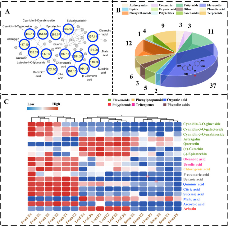
The metabolites were identified by comparing the precursor ions, MS2 fragmentation ions, accurate molecular weight and retention time with the standard databases of mzVault, mzCloud and BGI high resolution accurate mass plant metabolome database (BGI HRAM-PMDB). The GNPS platform is an open-access database of existing prevalent MS/MS spectral libraries that facilitate rapid compound identification based on MS/MS similarity networks by executing Proteowizard software to convert raw MS/MS files into mzXML data.18 After constructing a molecular network using raw MS/MS data of lingonberry extracts, data visualization was performed using Cytoscape 3.8.2 software, 1411 precursor ions of metabolites were observed, which were divided into 95 clusters (nodes ≥ 2) and 780 single nodes with a threshold cosine value of 0.7 (Fig. 3A).
Fig. 3. Identified metabolites in lingonberry (fruits, leaves and stems) and chemical marker selection. (A) The molecular network of primarily targeted metabolites; (B) classification of compounds; (C) cluster heat map target compound distribution.
After analysis of metabolites from various parts of lingonberries, a total of 95 metabolites were identified, as shown in Table S1.† Thereinto, 54 metabolites were identified through the GNPS library, and 23 known compounds were identified by comparison with the chemical standards. Additionally, in combination with the Compound Discovery 3.0 software mzCloud database, 20 metabolites were identified by comparing MS2 fragmentation patterns. The analysis results indicated that 37 flavonoids, 12 phenylethanoids, 11 fatty acids, 9 terpenoids, 5 phenolic acids, 5 organic acids, 4 saccharides, 3 coumarins, 3 anthocyanins, 2 lipids, 1 polyketide, and 3 compounds belonged to others (Fig. 3B).
In previous reports, investigations of lingonberry phytochemicals have focused on mixtures from localized sources and lacked criteria to assess their overall quality control.25 Based on this, this study conducted qualitative and quantitative analysis on lingonberry from different regions in China, with the aim of revealing the distribution characteristics of bioactive components in lingonberry and providing a basis for product development and biological breeding.
Quantification and distribution of the constituents in fruits, leaves and stems of lingonberry
Although 95 components were initially identified based on untargeted methods, quantification of all metabolites was challenging due to trace content and the absence of available reference substances. Therefore, to obtain accurate quantitative results, we used a targeted LC-QqQ-MS/MS method combined with chemical standards to quantify 18 target metabolites from 6 classes.
First, critical parameters of chromatographic separation, such as column, mobile phase and elution efficiency, were optimized to achieve favorable peak shapes and reproducible separations, thus improving sensitivity and reliability. In addition, the full-scan MS method was used to detect in both positive and negative ionization modes. Based on this, the highest and most stable MRM leap response signal was obtained with the combination of adjusting the standard solution fragmentation voltage and collision energy to suit all analytes. Finally, the analytes were scanned and identified after manually optimising the parameters of the system's MRM mode. The calibration curve of the signal intensity (peak area) of the MRM transition to the six concentration gradients of the standard solution was plotted. The limits of LOD (S/N = 3) and LOQ (S/N = 10) results for each analyte were below 0.3 ng mL−1 and 0.7 ng mL−1, respectively. The intra-day RSD of the peak area was less than 4.21%, and the daytime RSD was less than 3.45% (Table S2†). In conclusion, the established UPLC-QqQ-MS/MS method was satisfactory linearity, sensitivity, precision, accuracy, and stability for the simultaneous determination of 18 compounds in complex lingonberry-related matrices (fruits, leaves and stems).
To evaluate the distributional capacity of different parts, a clustering heat map model was developed. The results are shown in Fig. 3C, where triplicates from several same geographical regions were successfully clustered, further demonstrating the reliability of the method.
Obviously, the results showed that 7 flavonoids (cyanidin-3-O-glucoside, cyanidin-3-O-galactoside and cyanidin-3-O-arabinoside, quercetin, (+)-catechin, (−)-epicatechin and astragalin), 1 phenylpropanoid (chlorogenic acid), 2 triterpenes (ursolic acid and oleanolic acid), 2 phenolic acids (benzoic acid and p-coumaric acid) and 5 organic acids (succinic acid, quinic acid, malic acid, citric acid, and ascorbic acid) and 1 polyphenols (arbutin) were the dominant components in lingonberry, which could be used as marker compounds for quality control. To the best of our knowledge, aerial parts of lingonberry are enriched with bioactive components, which are utilized in functional food materials.26
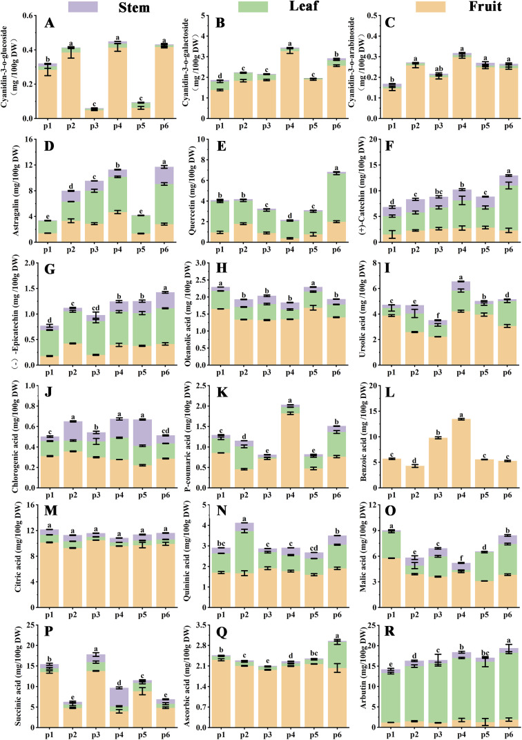
Previous findings have suggested that lingonberries contain a variety of bioactive phytochemicals including anthocyanins, flavonoids, flavanols, triterpenes and organic acids.27 The anthocyanins in lingonberry fruits are based on the parent nucleus of cyanidin with different glycoside compositions, as reported by Michiels et al.28 It has the highest content with strong antioxidant activity. In particular, we found that anthocyanidins accumulate most in fruits, with cyanidin-3-O-galactoside content up to 3.27 mg/100 g DW, and mature fruits show red cyanidin consistent with anthocyanidin causing.26 In addition, the content of triterpene acid in lingonberries had the highest percentage of the total triterpenoids and was significantly affected by the harvesting period.29 In the present study, we can conclude that the highest content of ursolic acid in the fruits (P4) was 4.23 mg/100 g DW, in contrast to chlorogenic acid (P2) which was the highest at 0.35 mg/100 g DW, indicating that the geographical location has a significant effect on the accumulation of active compounds (Fig. 4J). Furthermore, the organic acid content of the lingonberry fruits was important factor in the sensory characteristics of the fruits. It even has a significant influence on consumer acceptance.30,31 The results showed that phenolic and organic acids had the highest percentage of fruits composition, mainly dominated by citric acid (10.51 mg/100 g DW), quinic acid (1.92 mg/100 g DW), while benzoic acid (13.45 mg/100 g DW) was only detected in fruits, the main source of fruits acidity (Fig. 4L–N). In industrial production, acids are used as antioxidants, preservatives, acidulants and drug absorption modifiers.32 They are also used to maintain the quality and nutritional value of the fruits therefore, as a good source of acidic constituents for flavour and nutritional control, lingonberry fruits are an important indicator of berry quality parameters and a reasonable target for crop improvement.33
Fig. 4. The distribution of dominant compounds in lingonberry collected from different regions. (A) Cyanidin-3-O-glucoside; (B) cyanidin-3-O-galactoside; (C) cyanidin-3-O-arabinoside; (D) astragalin; (E) quercetin; (F) (+)-catechin; (G) (−)-epicatechin; (H) oleanolic acid; (I) ursolic acid; (J) chlorogenic acid; (K) P-coumaric acid; (L) benzoic acid; (M) citric acid; (N) quininic acid; (O) malic acid; (P) succinic acid; (Q) ascorbic acid; (R) arbutin. Bars labelled with different letters represent statistical differences (p < 0.05) between samples calculated from the sum of the total content of all fruit, leaf and stem compounds.
Lingonberry leaves contain a wide variety of chemical constituents compared to the fruits. Flavonoid glycosides are the most abundant phenolic compounds in lingonberry. They have astringent, antitussive, urinary antiseptic, diuretic, neuroprotective, antioxidant and anti-inflammatory effects, and possibly inhibitory effects on cancer cell growth.10 The results of this study showed that astragalin and quercetin, (+)-catechin and (−)-epicatechin were ranked in different parts: leaves > fruits > stems (Fig. 4D–G), with the highest content in the region P6. In addition, we found that the highest TFC was in the leaves (Fig. 2A), suggesting that the leaves are a potential site of application for flavonoid constituents, which correlates with the role of leaves in resistance defense.25,34 Interestingly, we found that the highest content of arbutin in leaves was up to 16.4 mg/100 g DW, which is 10 times higher than that in fruits. Lingonberry leaves are usually regarded as food waste and underutilised, with the advantage of being a potential low-cost source of food and medicine that avoids harming the plant.35
Compared to the fruits and leaves, the stems were found to contain fewer active compounds. As shown in Fig. S3A,† flavonoids were distributed in small amounts in the stems, while the chlorogenic acid content was similar to that of its parts. The bioactive compounds were mainly derived from the bark.25
In conclusion, the highest levels of anthocyanins, ursolic acid and phenolic acids were found in the fruits from the P4 region, while the leaves from the P6 region were more favourable for accumulating flavonoids and arbutin compounds, potentially serving as the best collecting point for the material.
Predominant chemical markers selection through multivariate statistical analysis
To visualize the comparison of quantitative data of the target compounds in fruits, leaves and stems, the PCA models were established (Fig. 5A). The results of the PCA indicated a strong separation trend of three parts (fruits, leaves and stems) from different geographical regions, which were clustered in three groups, indicating similarity in metabolite components and contents between the samples of the groups. Cyanidin-3-O-glucoside, cyanidin-3-O-galactoside, cyanidin-3-O-arabinoside, chlorogenic acid, ursolic acid, oleanolic acid, citric acid, quinic acid, p-coumaric acid, benzoic acid and ascorbic acid were responsible for the formation of the group I (fruits). Leaves in the group II were characterized by (+)-catechin, (−)-epicatechin, malic acid, quercetin, astragalin, succinic acid and arbutin.
Fig. 5. The PCA of lingonberry's fruits, stems and leaves from different regions (A) PCA score scatter plot; (B) loading plot.
Clustering heat maps and projection variable importance (VIP) values were also employed to identify differences between parts. The loading plot metabolites with VIP values > 1 combined with clustering heatmaps compound abundances were considered as potential chemical makers that could be characterised for differences in phytochemical composition between fruits, leaves and stems (Fig. 5B). Among these compounds, cyanidin-3-O-galactoside, ursolic acid, (+)-catechin, arbutin, benzoic acid, citric acid and astragalin as principal chemical markers for their high abundance in the various parts.
Antioxidant activity
As potential antioxidant active substances in various sites, DPPH, ABTS+ and FRAP assays were performed to monitor their activity. The results of DPPH, ABTS+ and FRAP showed a similar trend in the following order: fruits > leaves > stems (Table 2). It has been shown that catechins and flavonols contain a catechol structure in the b-ring and three hydroxyl groups in the c-ring.36 In the present study, although the flavonoid content of the leaves was high, the antioxidant value was lower than that of the fruits. They have strong antioxidant activity. The fruits were thought to contain other antioxidant substances such as anthocyanin. Interestingly, the highest values for DPPH, ABTS+ and FRAP were all observed at the fruits in the P4 region, with 269.34 mol Trolox/g DW, 363.16 mol Trolox/g DW and 102.13 mol Trolox/g DW respectively. On the other hand, the antioxidant activity of crude plant extracts is related to the bioactive ingredients and is influenced by both external biotic and abiotic factors.20 In addition, compared to stems and leaves, the fruits represent a food and medicinal source with the advantage of avoided plant damage. Therefore, the fruits of the P4 region could be the best potential collection site for antioxidant material.
Antioxidant properties of lingonberry's fruit, leaf and stem extractsa.
| Site | Fruits | Leaves | Stems | ||||||
|---|---|---|---|---|---|---|---|---|---|
| DPPH (mol Trolox/g DW) | ABTS (mol Trolox/g DW) | FRAP (mol Trolox/g DW) | DPPH (mol Trolox/g DW) | ABTS (mol Trolox/g DW) | FRAP (mol Trolox/g DW) | DPPH (mol Trolox/g DW) | ABTS (mol Trolox/g DW) | FRAP (mol Trolox/g DW) | |
| P1 | 226.88 ± 3.52b | 351.36 ± 5.66ab | 102.09 ± 0.93a | 79.07 ± 3.48d | 259.68 ± 6.75b | 78.04 ± 0.7b | 39.95 ± 8.91c | 89.15 ± 1.3c | 40.49 ± 0.58d |
| P2 | 93.14 ± 1.97e | 189.46 ± 4.3d | 64.38 ± 2.38d | 90.1 ± 9.13bc | 180.48 ± 6.65c | 62.41 ± 0.56d | 17.34 ± 4.87e | 127.95 ± 9.08ab | 32.77 ± 0.59f |
| P3 | 144.46 ± 3.97c | 349.96 ± 4.91b | 100.71 ± 0.34a | 102.73 ± 6.71ab | 336.11 ± 6.62a | 75.82 ± 0.58c | 27.26 ± 1.91d | 136.11 ± 6.66a | 43.1 ± 0.47c |
| P4 | 269.34 ± 9.74a | 363.16 ± 3.63a | 102.13 ± 1.08a | 89.41 ± 14.07bc | 345.36 ± 2.43a | 100.72 ± 0.26a | 71.45 ± 2.65a | 126.26 ± 2.3ab | 69.39 ± 0.46a |
| P5 | 109.85 ± 5.07d | 342.39 ± 6.64b | 92.91 ± 0.66b | 91.19 ± 9.38bc | 131.02 ± 9.7e | 75.21 ± 0.47c | 47.74 ± 9.09b | 107.48 ± 4.07bc | 50.38 ± 2.08b |
| P6 | 130.83 ± 3.98c | 225.42 ± 12.27c | 78.41 ± 0.56c | 120.49 ± 8.77a | 170.46 ± 6.69d | 74.87 ± 1.03c | 12.22 ± 3.27f | 140.61 ± 7.67a | 37.24 ± 0.97e |
Data are presented as mean ± SD (n = 3). Different lowercase letters in each column indicate significant differences (p < 0.05).
Antiproliferative activity
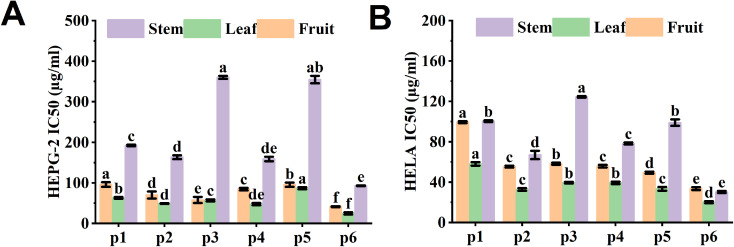
The antiproliferative activity of lingonberry from various origins was evaluated by MTT method. The results are shown in Fig. 6A and B, the lingonberry extracts showed strong antiproliferative effects on both HeLa and HepG-2 cell lines with IC50 values less than 400 μg mL−1. Notably, the leaves have the strongest antiproliferation effect, followed by the fruits and then the stems. It has been shown that arbutin and flavonoids are inhibitors of cancer cell proliferation.37 Of all the regions, we found the lowest IC50 values for leaves in the P6 region. This correlates with the high levels of arbutin and flavonoids in this region. As a result, potential pharmaceutical and healthcare products are also collected at the P6 site.
Fig. 6. Antiproliferative efficacy of lingonberry's fruits, leaves and stems of different origins. (A) HEPG-2 assay; (B) HELA assay. Bars marked with different letters indicate statistical differences (p < 0.05) in the content of compounds in fruits, leaves and stems between the different sites.
Pearson correlation between bioactive compounds and bioactivities
It has been suggested that polyphenol content in plants has a positive correlation with antioxidant capacity.14 In this study, Pearson correlation analysis was performed to reveal the potential correlation between bioactive compounds and antioxidant activity in order to characterise the important antioxidant properties in different parts. As shown in Table 3, TPC, TAC, and TFC were significantly and positively correlated with DPPH, ABTS+ and FRAP activities in fruits, leaves and stems, in agreement with previous reports.11
Pearson correlation analysis of bioactive compounds with antioxidant and antiproliferative activitiesa.
| Fruit | Leaf | Stem | |||||||||||||
|---|---|---|---|---|---|---|---|---|---|---|---|---|---|---|---|
| DPPH | ABTS | FRAP | HEPG2 (IC50) | HELA (IC50) | DPPH | ABTS | FRAP | HEPG2 (IC50) | HELA (IC50) | DPPH | ABTS | FRAP | HEPG2 (IC50) | HELA (IC50) | |
| TPC | 0.793** | 0.785** | 0.529* | 0.489* | 0.358 | 0.482* | 0.839** | 0.609** | 0.141 | 0.063 | 0.529* | 0.486* | 0.533* | 0.311 | 0.376 |
| TFC | 0.662** | 0.700** | 0.426 | 0.454 | 0.346 | 0.352 | 0.695** | 0.521* | −0.143 | −0.118 | 0.589* | 0.505* | 0.532* | 0.466 | 0.508* |
| TAC | 0.775** | 0.850** | 0.586* | 0.491* | 0.327 | 0.677** | 0.536* | 0.552* | 0.155 | 0.130 | 0.452 | 0.505* | 0.568* | −0.414 | −0.528* |
| Anthocyanin | 0.746** | 0.641** | 0.855** | −0.283 | −0.147 | 0.615** | 0.749** | 0.689** | −0.024 | −0.037 | −0.159 | −0.352 | −0.295 | 0.281 | 0.296 |
| Flavonols | 0.150 | 0.220 | 0.152 | 0.131 | 0.174 | 0.095 | 0.567* | 0.448 | −0.071 | −0.067 | 0.560* | 0.406 | 0.432 | 0.293 | 0.238 |
| Flavanols | 0.302 | 0.631** | 0.521* | −0.201 | −0.283 | 0.649** | 0.634** | 0.737** | −0.324 | −0.297 | −0.076 | −0.609 | −0.147 | −0.091 | −0.026 |
| Flavonoids | 0.548* | 0.430 | 0.594** | −0.129 | −0.010 | 0.550* | 0.756** | 0.556* | −0.544** | −0.448* | 0.549* | 0.530* | 0.534* | −0.082 | −0.018 |
| Phenylpropanoids | −0.118 | 0.178 | 0.104 | −0.032 | −0.369 | −0.235 | 0.183 | 0.148 | −0.161 | −0.116 | 0.479* | 0.216 | 0.463 | −0.323 | −0.207 |
| Phenolic acid | 0.477* | 0.597** | 0.476* | −0.046 | −0.147 | 0.435 | 0.125 | 0.234 | −0.415 | −0.360 | 0.207 | 0.082 | 0.199 | −0.114 | −0.075 |
| Polyphenols | 0.588* | 0.547* | 0.659** | −0.132 | −0.073 | 0.580* | 0.663** | 0.493* | −0.041 | −0.035 | 0.652** | 0.621** | 0.734** | −0.095 | −0.027 |
| Triterpene | 0.472* | 0.560* | 0.520* | 0.242 | 0.133 | −0.251 | 0.310 | 0.165 | −0.055 | −0.078 | 0.157 | 0.009 | 0.076 | −0.438 | −0.565* |
| Organic acid | 0.067 | 0.106 | −0.167 | 0.061 | 0.080 | −0.606** | −0.226 | −0.329 | −0.181 | −0.224 | 0.231 | 0.321 | 0.276 | −0.117 | −0.282 |
| TCM | 0.509* | 0.492* | 0.470* | 0.145 | 0.100 | −0.005 | 0.597** | 0.397 | −0.012 | −0.030 | 0.684** | 0.655** | 0.766** | −0.128 | −0.083 |
Asterisks indicate significant differences (*P < 0.05, **P < 0.01). Anthocyanins contain cyanidin-3-O-glucoside, cyanidin-3-O-galactoside and cyanidin-3-O-arabinoside; flavonols contain astragalin and quercetin; flavanols contain (+)-catechin and (−)-epicatechin; phenylpropanoids include chlorogenic acid; triterpene contain oleanolic acid and ursolic acid; phenolic acid contain p-coumaric and benzoic acid; organic acid contain quinic acid, citric acid, succinic acid and malic acid; flavonoids contain anthocyanins, flavonols and flavanols; polyphenols contain arbutin, flavonoids, phenylpropanoids and phenolic acids; TCM contains polyphenols, triterpenes and organic acids.
It has been shown that anthocyanins have good antioxidant effects and have a protective effect on plants.38 In the fruits, the levels of anthocyanins (cyanidin-3-O-glucoside, cyanidin-3-O-galactoside and cyanidin-3-O-arabinoside), phenolic acids (p-coumaric and benzoic acid) and triterpenes (oleanolic acid and ursolic acid) were high and significantly and positively correlated with antioxidant activity, indicated that lingonberry fruit has antioxidant activity associated with a variety of bioactive compounds, which is consistent with previous reports.14 Altitude is an important environmental factor affecting the accumulation of metabolic substances in plants. In this study, we found that antioxidant activity (DPPH, ABTS+, FRAT) was strongest in lingonberry fruits at P4 (Table 2), and correlation analyses showed that phenolic and anthocyanin components were significantly and positively correlated with antioxidant activity (DPPH, ABTS+, FRAT) (Table 3), suggesting that altitude affects antioxidant activity of resistant plants through the accumulation of phytochemicals, which is in accordance with previous reports.39
Similarly, the flavonoids in lingonberry leaves showed a significant positive correlation with antioxidant capacity, while the DPPH assay revealed a weak correlation (r = 0.352), indicating that there were other undetected compounds (Table 3). In comparison, flavanols (catechins and epicatechins) in the leaves were significantly positively correlated with antioxidant activity, in agreement with previous reports.40
Flavonoids found in berries have been shown to penetrate the cell membranes of cancer cells and have a powerful antiproliferative effect.41 Flavonoids exhibited a significant negative correlation with IC50 values of HeLa and HepG-2 cell lines (r = −0.544, −0.448), while flavanols showed a weak negative correlation (r = −0.324, −0.297), indicating the existence of other antiproliferative constituents in the leaves. Furthermore, arbutin in the leaves showed cytotoxic activity and non-toxicity.42
Conclusions
This study provides the first insight into the phytochemical composition and bioactivities of lingonberry fruits, leaves and stems from different regions based on an integrated strategy. The results of comparative analyses showed that habitat influenced the distribution of secondary metabolites in lingonberry. Flavanols and arbutin were higher in leaves, and anthocyanins, triterpenoids and organic acids were mainly present in fruits. In particular, the highest arbutin content was present in Huzhong (P6) at 15 mg/100 g DW. The highest content of ursolic acid and cyanidin-3-O-galactoside (4.5 mg/100 g DW and 3.2 mg/100 g DW, respectively) was found in the fruits of Tahe (P4). In addition, antioxidant and antiproliferative assays revealed that fruits (P4) exhibited the strongest antioxidant properties, while leaves (P6) had antiproliferative activity. In conclusion, this work identifies potential applications for lingonberries in the food and pharmaceutical industries. Further studies will continue to assess toxicity and explore key compounds with antioxidant activity.
Conflicts of interest
There are no conflicts to declare.
Supplementary Material
Acknowledgments
The authors gratefully acknowledge the financial supports by the National Natural Science Foundation of China (32271805), National Natural Science Foundation of China (31930076), Fundamental Research Funds for the Central Universities (2572020DR07), The 111 Project (B20088), Heilongjiang Touyan Innovation Team Program (Tree Genetics and Breeding Innovation Team).
Electronic supplementary information (ESI) available. See DOI: https://doi.org/10.1039/d3ra05698h
References
- Dhalaria R. Verma R. Kumar D. Puri S. Tapwal A. Kumar V. Nepovimova E. Kuca K. Antioxidants. 2020;9:1–38. doi: 10.3390/antiox9111123. [DOI] [PMC free article] [PubMed] [Google Scholar]
- Stefanescu B. E. Calinoiu L. F. Ranga F. Fetea F. Mocan A. Vodnar D. C. Crisan G. Antioxidants. 2020;9:649. doi: 10.3390/antiox9060495. [DOI] [PMC free article] [PubMed] [Google Scholar]
- Vilkickyte G. Raudone L. Plants. 2021;10:1986. doi: 10.3390/plants10101986. [DOI] [PMC free article] [PubMed] [Google Scholar]
- Mane C. Loonis M. Juhel C. Dufour C. Malien-Aubert C. J. Agric. Food Chem. 2011;59:3330–3339. doi: 10.1021/jf103965b. [DOI] [PubMed] [Google Scholar]
- Vyas P. Curran N. H. Igamberdiev A. U. Debnath S. C. Can. J. Plant Sci. 2015;95:663–669. [Google Scholar]
- Kostka T. Ostberg-Potthoff J. J. Starke J. Guigas C. Matsugo S. Mirceski V. Stojanov L. Velickovska S. K. Winterhalter P. Esatbeyoglu T. Antioxidants. 2022;11:467. doi: 10.3390/antiox11030467. [DOI] [PMC free article] [PubMed] [Google Scholar]
- Liu P. Z. Lindstedt A. Markkinen N. Sinkkonen J. Suomela J. P. Yang B. R. J. Agric. Food Chem. 2014;62:12015–12026. doi: 10.1021/jf503521m. [DOI] [PubMed] [Google Scholar]
- Szakiel A. Paczkowski C. Koivuniemi H. Huttunen S. J. Agric. Food Chem. 2012;60:4994–5002. doi: 10.1021/jf300375b. [DOI] [PubMed] [Google Scholar]
- Sanchez S. E. Kay S. A. Cold Spring Harbor Perspect. Biol. 2016;8:a027748. doi: 10.1101/cshperspect.a027748. [DOI] [PMC free article] [PubMed] [Google Scholar]
- Dragovic S. Dragovic-Uzelac V. Pedisic S. Cosic Z. Friscic M. Elez Garofulic I. Zoric Z. Food Technol. Biotechnol. 2020;58:303–314. doi: 10.17113/ftb.58.03.20.6662. [DOI] [PMC free article] [PubMed] [Google Scholar]
- Vilkickyte G. Raudone L. Foods. 2021;10:2243. doi: 10.3390/foods10102243. [DOI] [PMC free article] [PubMed] [Google Scholar]
- Vilkickyte G. Motiekaityte V. Vainoriene R. Raudone L. J. Food Compos. Anal. 2022;114:104796. [Google Scholar]
- Dragana M. V. Miroslav R. P. Branka B. R. G. Olgica D. S. Sava M. V. Ljiljana R. Č. Afr. J. Microbiol. Res. 2013;7:5130–5136. [Google Scholar]
- Bujor O. C. Le Bourvellec C. Volf I. Popa V. I. Dufour C. Food Chem. 2016;213:58–68. doi: 10.1016/j.foodchem.2016.06.042. [DOI] [PubMed] [Google Scholar]
- Gan R.-Y. Deng Z.-Q. Yan A.-X. Shah N. P. Lui W.-Y. Chan C.-L. Corke H. Lwt. 2016;73:168–177. [Google Scholar]
- Bai Z. Z. Tang J. M. Ni J. Zheng T. T. Zhou Y. Sun D. Y. Li G. N. Liu P. Niu L. X. Zhang Y. L. Food Res. Int. 2021;148:110609. doi: 10.1016/j.foodres.2021.110609. [DOI] [PubMed] [Google Scholar]
- Krithika S. J. Sathiyasree B. Theodore E. B. Chithiraikannu R. Gurushankar K. Crit. Rev. Food Sci. Nutr. 2020;62(9):1–8. doi: 10.1080/10408398.2020.1856772. [DOI] [PubMed] [Google Scholar]
- Wang M. Carver J. J. Phelan V. V. Sanchez L. M. Garg N. Peng Y. Nguyen D. D. Watrous J. Kapono C. A. Luzzatto-Knaan T. Porto C. Bouslimani A. Melnik A. V. Meehan M. J. Liu W. T. Crusemann M. Boudreau P. D. Esquenazi E. Sandoval-Calderon M. Kersten R. D. Pace L. A. Quinn R. A. Duncan K. R. Hsu C. C. Floros D. J. Gavilan R. G. Kleigrewe K. Northen T. Dutton R. J. Parrot D. Carlson E. E. Aigle B. Michelsen C. F. Jelsbak L. Sohlenkamp C. Pevzner P. Edlund A. McLean J. Piel J. Murphy B. T. Gerwick L. Liaw C. C. Yang Y. L. Humpf H. U. Maansson M. Keyzers R. A. Sims A. C. Johnson A. R. Sidebottom A. M. Sedio B. E. Klitgaard A. Larson C. B. Boya C. A. Torres-Mendoza D. Gonzalez D. J. Silva D. B. Marques L. M. Demarque D. P. Pociute E. O’Neill E. C. Briand E. Helfrich E. J. N. Granatosky E. A. Glukhov E. Ryffel F. Houson H. Mohimani H. Kharbush J. J. Zeng Y. Vorholt J. A. Kurita K. L. Charusanti P. McPhail K. L. Nielsen K. F. Vuong L. Elfeki M. Traxler M. F. Engene N. Koyama N. Vining O. B. Baric R. Silva R. R. Mascuch S. J. Tomasi S. Jenkins S. Macherla V. Hoffman T. Agarwal V. Williams P. G. Dai J. Neupane R. Gurr J. Rodriguez A. M. C. Lamsa A. Zhang C. Dorrestein K. Duggan B. M. Almaliti J. Allard P. M. Phapale P. Nothias L. F. Alexandrov T. Litaudon M. Wolfender J. L. Kyle J. E. Metz T. O. Peryea T. Nguyen D. T. VanLeer D. Shinn P. Jadhav A. Muller R. Waters K. M. Shi W. Liu X. Zhang L. Knight R. Jensen P. R. Palsson B. O. Pogliano K. Linington R. G. Gutierrez M. Lopes N. P. Gerwick W. H. Moore B. S. Dorrestein P. C. Bandeira N. Nat. Biotechnol. 2016;34:828–837. doi: 10.1038/nbt.3597. [DOI] [PMC free article] [PubMed] [Google Scholar]
- Yang Q. Q. Zhang D. Farha A. K. Yang X. Li H. B. Kong K. W. Zhang J. R. Chan C. L. Lu W. Y. Corke H. Gan R. Y. Ind. Crops Prod. 2020;143:111928. [Google Scholar]
- Bai Z. Z. Ni J. Tang J. M. Sun D. Y. Yan Z. G. Zhang J. Niu L. X. Zhang Y. L. Food Chem. 2021;343:128444. doi: 10.1016/j.foodchem.2020.128444. [DOI] [PubMed] [Google Scholar]
- Li D. Li B. Ma Y. Sun X. Lin Y. Meng X. J. Food Compos. Anal. 2017;62:84–93. [Google Scholar]
- Karppinen K. Zoratti L. Nguyenquynh N. Haggman H. Jaakola L. Front. Plant Sci. 2016;7:655. doi: 10.3389/fpls.2016.00655. [DOI] [PMC free article] [PubMed] [Google Scholar]
- Alam Z. Morales H. R. Roncal J. Botany. 2016;94:509–521. [Google Scholar]
- Rieger G. Muller M. Guttenberger H. Bucar F. J. Agric. Food Chem. 2008;56:9080–9086. doi: 10.1021/jf801104e. [DOI] [PubMed] [Google Scholar]
- Bujor O. C. Ginies C. Popa V. I. Dufour C. Food Chem. 2018;252:356–365. doi: 10.1016/j.foodchem.2018.01.052. [DOI] [PubMed] [Google Scholar]
- Kowalska K. Int. J. Mol. Sci. 2021;22:5126. [Google Scholar]
- Trivedi P. Nguyen N. Klavins L. Kviesis J. Heinonen E. Remes J. Jokipii-Lukkari S. Klavins M. Karppinen K. Jaakola L. Häggman H. Food Chem. 2021;354:129517. doi: 10.1016/j.foodchem.2021.129517. [DOI] [PubMed] [Google Scholar]
- Michiels J. A. Kevers C. Pincemail J. Defraigne J. O. Dommes J. Food Chem. 2012;130:986–993. [Google Scholar]
- Hajazimi E. Landberg R. Zamaratskaia G. Lwt. 2016;74:128–134. [Google Scholar]
- Silva B. M. Andrade P. B. Mendes G. C. Seabra R. M. Ferreira M. A. J. Agric. Food Chem. 2002;50:2313–2317. doi: 10.1021/jf011286+. [DOI] [PubMed] [Google Scholar]
- Ermis E. Hertel C. Schneider C. Carle R. Stintzing F. Schmidt H. Int. J. Food Microbiol. 2015;204:111–117. doi: 10.1016/j.ijfoodmicro.2015.03.017. [DOI] [PubMed] [Google Scholar]
- Daood H. G. Biacs P. A. Dakar M. A. Hajdu F. J. Chromatogr. Sci. 1994;32:481–487. [Google Scholar]
- Liang Z. Sang M. Fan P. Wu B. Wang L. Duan W. Li S. J. Food Sci. 2011;76:C1231–C1238. doi: 10.1111/j.1750-3841.2011.02408.x. [DOI] [PubMed] [Google Scholar]
- Tian Y. Liimatainen J. Alanne A. L. Lindstedt A. Liu P. Sinkkonen J. Kallio H. Yang B. Food Chem. 2017;220:266–281. doi: 10.1016/j.foodchem.2016.09.145. [DOI] [PubMed] [Google Scholar]
- Zhu X. Tian Y. Zhang W. Zhang T. Guang C. Mu W. Appl. Microbiol. Biotechnol. 2018;102:8145–8152. doi: 10.1007/s00253-018-9241-9. [DOI] [PubMed] [Google Scholar]
- Csepregi K. Neugart S. Schreiner M. Hideg E. Molecules. 2016;21:208. doi: 10.3390/molecules21020208. [DOI] [PMC free article] [PubMed] [Google Scholar]
- Sarli S. Ghasemi N. Eurasian Chem. Commun. 2020;2:302–318. [Google Scholar]
- Liang Z. Liang H. Guo Y. Yang D. Int. J. Mol. Sci. 2021;22:2261. doi: 10.3390/ijms22052261. [DOI] [PMC free article] [PubMed] [Google Scholar]
- Mikulic-Petkovsek M. Schmitzer V. Slatnar A. Stampar F. Veberic R. J. Sci. Food Agric. 2015;95:776–785. doi: 10.1002/jsfa.6897. [DOI] [PubMed] [Google Scholar]
- Zhang Y. Zhou X. Tao W. Li L. Wei C. Duan J. Chen S. Ye X. J. Funct. Foods. 2016;27:645–654. [Google Scholar]
- Rupasinghe V. Neir S. V. Parmar I. Funct. Foods Health Dis. 2016;6:754–768. [Google Scholar]
- Hazman Ö. Sarıova A. Bozkurt M. F. Ciğerci İ. H. Mol. Cell. Biochem. 2020;76:349–360. doi: 10.1007/s11010-020-03911-7. [DOI] [PubMed] [Google Scholar]
Associated Data
This section collects any data citations, data availability statements, or supplementary materials included in this article.


