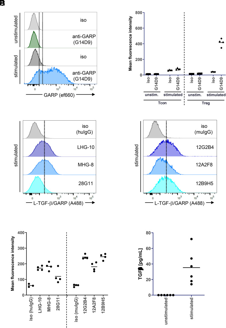
FIGURE 2.
Anti–L-TGF-β/GARP Abs bind to GARP expressed on activated Treg.
(A) Activated Treg express GARP. CD4+CD25+ T cells (Treg) were isolated from buffy coats and were activated with anti-CD3/CD28 beads for 3 d. GARP expression was determined by FACS directly after isolation (unstimulated) and after activation (stimulated) using the commercial anti-GARP AbG14D9. Histograms from one representative donor are shown. (B) Aggregate mean fluorescence intensity (MFI) data from four donors from the experiment described in (A) (Treg). In addition, Tcon from the same four donors were isolated, activated, and tested for GARP expression. (C) Anti–L-TGF-β/GARP Abs bind to activated Treg. Treg were isolated and activated as described in (A). Binding of the indicated anti–L-TGF-β/GARP Abs to activated Treg was determined by FACS. In the graph, histograms from the same donor as in (A) are shown and were grouped according to the Ab Fc part (left, human IgG; right, murine IgG). (D) Aggregate MFI data from four donors from the experiment described in (C). The experiment was done in parallel to (A) and (B) using the same donors. (E) Stimulated Treg release small amounts of TGF-β. CD4+CD25+ T cells were activated with anti-CD3/CD28 beads in serum-free medium for 3 d or left untreated. The concentration of TGF-β in supernatants was measured by ELISA (n = 6 donors).

