Abstract
Background
Dentists bend needles prior to certain injections; however, there are concerns regarding needle fracture, lumen occlusion, and sharps handling. A previous study found that a 30-gauge needle fractures after four to nine 90° bends. This fatigue life study evaluated how many 90° bends a 30-gauge dental needle will sustain before fracture when bent using a needle guide.
Methods
Two operators at Element Materials Technology, an independent testing, inspection, and certification company tested 48 30-gauge needles. After applying the needle guide, the operators bent the needle to a 90° angle and expressed the anesthetic from the tip. The needle was then bent back to a 0° angle, and the functionality was tested again. This process was repeated until the anesthetic failed to pass through the end of the needle due to fracture or obstruction. Each operator tested 24 needles (12 needles from each lot), and the number of sustained bends before the needle fracture was recorded.
Results
The average number of sustained bends before needle failure was 40.33 (95% confidence interval = 37.41–43.26), with a minimum of 20, median of 40, and a maximum of 54. In each trial, the lumen remained patent until the needle fractured. The difference between the operators was statistically significant (P < 0.001). No significant differences in performance between needle lots were observed (P = 0.504).
Conclusion
Our results suggest that using a needle guide increases the number of sustained bends before needle fracture (P < 0.000001) than those reported in previous studies. Future studies should further evaluate the use of needle guides with other needle types across a variety of operators. Furthermore, additional opportunities lie in exploring workplace safety considerations and clinical applications of anesthetic delivery using a bent needle.
Keywords: Dental Anesthesia, Needle Bending, Needle Fracture, Needle Guide, Needlestick Injury
INTRODUCTION
The needle is an essential tool for dental anesthesia, allowing precise application of anesthetic to desired targets. To access hard-to-reach injection sites, dentists may bend or curve their needles before injection [1]. When needle modification occurs, concerns may be raised regarding needle fracture, lumen occlusion, and sharps safety.
Fracturing dental needles using bends alone is difficult [2]. Robison et al. [3] found no needle fracture after applying 20 consecutive 25° angle bends to 90 needles of 25-, 27-, or 30-gauge. Oikarinen and Perkki [4] found no needle fracture when bending 13 needles 90° over a 10 mm rod or tightly winding the needles 360° around a 1.5 mm rod. Cooley and Robison [5] replicated their experiment using a total of 20 needles (10 27-gauge and 10 30-gauge) and reported no needle fractures. When applying bends directly to the hub, they discovered that the needles sustained only three bends before fracturing. When subjecting 60 needles to 10 consecutive mechanical bends at 30°, 60°, or 90° angles, Monteiro et al. [6] found no needle fracture in the 30° group and a range of 4–8 sustained bends prior to fracture in the 60°- and 90° group. The likelihood of fracture was lower in 30-gauge needles than in 25- or 27-gauge needles. To date, the effect of needle guides on needle bending has not been evaluated.
Existing needle evaluation studies have demonstrated that the lumen of a bent needle remains patent until fracture. Robison et al. [3] found that bending a needle 90° at 10 different locations along its shaft did not obstruct the lumen. In their evaluation of 60 needles bent 10 times at varying angles, Monteiro et al. [6] found that the lumen of each needle remained patent until needle fracture. While these studies did not detect a total obstruction of the anesthetic flow, a bent needle could subtly occlude the flow within its shaft. A characterization study evaluating the flow of the anesthetic through a bent needle shaft has yet to be conducted.
Sharps safety is an important consideration when bending needles. Currently, there are limited instructions and information on needle-bending protocols. Each practitioner uses their own methods and judgment when bending the needles for injections. Needles disfigured from bending may be difficult to recap, exposing dentists and their assistants to workplace safety hazards. To date, no studies have evaluated the workplace safety associated with needle bending in dentistry.
Malamed [1] mentions that a needle guide has been introduced to facilitate needle bending in clinical settings. Prior needle-bending studies employed mechanical methods of needle bending, which may not represent needle bending in clinical settings. This study aimed to evaluate the experience of a human operator bending a needle using a needle guide. The primary outcome was the number of sustained bends before failure, as defined by needle fracture or lumen obstruction.
METHODS
Testing was performed by Element Materials Technology (London, UK), an independent testing, inspection, and certification company. Two lots of 30-gauge Septoject® Evolution (Septodont, Lancaster, PA, USA) needles (#F04779AA manufactured in 2017 and #F02907AA manufactured in 2019) were evaluated using two TNN Needle Guides (TuttleNumbNow LLC, Provo, UT, USA). Articaine HCl and epinephrine anesthetic (#B16383AA; Septodont, Lancaster, PA, USA) was used.
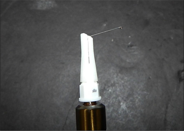
The Operator attached a needle guide to the needle hub while holding the syringe. The needle was bent to approximately 90° and released to allow springback. A few drops of anesthetic were then expressed from the tip (Fig. 1). Subsequently, the needle was bent back to approximately 0°, and the anesthetic was expressed again. The bending process was repeated until the anesthetic failed to pass through the tip of the needle due to a needle fracture or lumen occlusion. The number of sustained bends was recorded. Each operator tested 24 needles, 12 from each lot.
Fig. 1. Photograph of a needle bend using a needle guide.
A Kaplan–Meier survival analysis was performed. Log-rank tests were used to compare the results between the operators and needle lots. A two-tailed t-test for two independent means was used to compare the results of this study with those of Monteiro et al. [6].
RESULTS
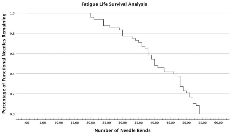
The average number of sustained bends prior to needle failure was 40.33 (95% confidence interval = 37.41–43.26), with a minimum of 20, median of 40, and a maximum of 54. In each instance, failure was due to needle fracture rather than lumen obstruction. Table 1 shows the number of sustained bends for each needle tested, grouped by lot and operator. Fig. 2 shows the fatigue life survival analysis, demonstrating the percentage of functional needles remaining over the course of needle bending. Needle operators did not detect any qualitative restriction of flow when expressing the anesthetic. In addition, no deterioration of the needle guide was observed. Statistical analysis revealed a significant difference between the operators (P < 0.001). No statistical difference in performance was observed between the needle lots (P = 0.504).
Table 1. Number of sustained bends for each needle specimen, grouped by lot and operator.
| Specimen | Operator | Lot | Sustained Bends |
|---|---|---|---|
| 1 | Operator 1 | F04779AA | 54 |
| 2 | Operator 1 | F04779AA | 39 |
| 3 | Operator 1 | F04779AA | 48 |
| 4 | Operator 1 | F04779AA | 41 |
| 5 | Operator 1 | F04779AA | 54 |
| 6 | Operator 1 | F04779AA | 53 |
| 7 | Operator 1 | F04779AA | 51 |
| 8 | Operator 1 | F04779AA | 40 |
| 9 | Operator 1 | F04779AA | 47 |
| 10 | Operator 1 | F04779AA | 48 |
| 11 | Operator 1 | F04779AA | 52 |
| 12 | Operator 1 | F04779AA | 54 |
| 13 | Operator 1 | F02907AA | 48 |
| 14 | Operator 1 | F02907AA | 39 |
| 15 | Operator 1 | F02907AA | 51 |
| 16 | Operator 1 | F02907AA | 40 |
| 17 | Operator 1 | F02907AA | 54 |
| 18 | Operator 1 | F02907AA | 43 |
| 19 | Operator 1 | F02907AA | 52 |
| 20 | Operator 1 | F02907AA | 40 |
| 21 | Operator 1 | F02907AA | 50 |
| 22 | Operator 1 | F02907AA | 52 |
| 23 | Operator 1 | F02907AA | 46 |
| 24 | Operator 1 | F02907AA | 49 |
| 25 | Operator 2 | F04779AA | 24 |
| 26 | Operator 2 | F04779AA | 36 |
| 27 | Operator 2 | F04779AA | 21 |
| 28 | Operator 2 | F04779AA | 20 |
| 29 | Operator 2 | F04779AA | 24 |
| 30 | Operator 2 | F04779AA | 20 |
| 31 | Operator 2 | F04779AA | 30 |
| 32 | Operator 2 | F04779AA | 38 |
| 33 | Operator 2 | F04779AA | 30 |
| 34 | Operator 2 | F04779AA | 48 |
| 35 | Operator 2 | F04779AA | 49 |
| 36 | Operator 2 | F04779AA | 38 |
| 37 | Operator 2 | F02907AA | 36 |
| 38 | Operator 2 | F02907AA | 33 |
| 39 | Operator 2 | F02907AA | 30 |
| 40 | Operator 2 | F02907AA | 24 |
| 41 | Operator 2 | F02907AA | 38 |
| 42 | Operator 2 | F02907AA | 48 |
| 43 | Operator 2 | F02907AA | 29 |
| 44 | Operator 2 | F02907AA | 35 |
| 45 | Operator 2 | F02907AA | 26 |
| 46 | Operator 2 | F02907AA | 37 |
| 47 | Operator 2 | F02907AA | 34 |
| 48 | Operator 2 | F02907AA | 43 |
Fig. 2. Fatigue life survival analysis representing the percentage of functional needles remaining over the course of needle bending.
DISCUSSION
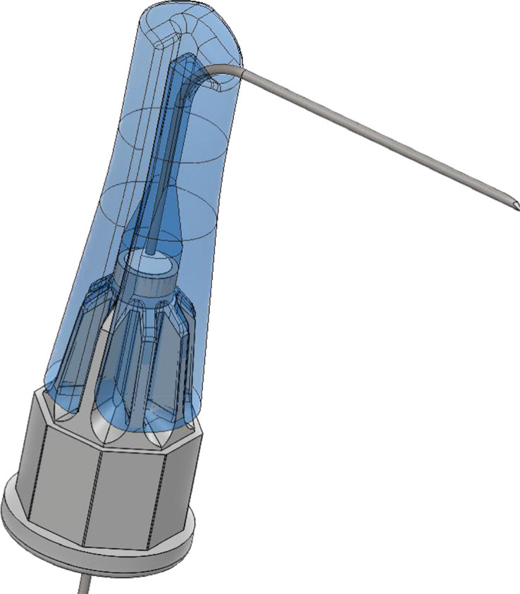
This study reported a greater number of sustained bends prior to needle fracture than those reported in previous studies on 30-gauge needles. The average number of sustained bends using the needle guide (40.33 bends) was significantly higher (P < 0.000001) than that reported by Montiero et al. [6] for the same needle gauge (5 bends). This difference could be attributed to the needle guide or differences in the bending methods. A needle guide is expected to extend the fatigue life by distributing the mechanical stress of each bend across the needle shaft (Fig. 3). The bending methods used by Monteiro et al. [6] differed in location and nature from the present study. In their study, a machine apparatus placed the bend 5 mm away from the hub. In contrast, the needle guide places the bend mid-shaft at 12.5 mm from the hub, possibly allowing the needle to exhibit greater compliance when bending. The human operators in this study might have applied a bending force less than that applied by the machine apparatus. Future studies could control for these intricacies to directly assess the impact of needle guide design, applied force, and bend location on needle bending.
Fig. 3. Graphical representation of a needle within the TNN needle guide. TNN, TuttleNumbNow.
For each needle, failure occurred due to needle fracture rather than lumen obstruction. Nonetheless, the flow of anesthetic through a bent needle may be partially obstructed. In our study, partial lumen occlusion was not evaluated, and anesthetic flow was not characterized. A needle bend may introduce turbulent flow or increase hydraulic pressure by reducing the luminal surface area. Turbulent flow and anesthetic pressure are known to play a role in anesthesia outcomes [7,8]. Therefore, future studies should more fully characterize the flow of anesthesia through a bent needle and evaluate its clinical implications.
The number of bends sustained in this experiment exceeds the requirements of routine clinical practice. For dental injections requiring a bent needle, a practitioner is likely to bend the needle one to three times. Bending a needle more than 20 times is extremely unlikely in clinical practice, which was the minimum number of sustained bends observed in this study. A previous study [6] without a needle guide demonstrated needle breakage after four 90° bends. Therefore, the use of a needle guide may place the risk of needle fracture far from the demands of clinical needle bending.
Our statistical analysis revealed a significant difference in the results between the operators (P < 0.001). This distinction could be attributed to the variation in the applied force between operators. Assessing a wide variety of practitioners may be helpful to ensure safe needle bending across different operators. Additionally, future studies should assess a wide variety of needle types that can be used in conjunction with a needle guide in clinical settings.
As discussed previously, sharps safety is an important consideration when bending needles. Each dentist has a personal responsibility to ensure the safety of needle bending in their practice. Future quality improvement studies could assess the safety of applying and removing a needle guide as well as workplace safety considerations for recapping bent needles.
The strengths of this study were as follows: a large sample size of needles tested, direct clinical relevance, novelty, and reliability of testing performed by an independent company. However, this study had some limitations, because it did not fully characterize the anesthetic flow, evaluate various needle types, or directly compare manual bending with and without the use of a needle guide.
In summary, this is the first study to evaluate the fatigue life of needle bending using a needle guide. The number of sustained bends prior to needle failure in this study is significantly higher than those observed in previous studies. With a minimum of 20 sustained bends, the results were well above the routine clinical demands for needle bending. Future studies will be helpful to further explore the impact of using a needle guide to bend needles.
ACKNOWLEDGEMENTS
Testing was performed at Element Materials Technology, an independent testing, inspection, and certification company. This study was financially supported by Septodont. Septodont provided materials for this study. Septodont and Element Materials Technology expressed permission for the release of the study data and were not involved in writing the report or its submission for publication. Gregory K. Tuttle, a coauthor of this study, holds a patent interest in the TNN Needle Guide. None of the other coauthors have any financial or proprietary interests to disclose.
Footnotes
- Jared Joseph Tuttle: Formal analysis, Writing – original draft, Writing – review & editing.
- Andrew Doran Davidson: Formal analysis, Writing – review & editing.
- Gregory Kent Tuttle: Conceptualization, Project administration, Supervision, Validation, Writing – review & editing.
References
- 1.Malamed SF. Handbook of Local Anesthesia. 7th edition. Amsterdam: Elsevier Health Sciences; 2019. p. 106. [Google Scholar]
- 2.Zimmerman RK. 27-gauge short. J Acad Gen Dent. 1975;23:37. [PubMed] [Google Scholar]
- 3.Robison SF, Mayhew RB, Cowan RD, Hawley RJ. Comparative study of deflection characteristics and fragility of 25-, 27-, and 30-gauge short dental needles. J Am Dent Assoc. 1984;109:920–924. doi: 10.14219/jada.archive.1984.0246. [DOI] [PubMed] [Google Scholar]
- 4.Oikarinen VJ, Perkki K. A metallurgic and bacteriological study of disposable injection needles in dental and oral surgery practice. Proc Finn Dent Soc. 1975;71:147–161. [PubMed] [Google Scholar]
- 5.Cooley RL, Robison SF. Comparative evaluation of the 30-gauge dental needle. Oral Surg Oral Med Oral Pathol. 1979;48:400–404. doi: 10.1016/0030-4220(79)90065-3. [DOI] [PubMed] [Google Scholar]
- 6.Monteiro MAO, Antunes ANDG, Basting RT. Physical, chemical, mechanical, and micromorphological characterization of dental needles. J Dent Anesth Pain Med. 2021;21:139–153. doi: 10.17245/jdapm.2021.21.2.139. [DOI] [PMC free article] [PubMed] [Google Scholar]
- 7.Teja KV, Ramesh S, Battineni G, Vasundhara KA, Jose J, Janani K. The effect of various in-vitro and ex-vivo parameters on irrigant flow and apical pressure using manual syringe needle irrigation: Systematic review. Saudi Dent J. 2022;34:87–99. doi: 10.1016/j.sdentj.2021.12.001. [DOI] [PMC free article] [PubMed] [Google Scholar]
- 8.Smith GN, Walton RE, Abbott BJ. Clinical evaluation of periodontal ligament anesthesia using a pressure syringe. J Am Dent Assoc. 1983;107:953–956. doi: 10.14219/jada.archive.1983.0357. [DOI] [PubMed] [Google Scholar]


